
Zoothamnium wangi Ji et al ., 2005
|
publication ID |
https://doi.org/10.5281/zenodo.278023 |
|
DOI |
https://doi.org/10.5281/zenodo.6190289 |
|
persistent identifier |
https://treatment.plazi.org/id/03E687ED-FF9C-885B-B3A6-8E69FDE7FE76 |
|
treatment provided by |
Plazi |
|
scientific name |
Zoothamnium wangi Ji et al ., 2005 |
| status |
|
Zoothamnium wangi Ji et al., 2005
( Fig. 3 View FIGURE 3 ; Table 1)
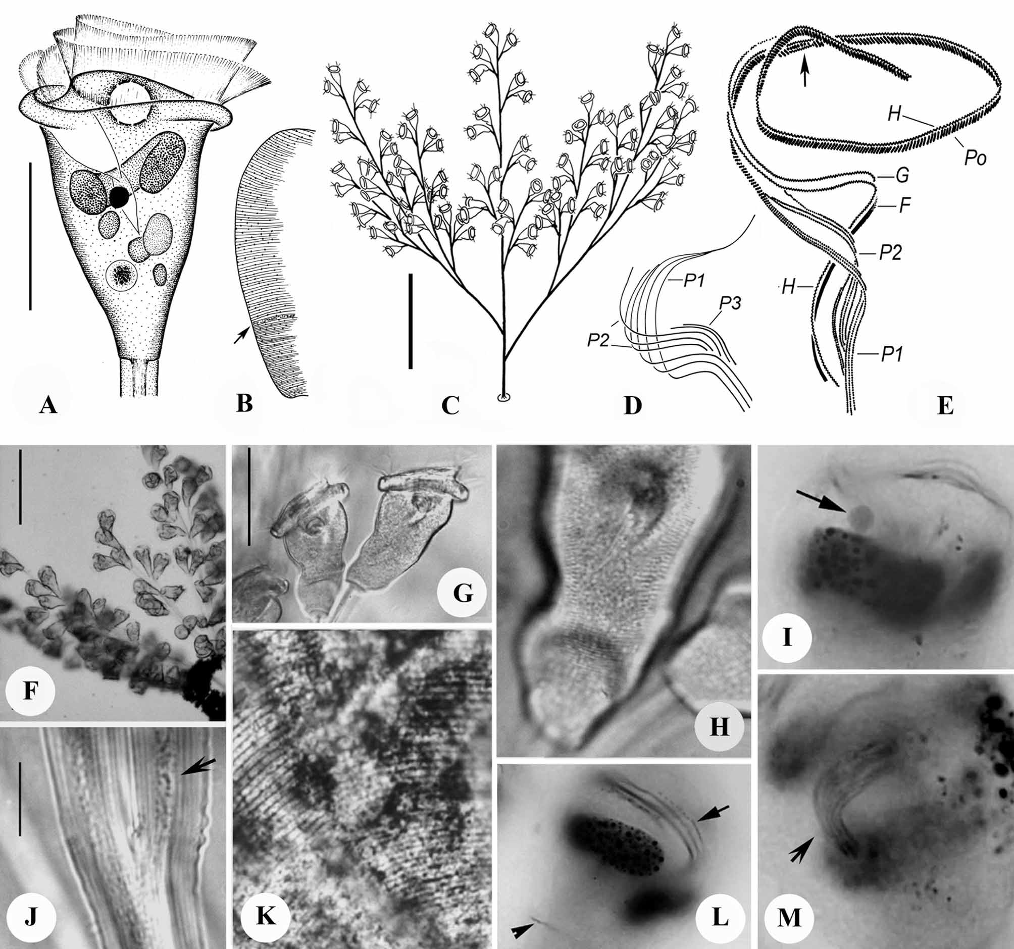
Emended diagnosis. Marine Zoothamnium with colony up to 1 mm high: alternately branched with few (2–4), very long secondary branches. Zooids campanulate to subconical, measuring. 65–90 × 45–55 µm in vivo. Peristomial lip thick, without medial, circumferential infolding when expanded. Macronucleus C-shaped, transversely oriented, located in oral half of cell. Pellicular striations closely spaced; 70–85 silverlines lying between peristomial lip and trochal band and 38–50 between trochal band and scopula. P3 consists of two ciliary rows, with row 2 offset slightly toward cytostome relative to row 1.
Redescription. Colony moderately large, up to 1 mm high and containing ca. 100 zooids, with broad, fanshaped outline ( Figs. 3 View FIGURE 3 E, 4A). Secondary stalks branching alternately from primary stalk, with most basal 2–3 secondary stalks growing to the same length as primary stalk ( Figs. 3 View FIGURE 3 E, 4A). Cortex of stalk colorless and transparent, with smooth surface and fine longitudinal striations in interior. Diameter of stalk ranging 20 µm in primary stalk to 10 µm in distal branches; spasmoneme measuring 4–10 µm in diameter, containing densely arranged mitochondria ( Fig. 3 View FIGURE 3 J).
Zooids campanulate to subconical, 65–90 µm (n=4) long, widest at peristomial lip, which measures 45–55 µm (n=4) in diameter when fully expanded ( Figs. 3 View FIGURE 3 A, G). Peristomial lip thick, without secondary circumferential infolding; epistomial disc moderately elevated above peristomial lip ( Figs. 3 View FIGURE 3 A, G). Pellicular striations closely spaced and not prominent, visible only above × 400 magnification ( Fig. 3 View FIGURE 3 H); surface of body appearing uniformly smooth at low magnifications.
Cytoplasm transparent and slightly grayish, occasionally containing a few gray or yellowish food vacuoles of uneven size (2–10µm) in center of body ( Figs. 3 View FIGURE 3 A, 4B, C). Single contractile vacuole in adoral position beneath epistomial disc and near dorsal wall of infundibulum. Macronucleus C-shaped, transversely oriented, surrounding micronucleus and lower half of infundibulum ( Figs. 3 View FIGURE 3 A, I, L). Micronucleus spherical, located adoral to center of macronucleus ( Figs. 3 View FIGURE 3 A, I, arrow).
Oral infraciliature as shown in Figures 3 View FIGURE 3 D, E, L, M. Haplo- and polykinety making one and one-quarter circuits around peristome and one additional circuit within infundibulum. Epistomial membrane short, located at entrance into infundibulum ( Fig. 3 View FIGURE 3 E, arrow). Germinal kinety running parallel to haplokinety in adoral half of infundibulum ( Fig. 3 View FIGURE 3 L, arrow). Infundibular polykineties 1 and 2 consisting of three rows of kinetosomes each; P3 consisting of two rows. All rows of Pl terminating adstomally at level of cytostome; rows of P2 terminating adstomally at adstomal curvature of P1. Rows of P2 terminating abstomally without merging with P1; abstomal 1/4 of row 3 of P2 diverging from other rows of P2 ( Figs. 3 View FIGURE 3 D, E). Row 2 of P3 displaced adstomally for short distance relative to row 1 ( Fig. 3 View FIGURE 3 D).
Trochal band consisting of band of dikinetids encircling cell at point 3/4 of distance from peristome to scopula ( Fig. 3 View FIGURE 3 L, arrowhead).
Silverline system consisting of closely spaced, parallel, transverse silverlines, which are spaced relatively wider apart near peristome ( Figs. 3 View FIGURE 3 B, K); 70–85 silverlines present between peristome and trochal band, 38–50 between trochal band and scopula. Pellicular pores staining faintly, numerous, randomly arranged along silverlines.
Remarks. Zoothamnium wangi is occasionally found in eutrophic marine waters and can be identified easily by the distinctive branching pattern of the colony, shape and size of zooids, number of silverlines, and the pattern of kinetosome rows in P3. Zoothamnium plumula , Z. commune , Z. alternans , and Z. xuianum resemble Z. wangi in having an alternately branched stalk and a relatively thin peristomial lip without a medial circumferential infolding. In the present study, it was observed that young colonies of Z. plumula very much resemble mature colonies of Z. wangi in outline as well as shape and size of zooids and, therefore, cannot be distinguished from the latter by characters visible in the living organisms. However, the two species can be distinguished easily in preparations stained with silver nitrate or protargol by the total number of silverlines ( 108–135 in Z. wangi vs. 72–87 in Z. plumula ) and the number of ciliary rows in P3 ( 2 in Z. wangi vs. 3 in Z. plumula ).
Zoothamnium commune resembles Z. wangi in most living characters. However, Z. wangi has more silverlines from the peristomial lip to the trochal band (70–85 vs. 59–70) and a different number of kinetosome rows in P3 ( 2 in Z. wangi vs. 3 in Z. commune ) ( Ji et al. 2006 b). Zoothamnium alternans has large macrozooids that form acetabuliform telotrochs on the primary stalk, which are lacking in Z. wangi , plus its microzooids are considerably smaller than zooids of Z. wangi (40–56 × 26–32 µm vs. 65–90 × 45–55 µm) ( Ji et al. 2006 b). Zoothamnium xuianum has also much smaller zooid size than that of Z. wangi (30–50 × 20–40 µm vs. 65–90 × 45–55 µm), thus it can be well distinguished from the latter (Sun et al. 2005)
Living colonies of two other congeners, Z. thiophilum Stiller, 1946 and Z. hentscheli Kahl, 1935 , also resemble Z. wangi , and their infraciliatures and silverline systems remain unknown, preventing a comparison with those of Z. wangi . However, both of these species are freshwater forms ( Kahl 1935; Stiller 1946) and Z. wangi seems to be found exclusively in marine habitats.
No known copyright restrictions apply. See Agosti, D., Egloff, W., 2009. Taxonomic information exchange and copyright: the Plazi approach. BMC Research Notes 2009, 2:53 for further explanation.
|
Kingdom |
|
|
Phylum |
|
|
Class |
|
|
Order |
|
|
Family |
|
|
Genus |
Zoothamnium wangi Ji et al ., 2005
| Ji, Daode, Shin, Mann Kyoon, Choi, Joong Ki, Clamp, John C., Al-Rasheid, Khaled A. S. & Song, Weibo 2011 |
Z. thiophilum
| Stiller 1946 |
Z. hentscheli
| Kahl 1935 |