
Zoothamnium nii Ji et al ., 2005
|
publication ID |
https://doi.org/10.5281/zenodo.278023 |
|
DOI |
https://doi.org/10.5281/zenodo.6190286 |
|
persistent identifier |
https://treatment.plazi.org/id/03E687ED-FF9E-8857-B3A6-88B6FCA4FC12 |
|
treatment provided by |
Plazi |
|
scientific name |
Zoothamnium nii Ji et al ., 2005 |
| status |
|
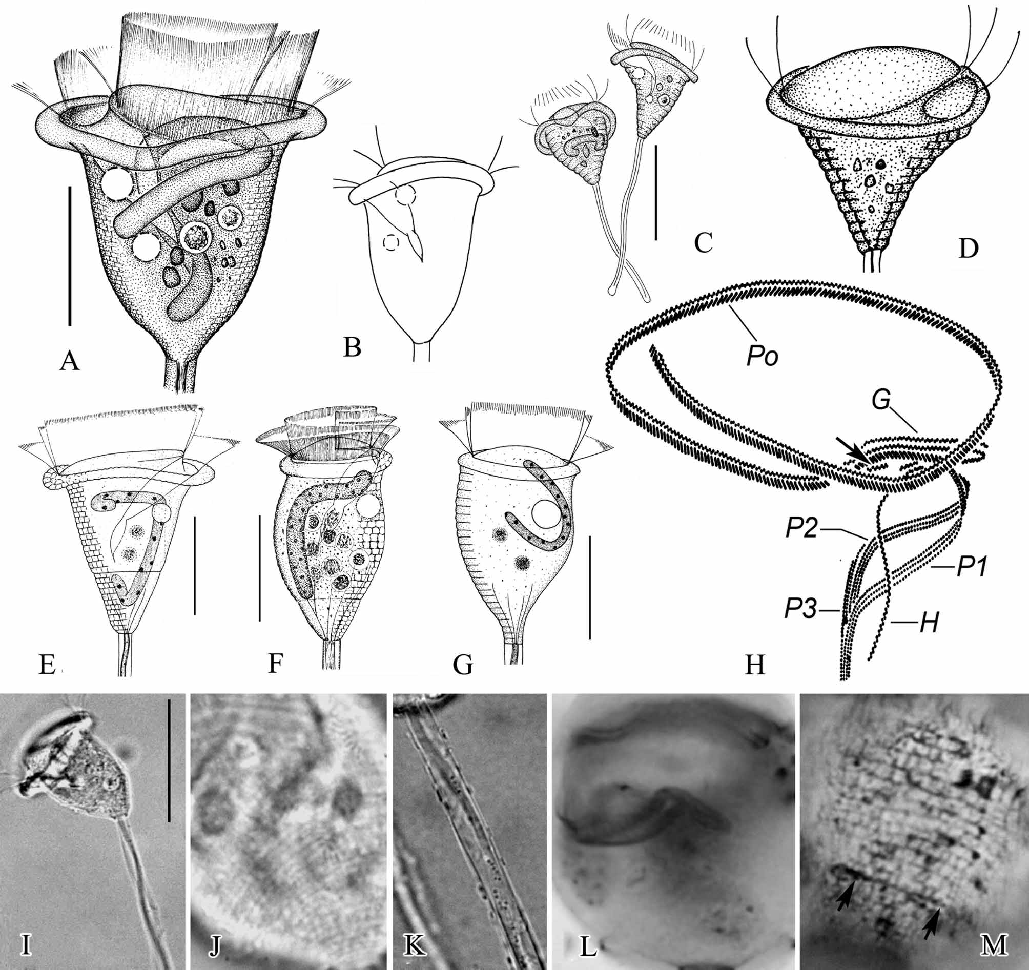
Zoothamnium nii Ji et al., 2005
( Fig. 2 View FIGURE 2 ; Table 1)
Emended diagnosis. Marine Zoothamnium with colony up to 1 mm high; moderately elongate, median primary stalk giving rise to secondary stalks in regular alternate series in single plane,. Zooids elongate, vase-shaped, measuring 70–80 × 40–50 µm in vivo. Peristomial lip extremely thick, with medial, circumferential infolding in peristomial lip when expanded (“double-layered”). Macronucleus C-shaped, transversely oriented, located in oral half of cell. Pellicular striations indistinct at lower magnifications; 45–60 silverlines lying between peristomial lip and trochal band and 20–30 between trochal band and scopula. P3 consists of three rows that are equal in length and parallel to each other, row 1 slightly separated from rows 2 and 3 for most of length, but all three rows converging adstomally.
Redescription. Colony moderately large, up to 1 mm high and containing 30–50 zooids, with broad, diamondshaped outline. Secondary stalks up to 500 µm long, branching off primary stalk in regular alternate series. Basal secondary stalks longest, with stalks decreasing progressively in length toward tip of colony; secondary stalks branching to form short, dichotomous tertiary branches bearing 2 zooids or single tertiary branches ( Figs. 2 View FIGURE 2 E, J, K). Stalk with smooth surface and diameter of 12 µm in basal portion of primary stalk, narrowing to 9 µm at distal ends of tertiary stalks. Spasmoneme with diameter of 3.5 µm in primary stalk and 2.5 µm at distal ends of tertiary stalks; band of mitochondria visible as dark granules measuring 0.5 × 0.8 µm, winding along helical path just beneath surface of spasmoneme.
Zooids elongate, bell-shaped, measuring 70–80 × 40–50 µm (n=12) ( Figs. 2 View FIGURE 2 A, I, L, O, P). Body deeply constricted below peristomial lip, which is indented by prominent, medial, circumferential infolding (“double-layered”). Maximum width of cell usually at peristomial lip, but occasionally in oral third of body as well; epistomial disc moderately elevated above peristomial lip ( Figs. 2 View FIGURE 2 A, I, O, P). Pellicular striations easily detectable above × 400 magnification ( Figs. 2 View FIGURE 2 O, P), but surface of body appears completely smooth at low magnifications ( Fig. 2 View FIGURE 2 L). Telotroch discoid, measuring 55–65 µm x 40 µm ( Fig. 2 View FIGURE 2 R).
Cytoplasm of zooids colorless or slightly gray, filled with tiny (0.5 µm diameter), dense granules and usually containing a few large (5–10 µm in diameter), transparent or gray food vacuoles typically located in center of body ( Figs. 2 View FIGURE 2 A, L). Single contractile vacuole in adoral position beneath epistomial lip and near dorsal wall of infundibulum. Macronucleus C-shaped, transversely oriented, surrounding micronucleus and lower half of infundibulum ( Figs. 2 View FIGURE 2 A, M, N).
Oral infraciliature as shown in Figures 2 View FIGURE 2 B, H, N. Haplo- and polykinety making one and one-quarter circuits around peristome and one additional circuit within infundibulum. Haplokinety and polykinety parallel on peristome, diverging within infundibulum to lie on opposite walls ( Fig. 2 View FIGURE 2 H). Epistomial membrane short, located at entrance into infundibulum ( Fig. 2 View FIGURE 2 H, arrow). Germinal kinety running parallel to haplokinety in adoral half of infundibulum ( Fig. 2 View FIGURE 2 N, arrow). Each of three infundibular polykineties consisting of three rows of kinetosomes. All rows of Pl terminating adstomally at level of cytostome; rows of P2 terminating adstomally at adstomal curvature of P1. Rows of P2 terminating abstomally without merging with P1; abstomal 1/4 of row 3 of P2 diverging from other rows of P2 ( Figs. 2 View FIGURE 2 B, H).. All rows of P3 terminating adstomally at point slightly beyond adstomal end of P2 and abstomally at point approximately 1/3 of distance from adstomal to abstomal end of P2. Row 1 of P3 converging with other tow reasons near adstomal end, separated slightly from rows 2 and 3 along remainder of length ( Fig. 2 View FIGURE 2 B).
Trochal band consisting of band of dikinetids encircling cell at point 2/3 of distance from peristome to scopula ( Fig. 4 View FIGURE 4 H, arrows).
Silverline system consisting of closely spaced, parallel, transverse silverlines ( Fig. 2 View FIGURE 2 Q); 45–60 silverlines present between peristome and trochal band, 20–30 between trochal band and scopula. Silverlines near peristome spaced farther apart than those around scopula, and pellicular pores sparsely distributed alongside silverlines ( Fig. 2 View FIGURE 2 Q).
Remarks: In general, zooids of alternately branched species of Zoothamnium such as Z. alternans , Z. niveum and Z. plumula , lack a medial, circumferential infolding on the peristomial lip (“single-layered”), and zooids with an infolding around the peristomial lip (“double-layered”) are more typical of dichotomously branched species such as Z. duplicatum , Z. mucedo , and Z. maximum ( Bauer-Nebelsick et al. 1996; Ji & Song 2004; Ji et al. 2005 a, 2006 b). Therefore, Z. nii has an unusual complex of characters (i.e. the alternately branched stalk and the infolded peristomial lip of the zooid), by which it can be distinguished easily from all the congeners mentioned above.
Another species described very recently, Z. alrasheidi Ji et al., 2009 , also possesses this set of characteristics during its early development because its zooids have an infolded peristomial lip and its young colonies are alternately branched. However, a colony of Z. alrasheidi in its early stages of development can be distinguished from Z. nii by its larger zooids (80–120 × 50–60 µm vs. 70–80 × 40–50 µm) and a differing pattern of rows in P3 of the infundibular infraciliature (rows 2 and 3 combined into a single band and divergent from row 1 vs. all three rows distinct and parallel) (Ji et al. 2009).
Kahl (1933, 1935) reported two populations of Zoothamnium with an infolded peristomial lip as Z. duplicatum ( Figs. 2 View FIGURE 2 C, D) and Zoothamnium sp. ( Figs. 2 View FIGURE 2 F, G) respectively. However, both of them had an alternately branched stalk, which is distinctly different from the dichotomous branching pattern characteristic of Z. duplicatum ( Kahl 1933; Ji et al. 2005 a), but very similar to present species. Accordingly, we suggest that both of these Zoothamnium populations described by Kahl be regarded as synonyms of Z. nii .

No known copyright restrictions apply. See Agosti, D., Egloff, W., 2009. Taxonomic information exchange and copyright: the Plazi approach. BMC Research Notes 2009, 2:53 for further explanation.
|
Kingdom |
|
|
Phylum |
|
|
Class |
|
|
Order |
|
|
Family |
|
|
Genus |