
Sayimys, WOOD, 1937
|
publication ID |
https://doi.org/10.2478/if-2019-0023 |
|
persistent identifier |
https://treatment.plazi.org/id/03E6B817-8C37-617D-FC54-FD337E66FB54 |
|
treatment provided by |
Diego |
|
scientific name |
Sayimys |
| status |
|
Sayimys giganteus LÓPEZ- ANTOÑANZAS, SEN et SARAÇ, 2004
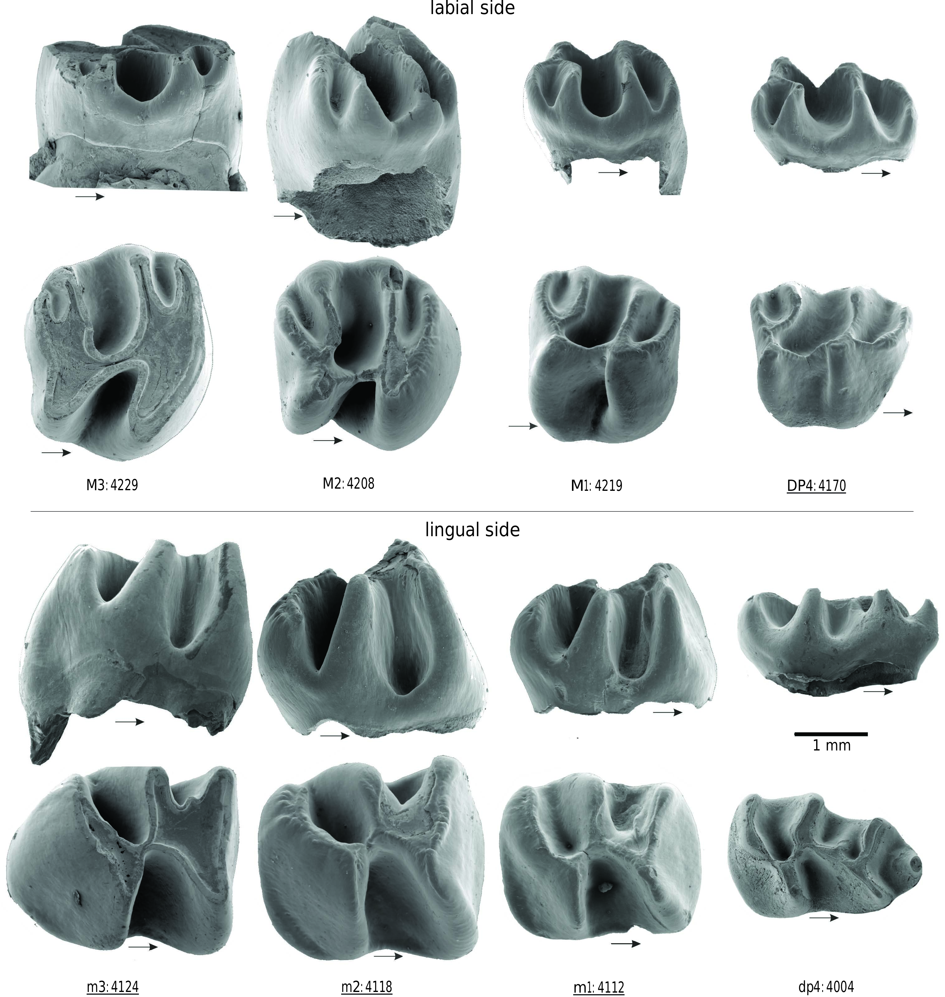
Text-figs 9–11 View Text-fig View Text-fig View Text-fig
H o l o t y p e. KSK1–100, a fragmentary left maxilla with P4–M1 ( López-Antoñanzas et al. 2004: fig. 5C, D).
Ty p e l o c a l i t y. Keseköy 1 (Kizilcahamam, Ankara,
Turkey).
Ty p e h o r i z o n. Güvem Formation.
A g e. Early Miocene, MN 3 ( López-Antoñanzas et al. 2004), 20.2 ± 2.2 Ma ( Krijgsman et al. 1996, Wessels et al. 2001).
M a t e r i a l. 335 cheek teeth, 261 isolated and 74 in partial jaws.
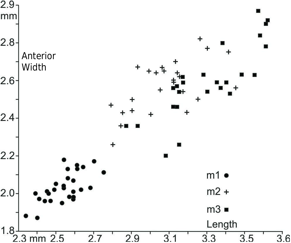
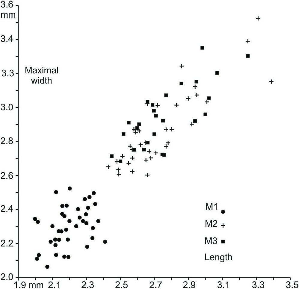
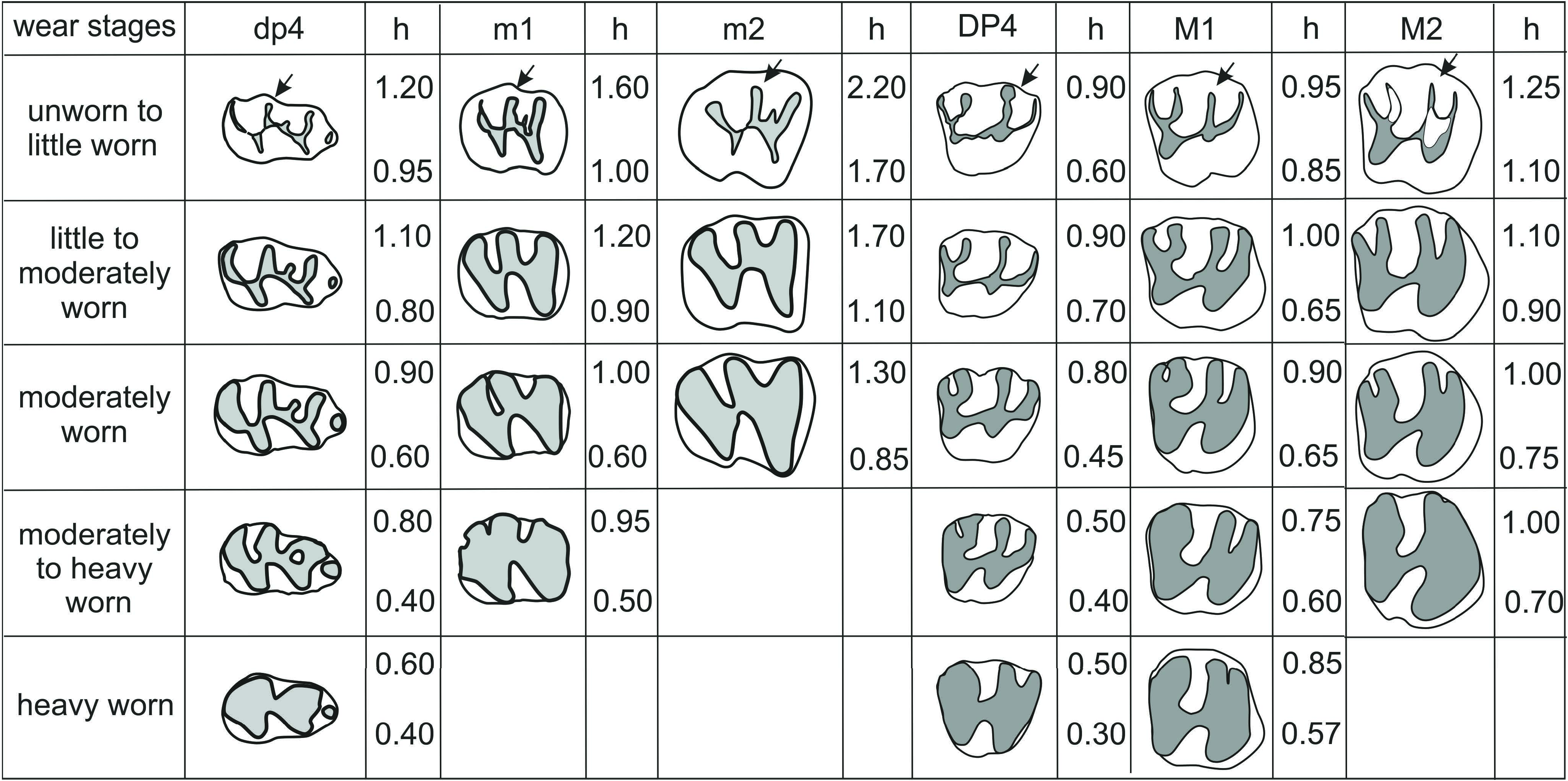
M e a s u r e m e n t s. Length-width measurements in Tab. 1, Text-figs 4 View Text-fig and 5 View Text-fig , crown-height measurements in Text-fig. 8. View Text-fig
O r i g i n a l d i a g n o s i s. “ Sayimys species of large size with the m3 lacking a constriction in the posterolophid; dp4 with a metalophulid II [= the mesolophid of Text-fig. 3 View Text-fig ] connecting with the metaconid or nearly reaching it. DP4 without metaflexus, but with a well-developed paraflexus; P4 with a long posteroloph connecting to the paracone and with the anteroloph joining the protocone; upper molars with a paraflexus longer than the metaflexus” (in López- Antoñanzas et al. 2004).
E m e n d e d d i a g n o s i s. Large-sized species of Sayimys . On the dp4 the mesolophid can be: 1) absent, 2) present and half the length of the metalophulid or, 3) long and connected to a mesostylid. The rounded anteroconid is isolated; protoconid is extending less far labially than the hypoconid, mesolophid connecting at right angles to the ectolophid. The hypolophid connected to ectolophid at obtuse or about right angle.
Some p4 have a posterior spur at the junction of the hypolophid and the ectolophid, a stylid can be present on the postero-labial cingulid (lower arrow on Text-fig. 9 View Text-fig , no 4058), as well as a vestigial mesolophid (upper arrow on Text-fig. 9 View Text-fig , no 4058). The metalophulid of lower molars is wide near the protoconid, the mesoflexid is short and v-shaped. The m2 and m3 are lacking a constriction in the posterolophid; a vestigial mesolophid is present in some lower molars (upper arrows on Text-fig. 9 View Text-fig , no 4064 and 4084); m1 and m2 with a distinct postero-labial cingulid.
DP4 with paraflexus reaching the protocone, paraflexus longer than metaflexus; metaloph connecting at right angles to the posteroloph. P4 with metaloph of variable length and connected to the protoloph; a small hypocone or a hypocone spur is present, protocone is large.
Upper molars with a well-developed paraflexus and metaflexus, paraflexus longer than metaflexus reaching the protocone; M1 with a metaloph connecting at right angles to the posteroloph, M2 and M3 with a metaloph connecting not at right angles to the posteroloph.
D e s c r i p t i o n. dp4. The dp4 has a low crown, its anteroconid is isolated, circular to slightly ovoid and positioned on the mid-line of the tooth. The metaconid is located slightly anterior to the protoconid. The metalophulid of some slightly worn specimens appears to be vaguely S-shaped ( 4023 in Text-fig. 9 View Text-fig ) or can have a convex anterior side ( 4013 in Text-fig. 9 View Text-fig ). A mesolophid is present in 43 out of 48 specimens, but vestigial in 12 of these ( 4006 in Text-fig. 9 View Text-fig ). In the other 31 it is long, that is, extending halfway or more toward the lingual tooth margin. In six of the specimens, a small mesostylid is present; in three of these specimens a (vestigial) mesolophid is also present. In slightly worn specimens, the lingual end of the mesolophid is wide, suggesting a mesostylid. With increasing wear, the mesolophid eventually connects to the metalophulid to form an enamel lake (see the line drawings in Text-fig. 8 View Text-fig ). The mesolophid basin extends to the mid-line of the tooth or slightly beyond, but withdraws toward the lingual border with increasing wear. The mesoflexid has no sill at its entrance; it is deeper and wider than the metaflexid (Textfig. 11), whereas the mesolophid basin is shallower than the mesoflexid. The hypolophid connects at approximately a right angle to the ectolophid. In unworn condition, the connection of the hypolophid to the ectolophid is narrow, but it widens with increasing wear. The constriction at the connection of the hypolophid to the anterior arm of the hypoconid is more persistent. The hypoflexid is deep; it has no sill at the tooth margin. The posterolophid is curved. It is narrow at its connection with the hypoconid. An elevation in the posterolophid of unworn dp4 marks a hypoconulid. In some specimens, a low sill at the lingual border is present in the metaflexid, and an enamel lake appears with increasing wear. The metaflexid is deepest close to the hypoconid. The development of the postero-labial cingulid varies from weak to distinct.
p4. In little worn specimens, protoconid, metaconid and entoconid form a V-shape. The metalophid connects at right angles to the posterior side of the protoconid. In some premolars, the metalophid is constricted where it meets the protoconid. In five out of 18 specimens, a vestigial mesolophid is present ( Text-fig. 9 View Text-fig , 4058, arrowed). The ectolophid plus a short and a transversely oriented hypolophid connect to the entoconid; a posterior spur may be present. The postero-labial cingulum is distinct in most teeth; in eight a prominent cingulid-stylid is present (4058 on Text-fig. 9 View Text-fig , lower arrow).
m1. The m1 has a high crown. The anterior margin of the occlusal surface is slightly concave, and becomes straighter and a bit convex with increasing wear. The metalophulid is broad at the protoconid-side and tapers lingually. In general, the metalophid extends less far lingually than the hypolophid. In some slightly worn specimens a small short mesolophid is present ( Text-fig. 9 View Text-fig , 4064, arrowed), it is separated from the metalophulid by a narrow, very shallow valley, and fuses with the metalophulid at an early stage of wear. The mesoflexid does not reach the mid-line of the tooth; in occlusal view it is V-shaped when slightly worn, changing into U-shape with wear. The mesoflexid is shorter but deeper than the metaflexid ( Text-fig. 11 View Text-fig ). The metaflexid may have a low sill at the tooth border. The connection between hypolophid and ectolophid becomes more confluent with wear, but the posterior margin of the hypolophid remains offset with respect to the ectolophid. The connection of the hypolophid to the anterior arm of the hypoconid is narrow. The hypoflexid is deeper than the mesoflexid and metaflexid. In two specimens, a low sill is present in the hypoflexid. In little-worn specimens, the posterolophid is slightly curved; in unworn specimens, a hypoconulid can be recognized as an elevation of the posterolophid. The metaflexid extends to the mid-line of the tooth, or slightly beyond it. In about half of the specimens, a prominent sill is present and connects to the posterolophid and hypolophid with wear, to form an enamel lake. The postero-labial cingulid is distinct in most specimens, weak in some specimens.
m2. The m2 and m1 are similar in morphology, but differ in length and width ( Text-fig. 4 View Text-fig ), moreover, in unworn condition the crown of the m2 is higher ( Text-fig. 11 View Text-fig ). Also, there are some minor morphological differences. On m2, the connection between ectolophid and hypolophid is wider, and these lophids are oriented more in line than on m1. As on m1, a short mesolophid branch may be present in unworn specimens, but it fuses with the metalophulid in an early stage of wear. The mesoflexid is similar to that in the m1, but a weak sill is present in more specimens. In m2, the hypoflexid is more in line with the metaflexid compared to the m1. The connection between hypolophid and anterior arm of the hypoconid is narrow. The posterolophid is less curved than in the m 1 in early stages of wear, and when moderately worn, it is straight and obliquely oriented. The posterolophidhypoconid connection is wide. The sill at the lingual tooth border in the metaflexid is similar to m1; with increasing wear, the metaflexid becomes narrower at its lingual margin.
m3. The m3 is somewhat larger ( Text-fig. 4 View Text-fig ) and higher than m2. It can be distinguished by its narrow convex posterior margin; its posterior width is smaller than its anterior width. The metaconid lies anterior to the labial part of the protoconid. The apex of the unworn protoconid lies slightly more toward the mid-line of the molar relative to that of the hypoconid. The protoconid is oblique and the hypoconid is transverse. In little-worn m3, a short mesolophid is present, but it merges with the metalophulid at an early wear stage. The mesoflexid has a V-shape, which does not reach the mid-line of the tooth. In lingual view, the mesoflexid is deeper than the metaflexid ( Text-fig. 11 View Text-fig ); a minor sill may be present. The metaflexid is long and has a prominent sill. The straight hypolophid is confluent, in line with the ectolophid. The connection of the hypoconid to the ectolophid is narrow. The hypoflexid is deeper than the meso- and metaflexid. The posterolophid is more or less straight and directed obliquely. The postero-labial cingulid
can be absent, weak or distinct, with a stylid positioned on it
in some specimens ( Text-fig. 9 View Text-fig , 4084, arrowed).
DP3. This small low-crowned element has a variable morphology. A complex specimen is in Text-fig. 10 View Text-fig (no 4292); it shows an obliquely oriented loph, which is wider
and highest at its labial end. An anterocone is present. Posteriorly, a low cingulum is separated from the oblique loph by a valley. Other specimens show a single sharp cusp with traces of a subsidiary cusp and cingulum.
DP4. The DP4 has a low crown, with morphology showing minor variation ( Text-fig. 10 View Text-fig ). Some DP4 have a thickening in the anteroloph at the anterocone position, in others, this is absent. The labial end of the anteroloph shows a thickening in some specimens, in others it tapers. In some unworn specimens, the paracone seems isolated, due to a low protoloph. Unworn specimens show that the apex of the protocone is located more labially than the hypocone; protocone and hypocone are higher than paracone and metacone. With increasing wear, the labial end of the posteroloph connects to the metacone, due to the presence of a sill in the metaflexus. In late stages of wear, the metaloph merges with the posteroloph, and the protoloph with the anteroloph. In such specimens, the anterior fused loph is wider than the posterior fused loph.
P4. We are not certain about the homology of the elements of the P4; we follow here Text-fig. 3 View Text-fig , where a relatively unreduced specimen is shown. Text-fig. 10 View Text-fig shows the morphological variety from strongly reduced (no 4192) to relatively weakly reduced (no 4203). The strongly-reduced specimens show two cusps, the protocone and the paracone, connected by a transversely oriented loph and a small posterior spur. Its hypoflexus is at the posterior side, while the paraflexus is absent. In the weakly-reduced specimen, the mesoflexus and paraflexus are both present, the hypoflexus is located lingually. Protoloph and posteroloph (= metaloph) are both well-developed.
M1. The M1 is high-crowned. Its protoloph is straight or slightly curved; the posterior rim is convex and confluent with the labial margin of the endoloph. The apex of the protocone of unworn specimens is located more internally than that of the hypocone; protocone and hypocone are higher than metacone and paracone. The posteroloph is straight and obliquely oriented; its tip at the labial end is often slightly curved. The metaloph is connected about halfway to the posteroloph. The straight paraflexus extends to the protocone, decreasing in length with wear. In labial view ( Text-fig. 11 View Text-fig ), the paraflexus is as low and wide as the metaflexus. In heavily worn specimens, protoloph and anteroloph are fused ( Text-fig. 8 View Text-fig ). The mesoflexus extends beyond the mid-line of the tooth, is deepest internally, and a low sill is present near the tooth margin. The sill in the short metaflexus is not prominent; an enamel lake does not form in all worn specimens. The metaloph and posteroloph are completely fused in worn specimens ( Text-fig. 8 View Text-fig ). The fused anterior loph is wider than the fused posterior loph.
M2. The M2 is distinctly wider and longer, and in unworn condition, higher than the M1 ( Text-figs 5 View Text-fig , 6 View Text-fig , 11 View Text-fig ), and it is widest anteriorly. The lophs and cusps are confluently fused into ridges that are arc-shaped. Anteroloph and protocone form the smoothly bow-shaped anterior side of the molar. The protoloph is transverse or slightly oblique. The posterior side of the protoloph curves smoothly and confluently into the labial margin of the endoloph. There is some variation in the configuration of the posteroloph and metaloph, related to stages of wear. The posteroloph seems an offshoot from the curved loph formed by metacone, metaloph and hypocone. In other specimens, the posteroloph seems to split smoothly from the metaloph. Unlike in M1, the metaloph and posteroloph do not meet at a right angle. The paraflexus extends to the protocone in lightly worn specimens, but decreases in length with wear ( Text-fig. 8 View Text-fig ). Usually a low sill is present, and the paraflexus can form an enamel lake in later wear stages. The paraflexus and metaflexus are about equally deep and wide ( Text-fig. 11 View Text-fig ); both are shallower than the mesoflexus and hypoflexus. The mesoflexus extends beyond the mid-line. A sill is present in the mesoflexus; the deepest part of the mesoflexus lies internally. The hypoflexus is deeper than the mesoflexus, but it is shorter, not reaching the mid-line of the tooth. A sill is present in the metaflexus of most specimens; in advanced stages of wear, the metaflexus forms an enamel lake, in later stages of wear the metaloph and posteroloph are completely fused.
M3. The isolated M3 is similar in length and width to the M2 ( Text-fig. 5 View Text-fig ), but higher in an unworn condition. The posterior side of the M3 is relatively narrow, resulting in an anterior/posterior width ratio that is larger in the M3 than in the M2 ( Text-fig. 7 View Text-fig ). The M3 has a smooth convex anterior margin, and a concave posterior margin. Similar to the M2, the ridges formed by the confluently fused lophs and cusps are more or less arc-shaped, but in the M3 arcs are stronger bowed, the labial tips more posteriorly located. The metaloph meets halfway to the posteroloph, the posteroloph seems a posterior offshoot from the curved ridge formed by the metacone, metaloph and hypocone. In some specimens and stages of wear, the metaloph seems an anterior offshoot of a confluent arc formed by posteroloph and hypocone. The morphology of the occlusal surface and depth of flexi are like those in the M2.
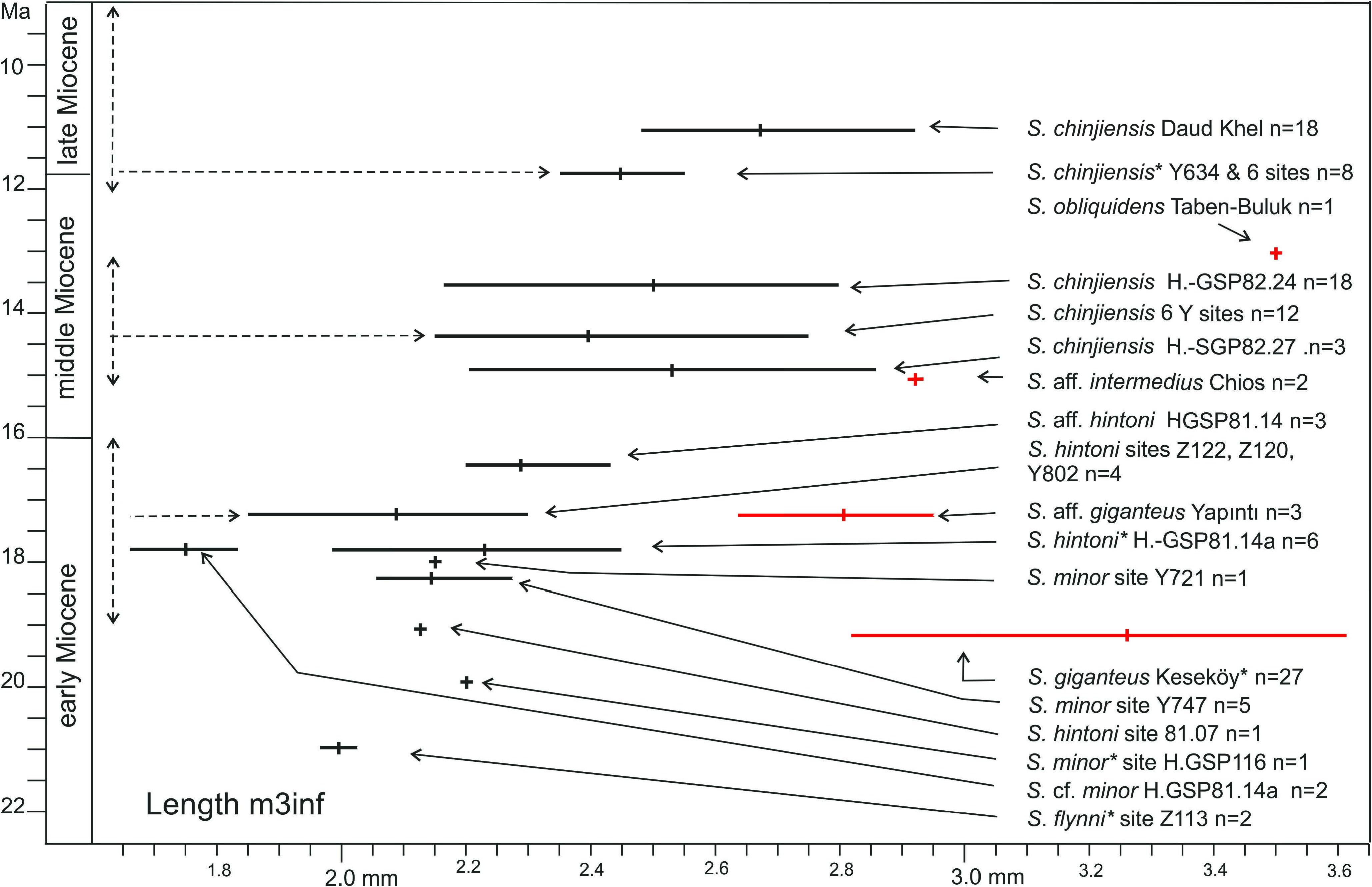
Va r i a t i o n i n s i z e. Most samples of Sayimys are small. The two large samples of Sayimys previously known are Daud Khel and H.-GSP 82.24 in Pakistan ( Munthe 1980, de Bruijn et al. 1989). Together with the Keseköy assemblage, these allow a reliable estimate of the variation in length and width. Length-width variation (size) of rodent teeth is discussed in Freudenthal and Cuenca Bescos (1984). These authors introduced a simple coefficient of variation, V’ = 100r/m in which r is the range (the difference between the maximum and minimum) and m is the midpoint between maximum and minimum. Tab. 2 shows values for V’ for the three large samples of Sayimys ; note that the value of V’ for undivided assemblages of first and second molars is large.
The values for V’ calculated from the three large Sayimys populations may be used to estimate a size range if only a small sample is available. To show this, we have estimated a size range for the dp4 and m1–2 of three small samples: Sayimys intermedius from its type locality Al Jadidah, S. assarrarensis from As Sarrar and Sayimys aff. intermedius from Chios (data in López-Antoñanzas and Sen 2004, López- Antoñanzas et al. 2005). Tab. 3 shows that the estimated ranges for the m1–2 and dp4 of Al Jadidah and As Sarrar are largely overlapping. The Chios m1–2 is similar in size to that from Al Jadidah and As Sarrar, but its dp4 is much larger and outside the range of the other two sites.
The calculated ranges are indicative; in this exercise the available measurements are assumed to be close to the midpoint of the constructed range, a presumption not necessarily true.
Text-figs 12 View Text-fig and 13 View Text-fig show the ranges and averages of the length of dp4 and m3 of most species of Sayimys compiled from the literature; length ranges of DP4 and M3 show a similar picture, but are not shown here. Diagrams of first and second molars are not helpful, because these molars are rarely separated in the literature, though different in size: large assemblages have therefore very large ranges, whereas those of small assemblages are erratic.
The height of the crown has been measured in the Keseköy assemblage; for this measure, the height of the entoconid in the lower teeth and of the paracone in the upper teeth has been selected. Results are shown in Text- fig. 8 for different wear classes. Even in the large Keseköy assemblage, the number of un- or lightly worn cheek teeth is low.
Va r i a t i o n i n m o r p h o l o g y.Presenceandlength of the mesolophid in the dp4 is considered an important species-specific character. This feature varies strongly among the 47 specimens of Sayimys from Keseköy; it can be complete, extending to the lingual tooth margin, relatively short, or absent. Absence of a mesolophid in a single or a few dp4 is thus not a species-specific character. A mesostylid is present in some dp4 from Keseköy either as a separate tubercle or recognizable as a thickening of the lingual end of the mesolophid.
The development of the anteroloph in P4, considered species-specific by some authors, occurs in the Keseköy assemblage with four character stages: a) well-developed, extending from the protocone to halfway the anterior tooth margin, paraflexus well-developed ( Text-fig. 10 View Text-fig , no 4203); b) short, extending to about a quarter of the anterior tooth margin, paraflexus short; c) anteroloph and paraflexus absent ( Text-fig. 10 View Text-fig , nos 4192 and 4193) and d) anteroloph absent, but with a small isolated cusp in front of the anteroloph (similar to YAP 1037 in Text-fig. 10 View Text-fig ). This suggests that this character has little value if only a few premolars are available. In general, the morphology of the P4 is rather variable ( Text-fig. 10 View Text-fig ), and better not used as a species diagnostic element.
A variable character is the postero-labial cingulid of lower molars ( Text-fig. 3 View Text-fig ). In the Keseköy material it is present in most m1 and m2, but variable in size. In general, it is better developed in m1 than in m2, and weak or absent in m3. A stylid on the postero-labial cingulid is present in 11 of the 18 p4 from Keseköy, and three specimens of Sayimys baskini (= S. minor) from Pakistan ( Baskin 1996); it seems present in the single specimen of S. obliquidens from Taben- Buluk and in ‘ S. perplexus ’ (= S. sivalensis) from Hari Talyangar, India ( Wood 1937), but it has not been described in other species.


No known copyright restrictions apply. See Agosti, D., Egloff, W., 2009. Taxonomic information exchange and copyright: the Plazi approach. BMC Research Notes 2009, 2:53 for further explanation.