
Trachelissa Aurivillius, 1912
|
publication ID |
https://doi.org/ 10.11646/zootaxa.3793.5.1 |
|
publication LSID |
lsid:zoobank.org:pub:5DD6186E-B04B-481F-9EEC-176C6A683C45 |
|
DOI |
https://doi.org/10.5281/zenodo.6143107 |
|
persistent identifier |
https://treatment.plazi.org/id/03E6D21C-FF8F-3965-A0E7-FA1BFA88FCC3 |
|
treatment provided by |
Plazi |
|
scientific name |
Trachelissa Aurivillius, 1912 |
| status |
|
Trachelissa Aurivillius, 1912 View in CoL
Trachelissa Aurivillius, 1912: 449 View in CoL nomen novum pro Trachelia Audinet-Serville, 1834 non Trachelia Scopoli, 1769 , Aves [ Quintino & Monné, 2010: 67 nomen protectum]; Monné, 2005: 646; Monné, 2012: 62; Bezark & Monné, 2013: 196.
Trachelia Audinet-Serville, 1834: 25 .
Hippias Gistel, 1848 , nomen oblitum.
Loxodromus Gistel, 1848 , nomen oblitum.
Type-species. Trachelissa pustulata ( Audinet-Serville, 1834) View in CoL by subsequent designation by Chevrolat in D`Orbigny, 1848: 625.
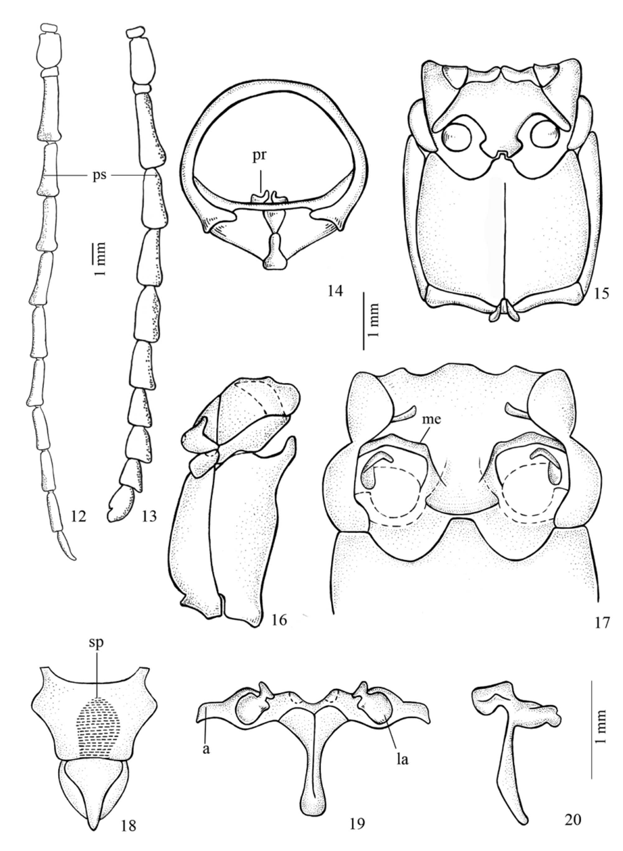
Body narrowed, moderately elongate. Head ( Figs 1–4 View FIGURES 1 – 11 ) as wide as long; sides of head slightly converging and narrowing at apical third; evident to indistinct groove present behind the eyes. Frons short, vertical, transverse and slightly depressed, with irregularly distributed, dense punctures and short dense setae. Vertex slightly depressed, with longitudinal carina between the antennal tubercles. Antennal alveoli near insertion of mandibles. Antennal tubercles distant from each other, bloated at base, prominent at apex and rounded. Submentum with fine dense punctures, and long erect setae. Eyes finely faceted; lower ocular lobe subtriangular. Gena projected, triangular, shorter than mandibles, rounded at apex and densely punctate. Mandibles robust with bifid apices. Apical segment of palps ( Figs 7, 8 View FIGURES 1 – 11 ) cylindrical and rounded at apex. Labium: apical segment longest. Antennae ( Figs 12, 13 View FIGURES 12 – 20 ) distinctly sexually dimorphic; male with antennae filiform, 11- or 12–segmented; females ( Fig. 13 View FIGURES 12 – 20 ) with antennae serrate (more evident in apical segments), 11–segmented. Outer margin of segments III–XI with sensory system ( Figs 12, 13 View FIGURES 12 – 20 ), divided by longitudinal carina. Scape short, cylindrical, slightly enlarged apically; scar at base, fine, shallow, dense punctures at base, more sparsely punctate at apex. Pedicel short, transverse, without projections. Antennomere III depressed or with longitudinal carina, more evident in males. Antennomere III in females about 2 times length of scape. Antennomere XI in females appendiculate. Antennomere XII in males curved at apex.
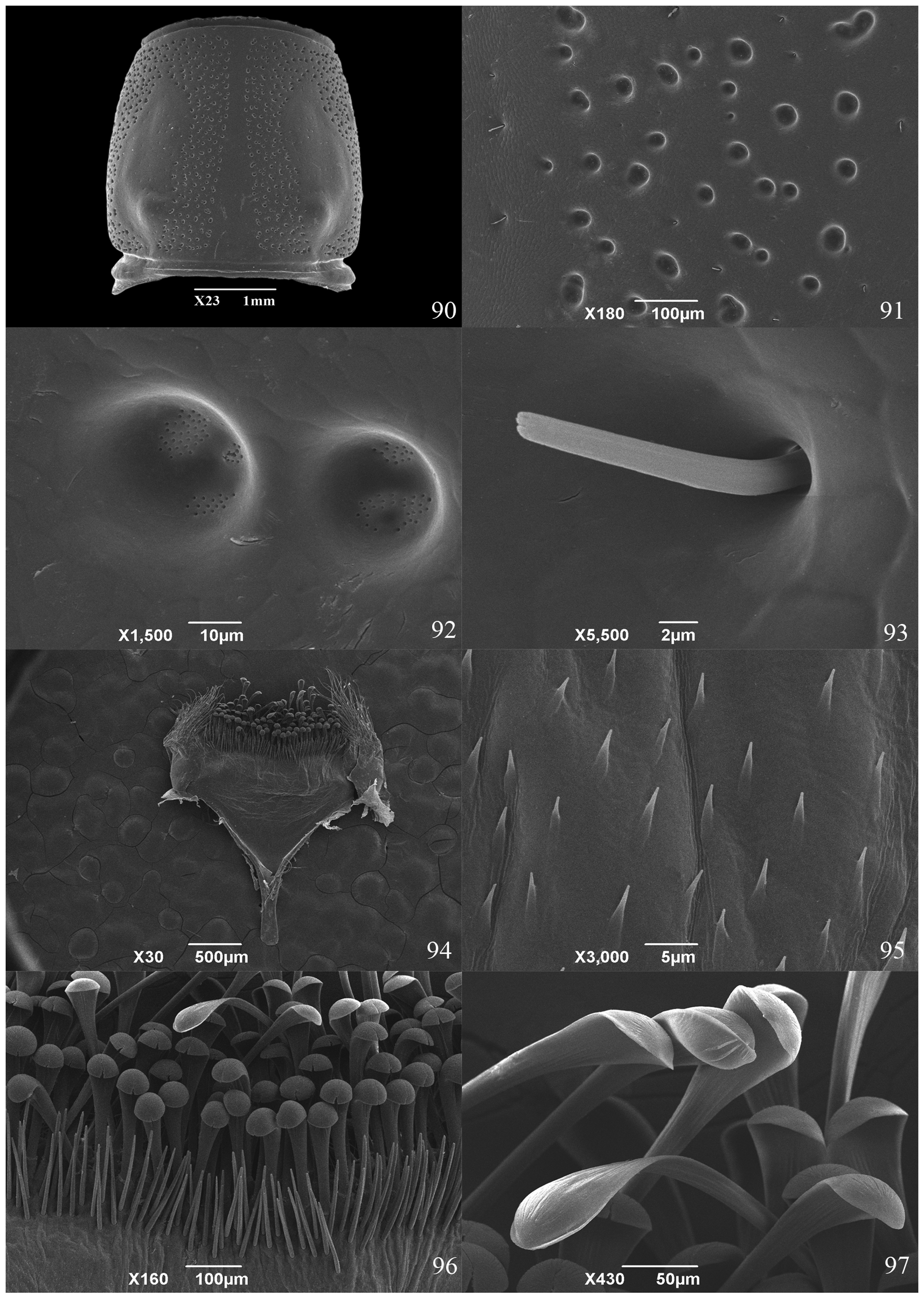
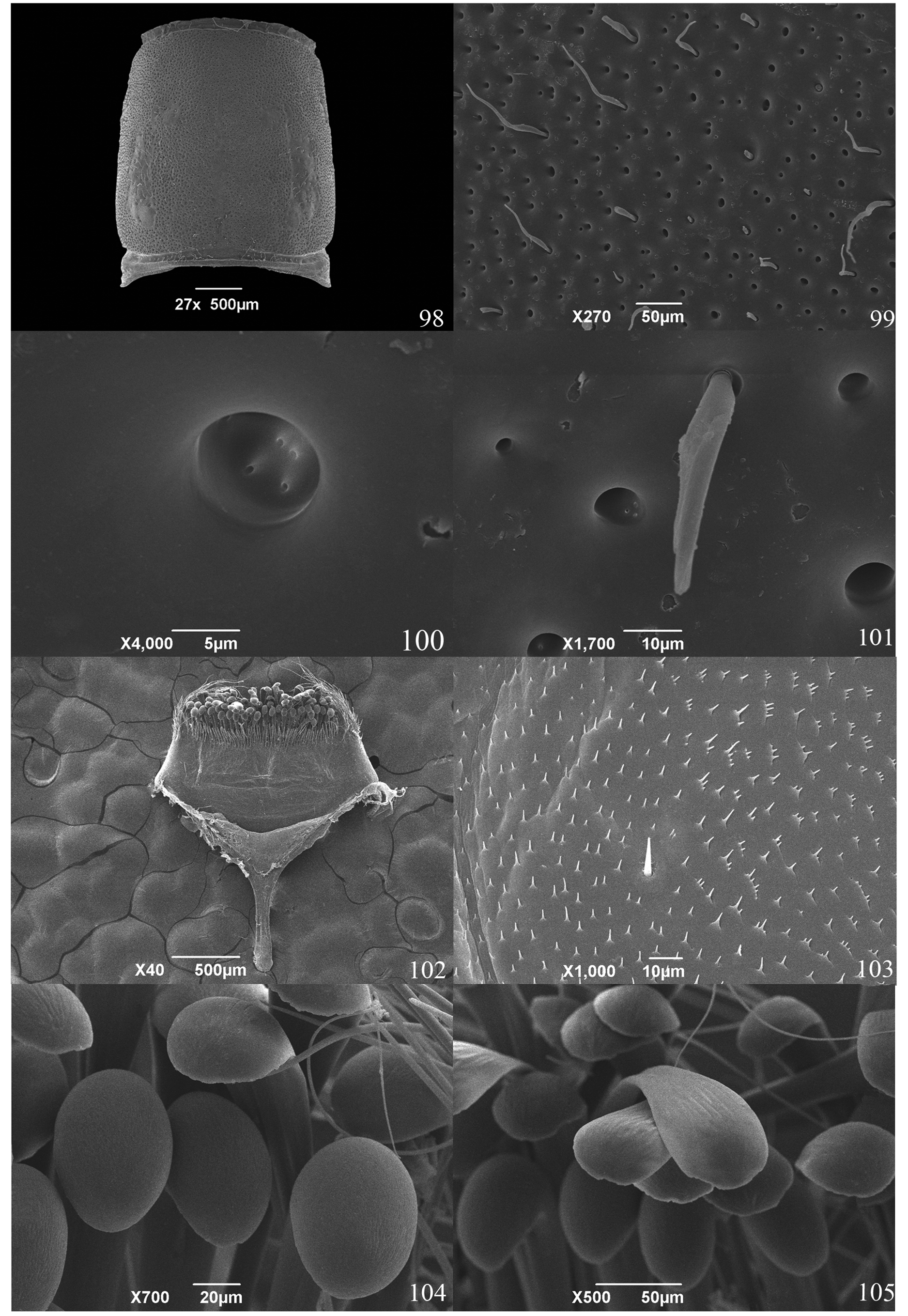
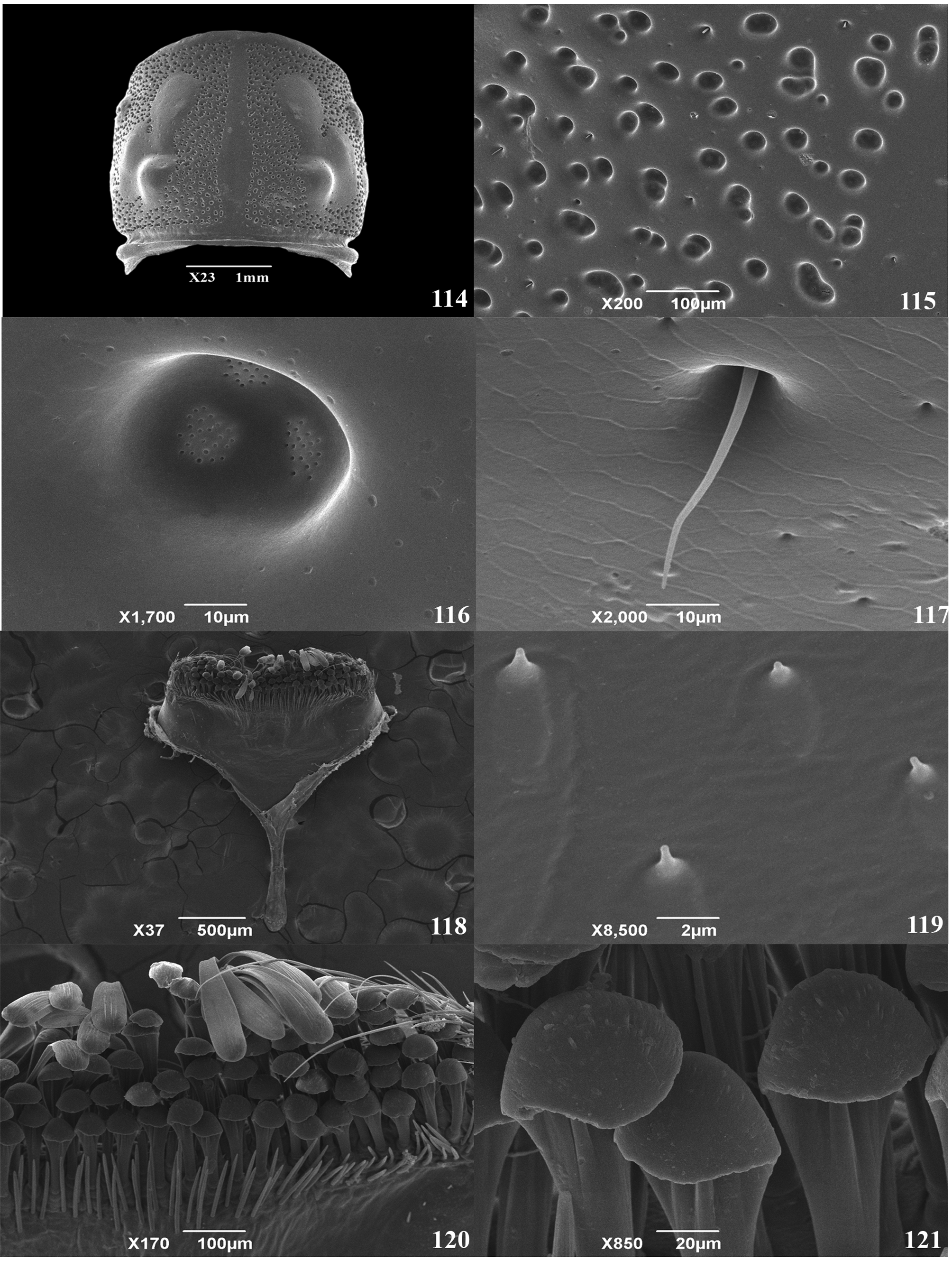
Prothorax ( Figs 14 View FIGURES 12 – 20 , 90 View FIGURES 90 – 97 , 98 View FIGURES 98 – 105 , 106 View FIGURES 106 – 113 , 114 View FIGURES 114 – 121 ) about as long as wide, subparallel at sides or slightly rounded. Basal and apical margin slightly narrowed; basal margin as wide as apical margin. Prosternum with basal margin slightly raised, smooth, with fine transverse striae; long, erect yellowish setae; intercoxal process as broad as 1/3 of procoxal cavity. Anterior coxal cavities rounded, slightly angulate at sides, with longitudinal grooves, open behind. Mesosternum with fine dense punctures; short dense whitish setae. Intercoxal process as wide as mesocoxa, with lateral projections that fit into notches on coxae, and medially notched at apex to fit metasternum. Intermediate coxal cavities open laterally. Scutellum small, triangular, flattened or slightly depressed. Mesepisternum, mesepimeron, metasternum, and metepisternum with long dense whitish setae. Mesepimeron transverse, with elevated lateral margin. Metasternum ( Figs 15, 16 View FIGURES 12 – 20 ) convex with fine dense punctures; metasternal suture almost reaching anterior margin. Metepisternum narrowed and with a little-developed glandular pore. Elytra about 3 times as long as prothorax; slightly wider than prothorax on the base. Humeri rounded, not projected, and slightly depressed.
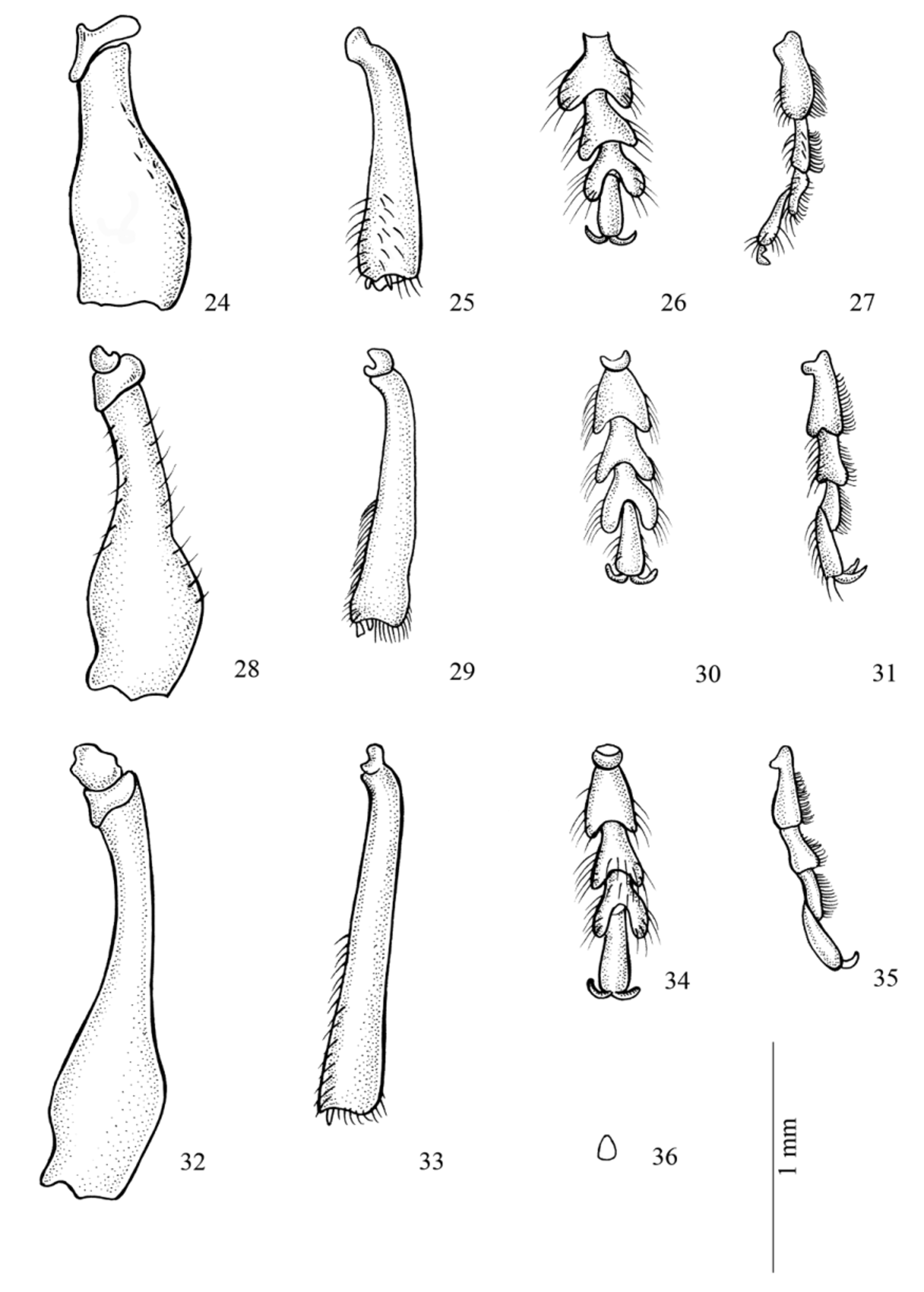
Legs ( Figs 24–36 View FIGURES 24 – 36 ) short; hind legs about 1/3 as long as prolegs. Coxa with fine, shallow dense punctures and long dense whitish setae; procoxa and mesocoxa rounded. Trochanter with tuft of whitish setae. Femora ( Figs 24, 28, 32 View FIGURES 24 – 36 ) pedunculate, clavate, with curved spine at inner apex. Tibiae ( Figs 25, 29, 33 View FIGURES 24 – 36 ) depressed, slightly expanded to apex. Tibial spurs short and subequal in length. Tarsi short ( Figs 26, 27, 30, 31, 34, 35 View FIGURES 24 – 36 ); metatarsomere I about 1/ 3 longer than II. Empodium ( Fig. 36 View FIGURES 24 – 36 ) reduced. Sternites I–V with fine dense punctures; long and short, dense whitish setae; sternite V with short setae near apical margin.
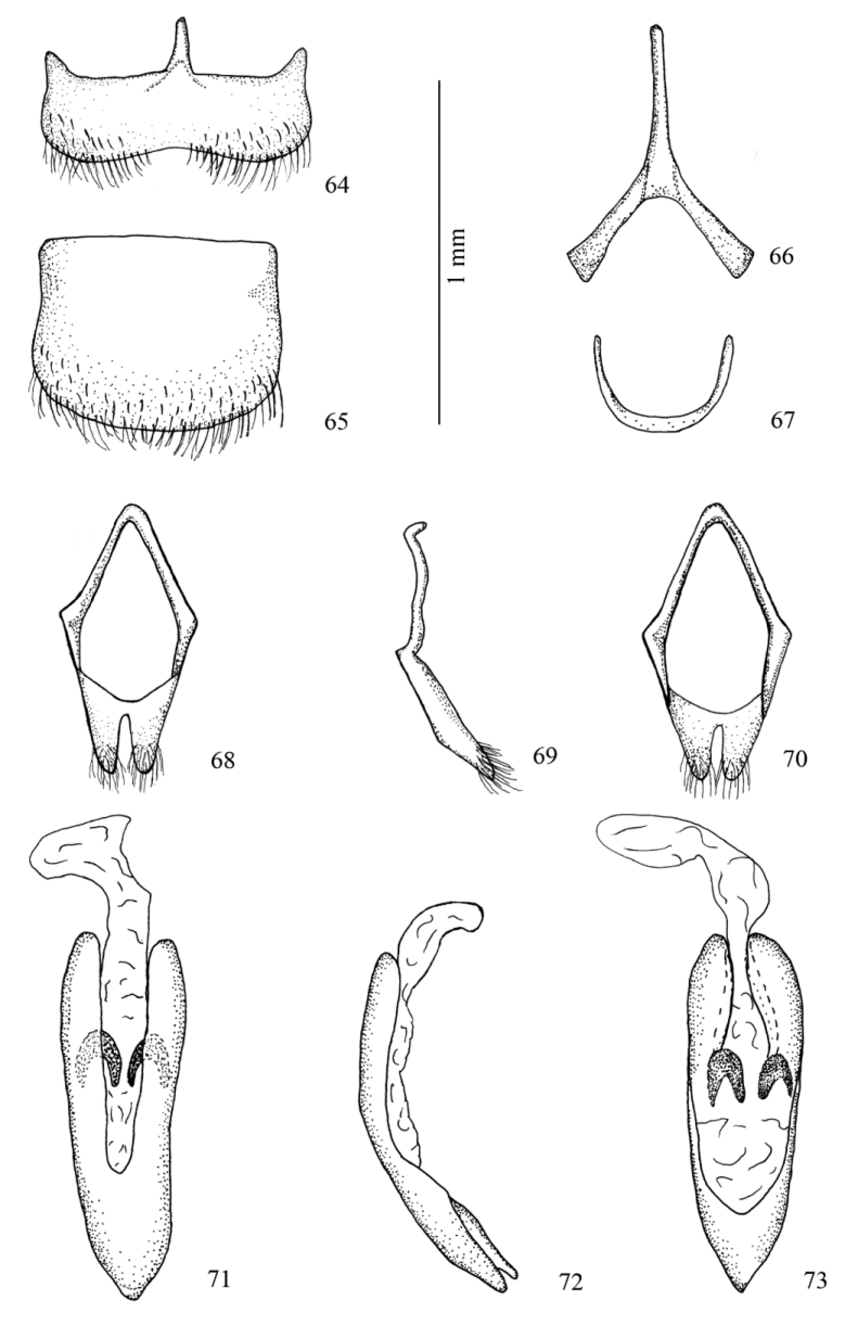
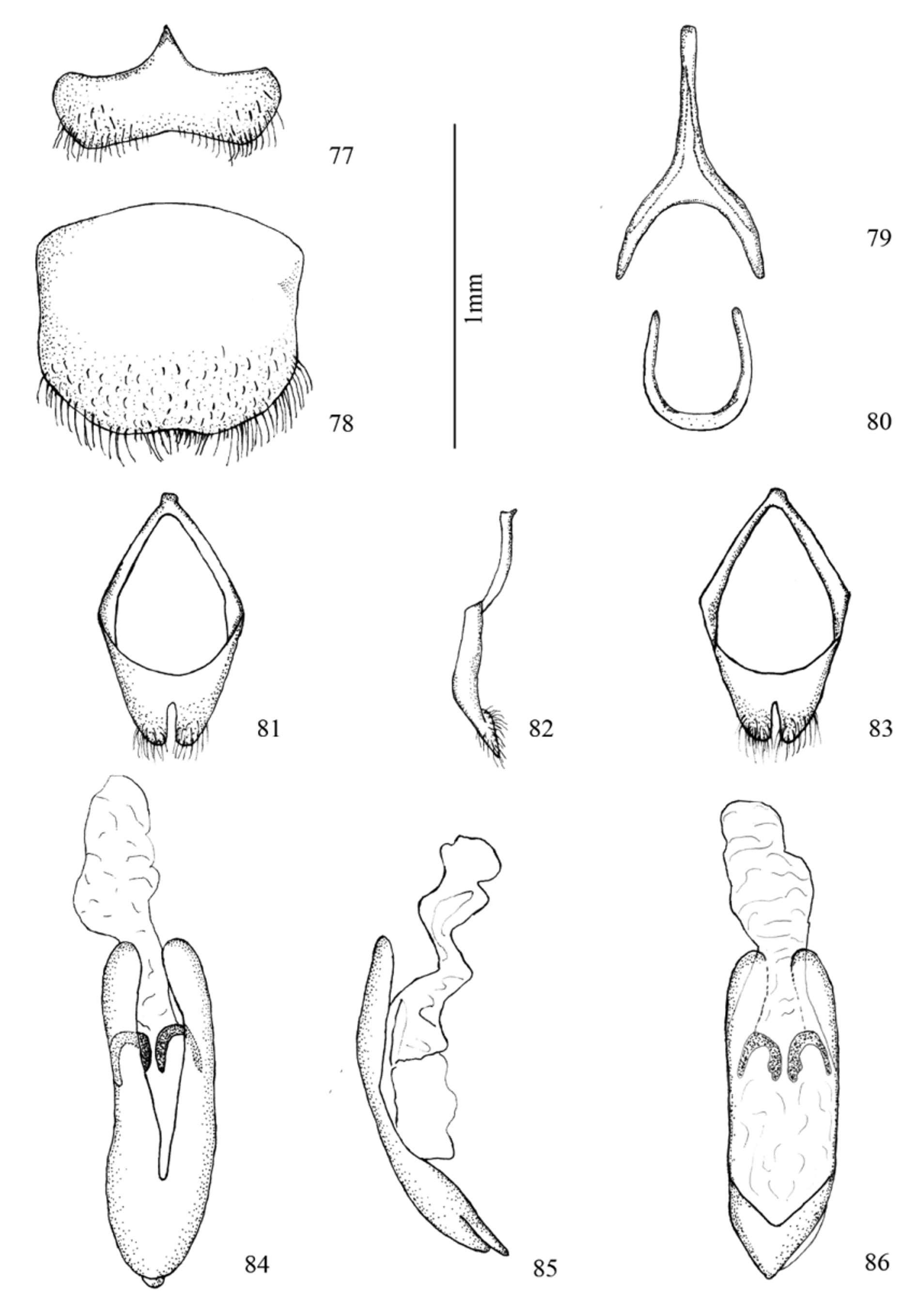
Male terminalia: tergite VIII ( Figs 39 View FIGURES 37 – 47 , 52 View FIGURES 51 – 60 , 65 View FIGURES 64 – 73 , 78 View FIGURES 77 – 86 ) subrectangular; sternite VIII ( Figs 38 View FIGURES 37 – 47 , 51 View FIGURES 51 – 60 , 64 View FIGURES 64 – 73 , 77 View FIGURES 77 – 86 ) transverse, apical third with long setae, setae decreasing in length to middle region; ventral arc ( Figs 40 View FIGURES 37 – 47 , 53 View FIGURES 51 – 60 , 66 View FIGURES 64 – 73 , 79 View FIGURES 77 – 86 ) forkshaped; dorsal arc ( Figs 41 View FIGURES 37 – 47 , 54 View FIGURES 51 – 60 , 67 View FIGURES 64 – 73 , 80 View FIGURES 77 – 86 ) “U”-shaped; tegmen ( Figs 42–44 View FIGURES 37 – 47 , 55–57 View FIGURES 51 – 60 , 68–70 View FIGURES 64 – 73 , 81–83 View FIGURES 77 – 86 ) with distal region divided into slender cylindrical parameres with rounded apex and dense setae; median lobe ( Figs 45–47 View FIGURES 37 – 47 , 58–60 View FIGURES 51 – 60 , 71–73 View FIGURES 64 – 73 , 84–86 View FIGURES 77 – 86 ) slightly curved in lateral view.
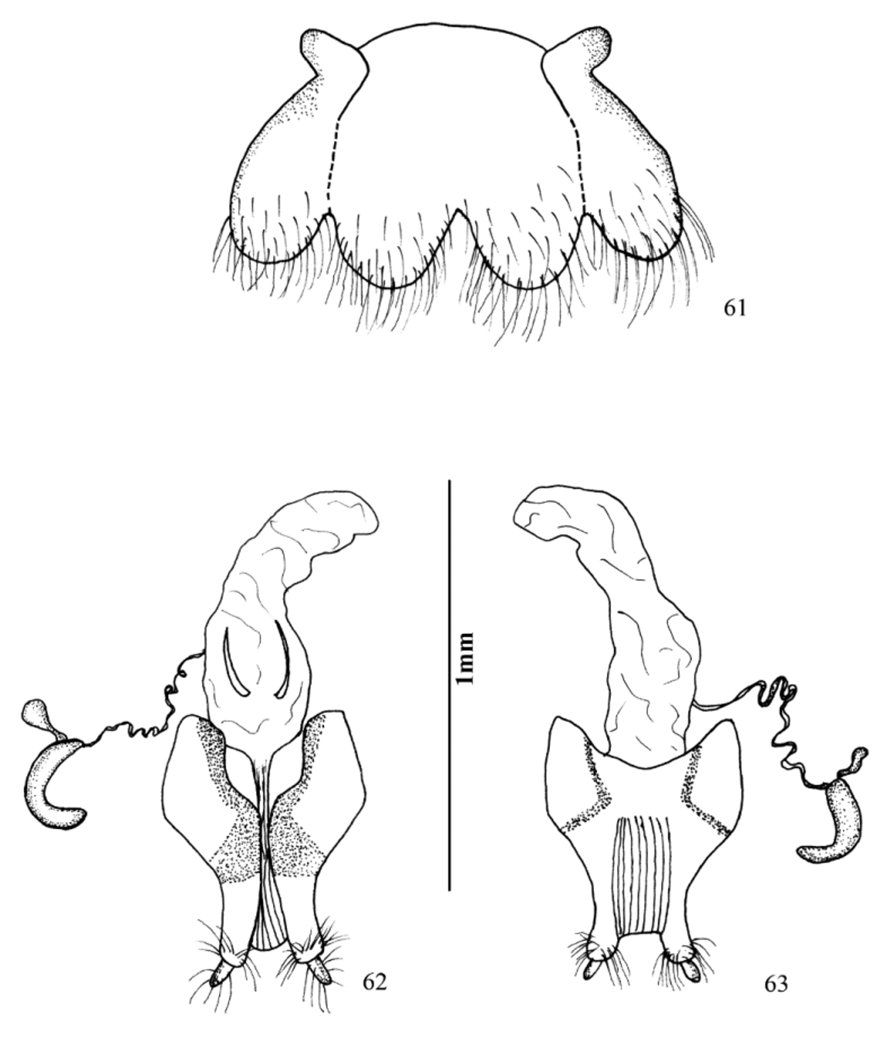
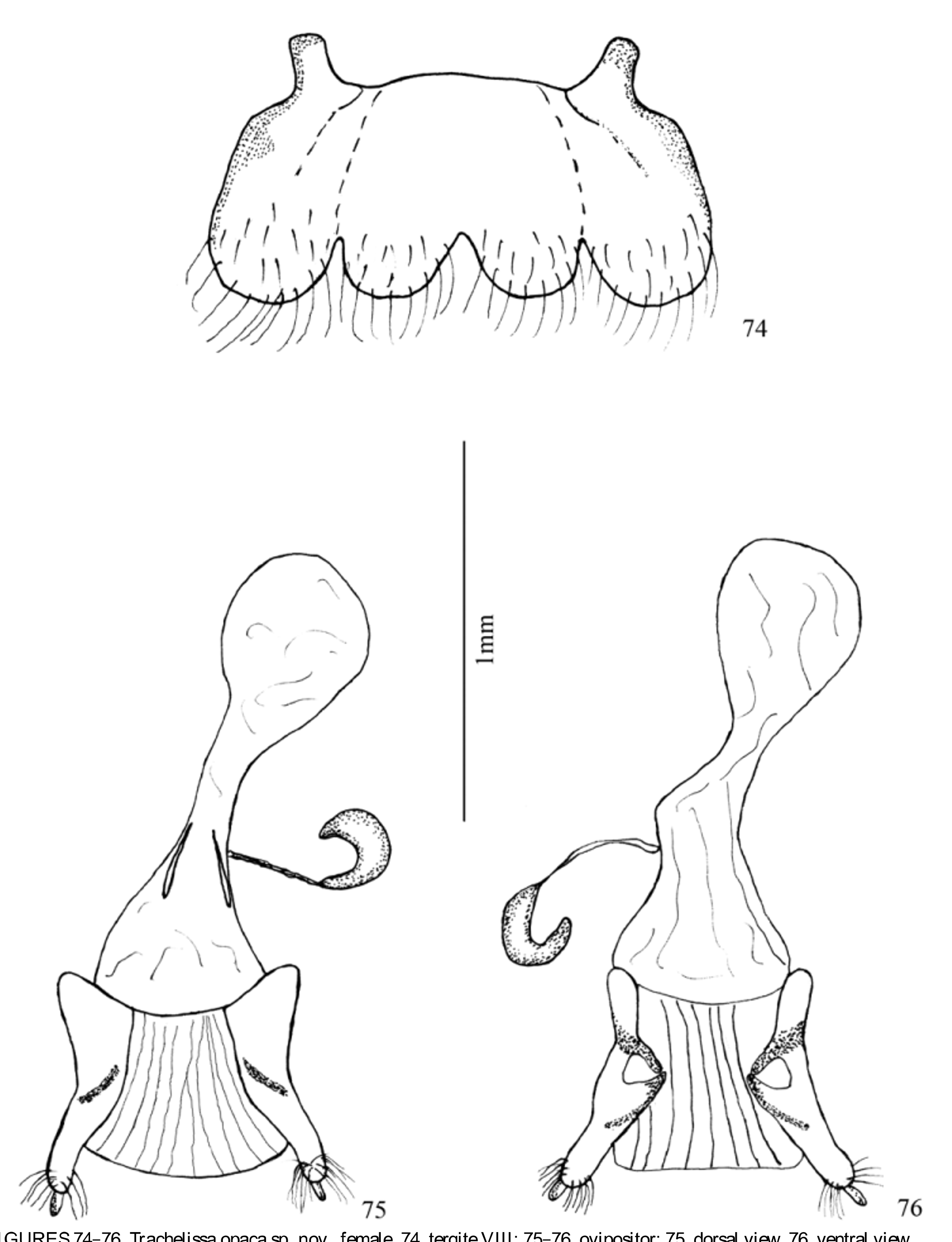
Female terminalia: purpuriceniform (sensu Fragoso et al., 1987). Tergite VIII ( Figs 48 View FIGURES 48 – 50 , 61 View FIGURES 61 – 63 , 74 View FIGURES 74 – 76 , 87 View FIGURES 87 – 89 ) transverse, about twice as long as broad, with four lobes on apical margin, these bearing long dense setae. Posterior region of sternite VIII ( Figs 94, 96 View FIGURES 90 – 97 , 102 View FIGURES 98 – 105 , 110 View FIGURES 106 – 113 , 118, 120 View FIGURES 114 – 121 ) with a brush formed by aciculate ( Figs 96 View FIGURES 90 – 97 , 112 View FIGURES 106 – 113 , 120 View FIGURES 114 – 121 ), conchoidal ( Figs 96 View FIGURES 90 – 97 , 104 View FIGURES 98 – 105 , 112 View FIGURES 106 – 113 , 120, 121 View FIGURES 114 – 121 ) and petiolate setae ( Figs 97 View FIGURES 90 – 97 , 105 View FIGURES 98 – 105 , 113 View FIGURES 106 – 113 , 120 View FIGURES 114 – 121 ) arranged in several rows. Conchoidal and petiolate setae elongated and with dorsal grooves; aciculate setae shorter than conchoidal setae. Ovipositor ( Figs 49, 50 View FIGURES 48 – 50 , 62, 63 View FIGURES 61 – 63 , 75, 76 View FIGURES 74 – 76 , 88, 89 View FIGURES 87 – 89 ) short, with cylindrical and slightly divergent lateral lobes in distal region; short setae moderately dense in whole lobe and apices with row of long dense setae; apical styli cylindrical; vulva membranous, longitudinally plicate. Spermatheca “C”-shaped, curved and rounded apically. Apodemes at base of common oviduct moderately elongated.
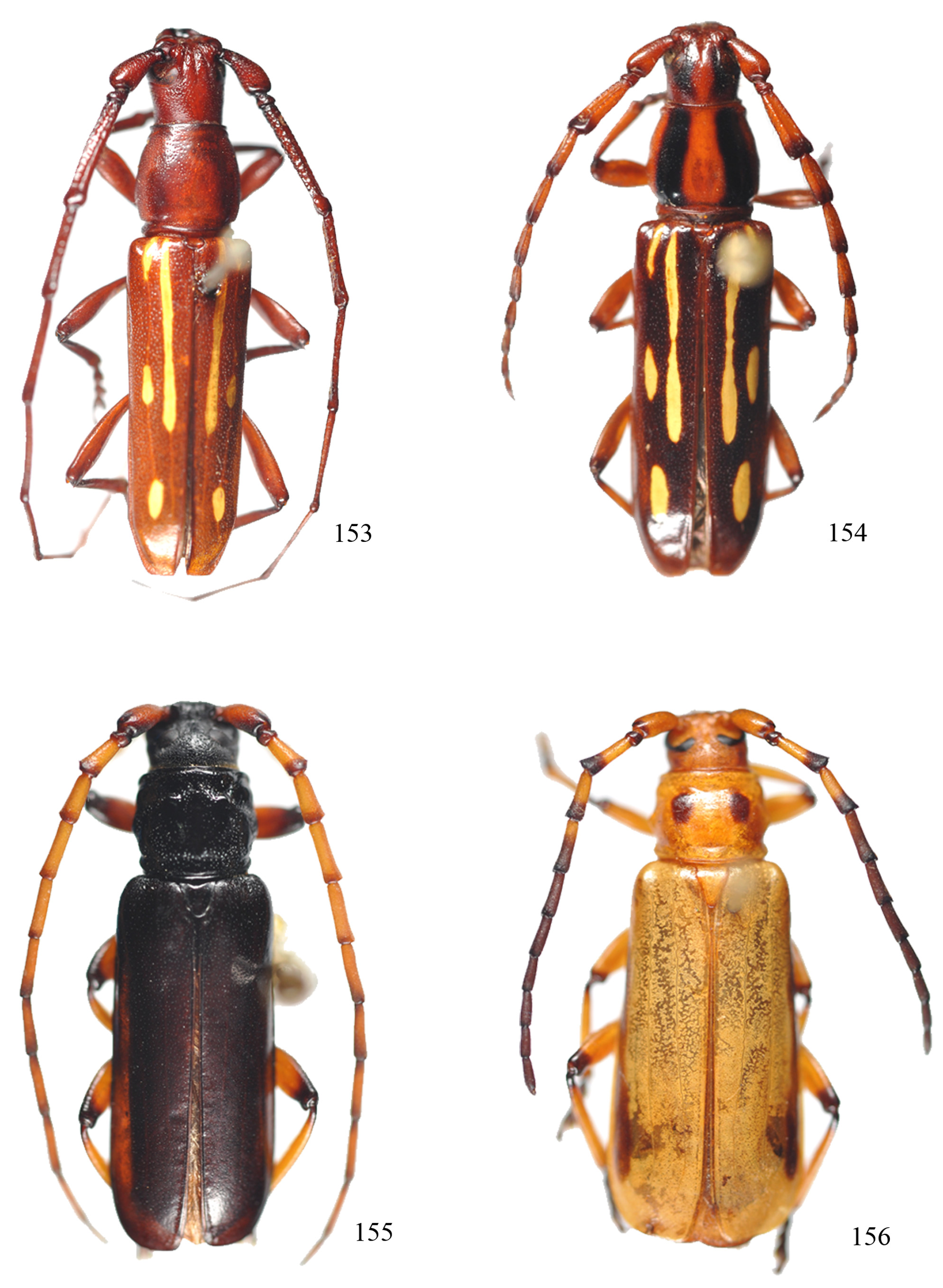
Comments. Trachelissa is similar to Cervilissa Monné & Monné, 2000 ( Figs 153, 154 View FIGURES 153 – 156 ) and Pseudophimosia Delifino, 1990 ( Figs 155, 156 View FIGURES 153 – 156 ). Monné & Monné (2000) described Cervilissa and emphasized several features in common with Trachelissa , including antennomere III longer than IV in males; the surfaces of the mesepisternum, mesepimeron and metepisternum with fine, shallow and confluent sculpturing; the humeri rounded and not projected; legs short; and metafemora reaching the apical third of the elytra. Cervilissa differs from Trachelissa by having antennomere III as long as the scape and with a transverse carina at the apex; the antennae, in males, exceeding the elytral apices at antennomere XI; the prosternal process without a projection; and the femora fusiform and without an apical inner spine. In Trachelissa antennomere III is grooved, without a transverse carina and about 1.5 times as long as the scape; the antennae, in males, exceed the elytral apices by at least three segments; the prosternal process has an apical projection; and the apical projection and the femora are pedunculateclavate, with an inner apical spine. The characteristics cited by Monné & Monné (2000) for Trachelissa , the antennomere III about 1.5 times as long as the scape, and the antennae, in males, exceeding the elytral apices by at least three segments, are not supported for all members of the genus.
Delfino (1990) transferred Trachelissa eburiodes to Pseudophimosia but did not provide a discussion of these genera. Trachelissa is similar to Pseudophimosia with respect to the general body appearance, the antennomere III dorsally sulcate, the prothorax as long as wide, and the clavate femora with the inner apical margin projected in a short spine. However, we noted that in Pseudophimosia , antennal segments II–XI have a carina on the outer margin, segments III–X dorsally sulcate, and the prothorax without tubercles.
Usually in members of Cerambycidae , the antennae are composed of 11 segments, with some exceptions such as Prionus (Neopolyarthon) imbricornis (Linnaeus, 1767) , which has 15–20 segments in females and 18–20 in males. In Trachyderini , some species, for instance Batus hirticornis (Gyllenhal, 1817) , have a 12-segmented antenna. In Trachelissa the number of antennal segments may be sexually dimorphic, with 11 in males and 12 in females of the same species. This dimorphism occurs in all species except Trachelissa rugosipennis , where both sexes have the antenna composed of 11 segments.
The use of a scanning electron microscope (SEM) revealed useful structures to distinguish the species, for example the brush of setae, with very short setae with rounded or tapered tips, which may be arranged in rows. In the pronotum, we observed that the sexually dimorphic punctation in males with deep, rounded or ovate indentations with a variable number of pores resembles the sexually dimorphic punctation observed in other groups of Cerambycidae (e.g., Noldt et al., 1995; Monné, M. L., 2005; Nearns & Ray, 2006; Ray et al., 2006; Botero & Monné, 2012).
Trachelissa can be differentiated from the other genera by the antennomere III with a longitudinal groove; the pronotum with a pair of tubercles near the posterior margin; the prosternal process with a projected apex; the prothorax, in males, with evident sexually dimorphic punctation; and the femora pedunculate clavate.


No known copyright restrictions apply. See Agosti, D., Egloff, W., 2009. Taxonomic information exchange and copyright: the Plazi approach. BMC Research Notes 2009, 2:53 for further explanation.
|
Kingdom |
|
|
Phylum |
|
|
Class |
|
|
Order |
|
|
Family |
Trachelissa Aurivillius, 1912
| Quintino, Hingrid Yara S. & Monné, Marcela L. 2014 |
Trachelissa
| Bezark 2013: 196 |
| Aurivillius 1912: 449 |
Trachelia
| Audinet-Serville 1834: 25 |