
Acentroptera cf. tessellata Baly, 1958
|
publication ID |
https://doi.org/ 10.11646/zootaxa.4243.3.6 |
|
publication LSID |
lsid:zoobank.org:pub:879A07F6-395D-41D9-86C1-1972B2E99DF5 |
|
DOI |
https://doi.org/10.5281/zenodo.5999085 |
|
persistent identifier |
https://treatment.plazi.org/id/03E84957-FFE3-0528-2FE5-F954D88991AE |
|
treatment provided by |
Plazi |
|
scientific name |
Acentroptera cf. tessellata Baly, 1958 |
| status |
|
Acentroptera cf. tessellata Baly, 1958 View in CoL
( Figs 53–95 View FIGURES 53 – 61 View FIGURES 62 – 65 View FIGURES 66 – 70 View FIGURES 71 – 76 View FIGURES 77 – 81 View FIGURES 82 – 86 View FIGURES 87 – 95 )
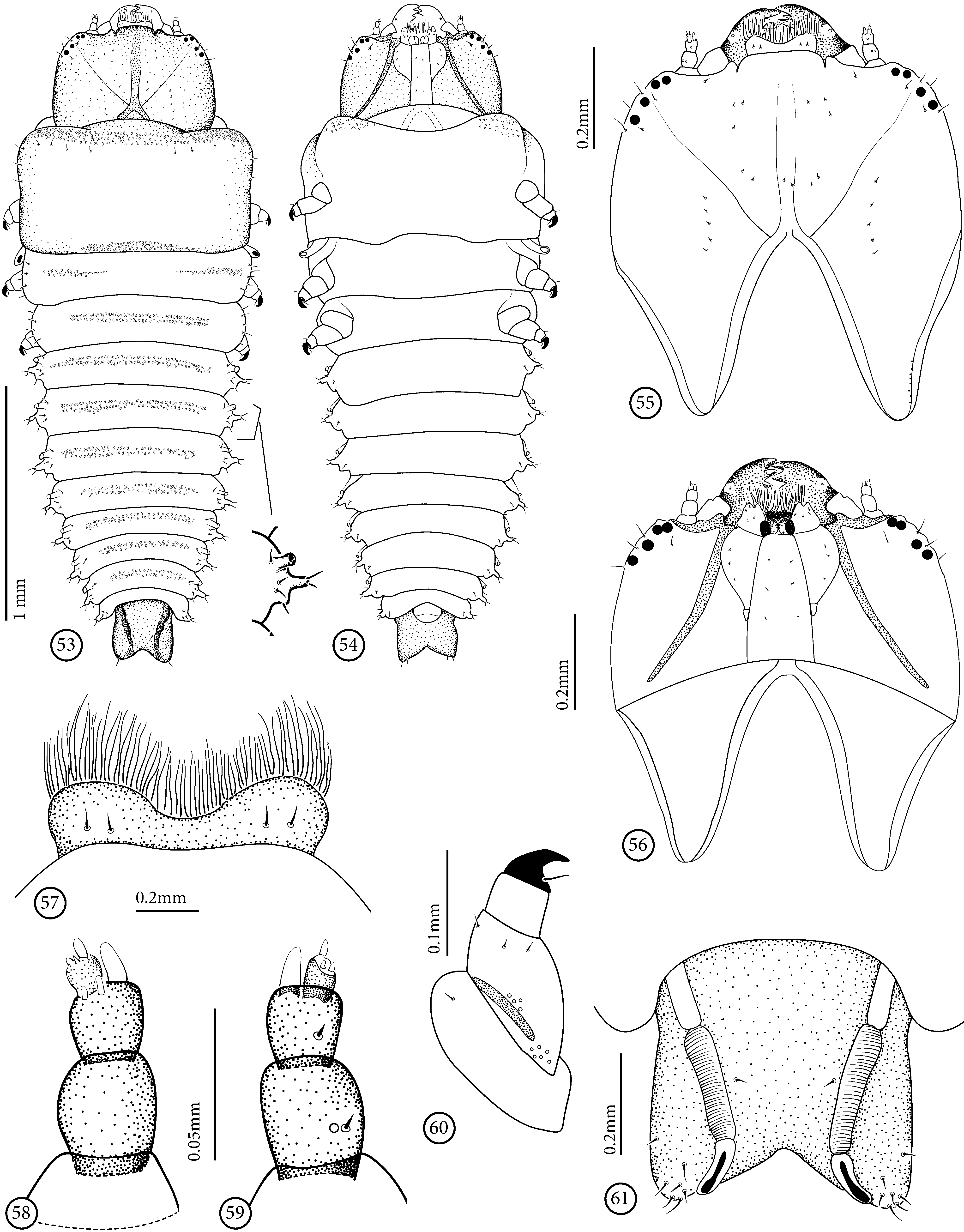
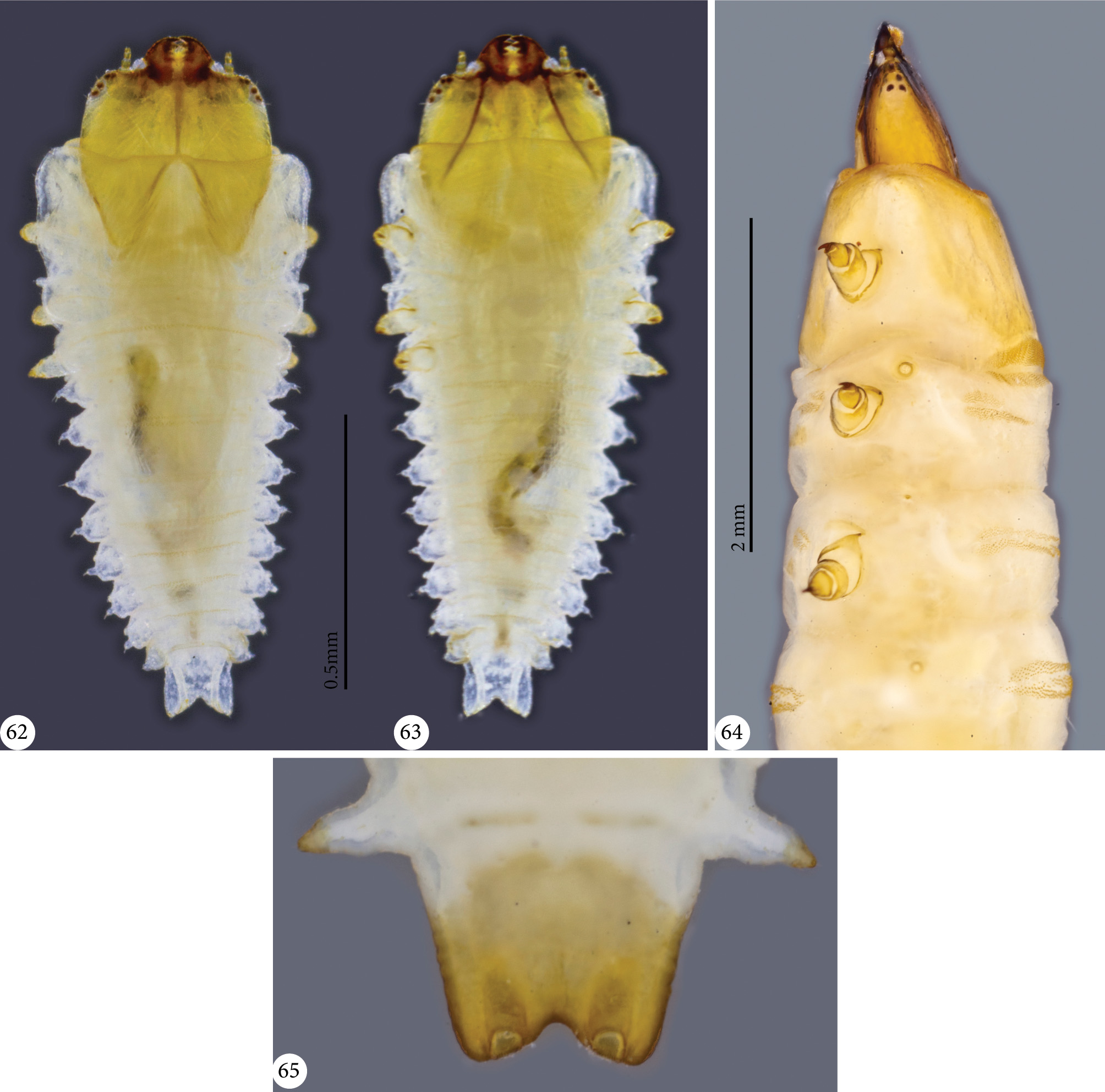
Description of first instar larva ( Figs 53–63 View FIGURES 53 – 61 View FIGURES 62 – 65 , 88, 89 View FIGURES 87 – 95 ). Length: 2.5 mm.
Coloration yellowish-white; head ( Figs 55, 56 View FIGURES 53 – 61 , 62, 63 View FIGURES 62 – 65 ) yellowish with anterior margin of head capsule, mandibles, sagittal line, hypostomal rods and stemmata dark–brown or brown. Prothorax with yellowish rectangular area dorsally and ventrally. Legs ( Figs 60 View FIGURES 53 – 61 , 63 View FIGURES 62 – 65 ) partially yellowish with tarsunguli dark-brown. Thorax and abdomen with granulated dorsal areas. Antennae ( Figs 58, 59 View FIGURES 53 – 61 , 62, 63 View FIGURES 62 – 65 ) with 3 elongate antennomeres: basal bearing one short seta and one campaniform sensillum ventrally, near base; median shorter than basal, bearing one short seta ventrally, near middle, 2 stout and short setae dorsally, near apex, and one membranous sensorial appendix at apex, shorter than distal antennomere; distal, shorter and narrower than previous, bearing at apex 3 stout and short setae, one longest.
Head and mouthparts similar to “mature” larva.
Pronotum dorsally granulated in anterior band and in median posterior elliptical area; ventrally each side with granulated anterior fold. Prothoracic spiracle at apex of a tubular projection. Mesonotum grooved transversely slightly granulated on each extremity. Metanotum and segments I–VII grooved transversely, each side of groove granulated. Segments I–VIII with one lateral median elongate projection on each side, each with 2 setae at apex and 2–3 setae near base; segments I–VII with a tubular lateroanterior projection on each side, with spiracular opening at apex; spiracular opening elliptical. Segment IX ( Fig. 61 View FIGURES 53 – 61 ) narrow than previous, almost as long as wide, with V-shaped apex and two dorsally placed spiracular openings, innerly to distal angles; distal angles setose; segment X ( Fig. 54 View FIGURES 53 – 61 ) reduced, rounded, placed ventrally.
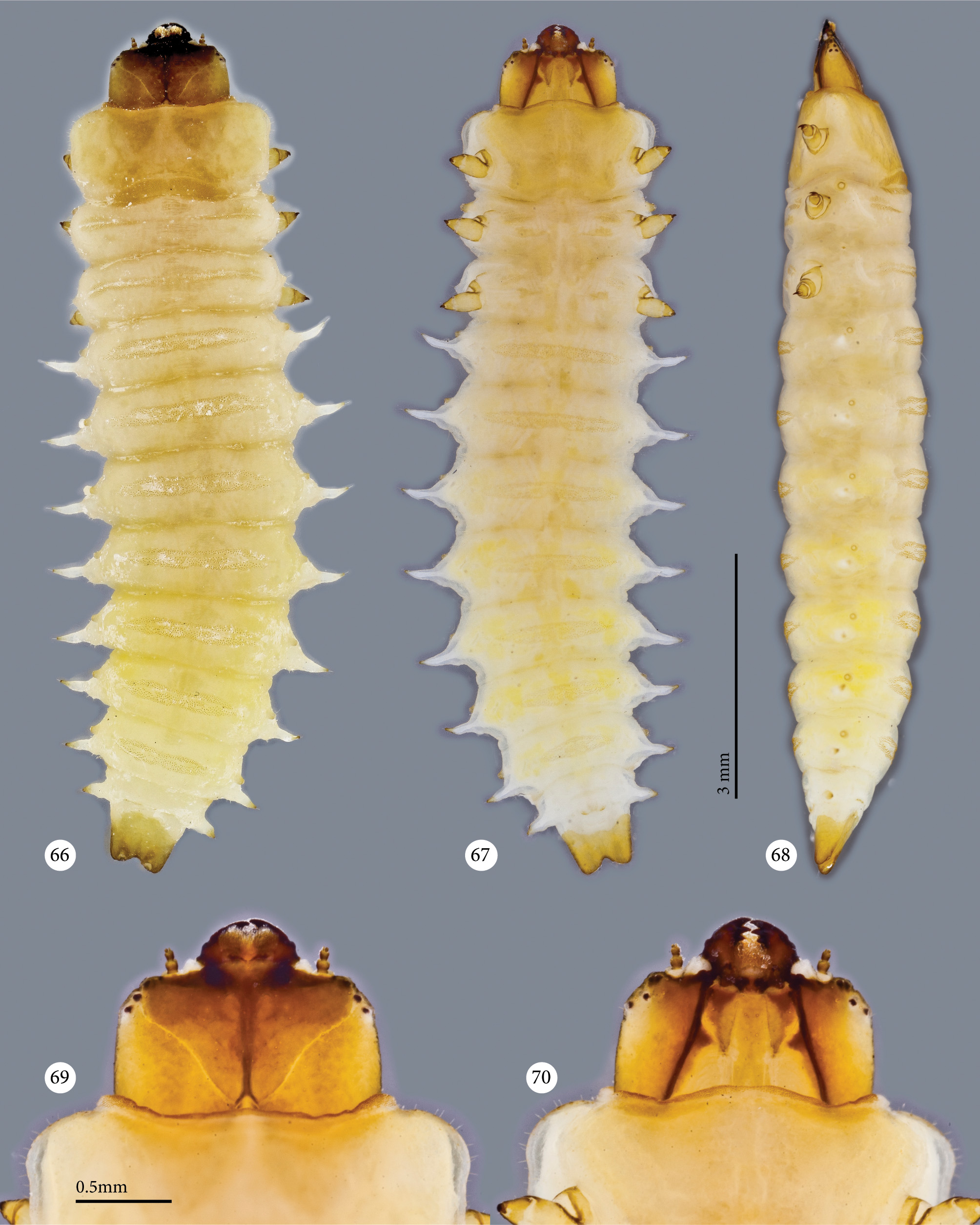
Description presumably mature larva ( Figs 64–81 View FIGURES 62 – 65 View FIGURES 66 – 70 View FIGURES 71 – 76 View FIGURES 77 – 81 ).
Length: 10–11 mm (n=2)
Body ( Figs 64–68 View FIGURES 62 – 65 View FIGURES 66 – 70 ) elongate, wide, strongly dorso-ventrally flattened; abdomen with lateral elongate projections. Coloration cream after fixation; head strongly sclerotized, yellowish with frons darker; mandibles, endocarina and hypostomal rods brown; pronotum yellowish–cream; legs yellowish, partially brown; tarsunguli brown. Body almost glabrous; short setae present laterally in whole length. Thorax and abdomen with granulated elliptical areas; smaller areas ventrally.
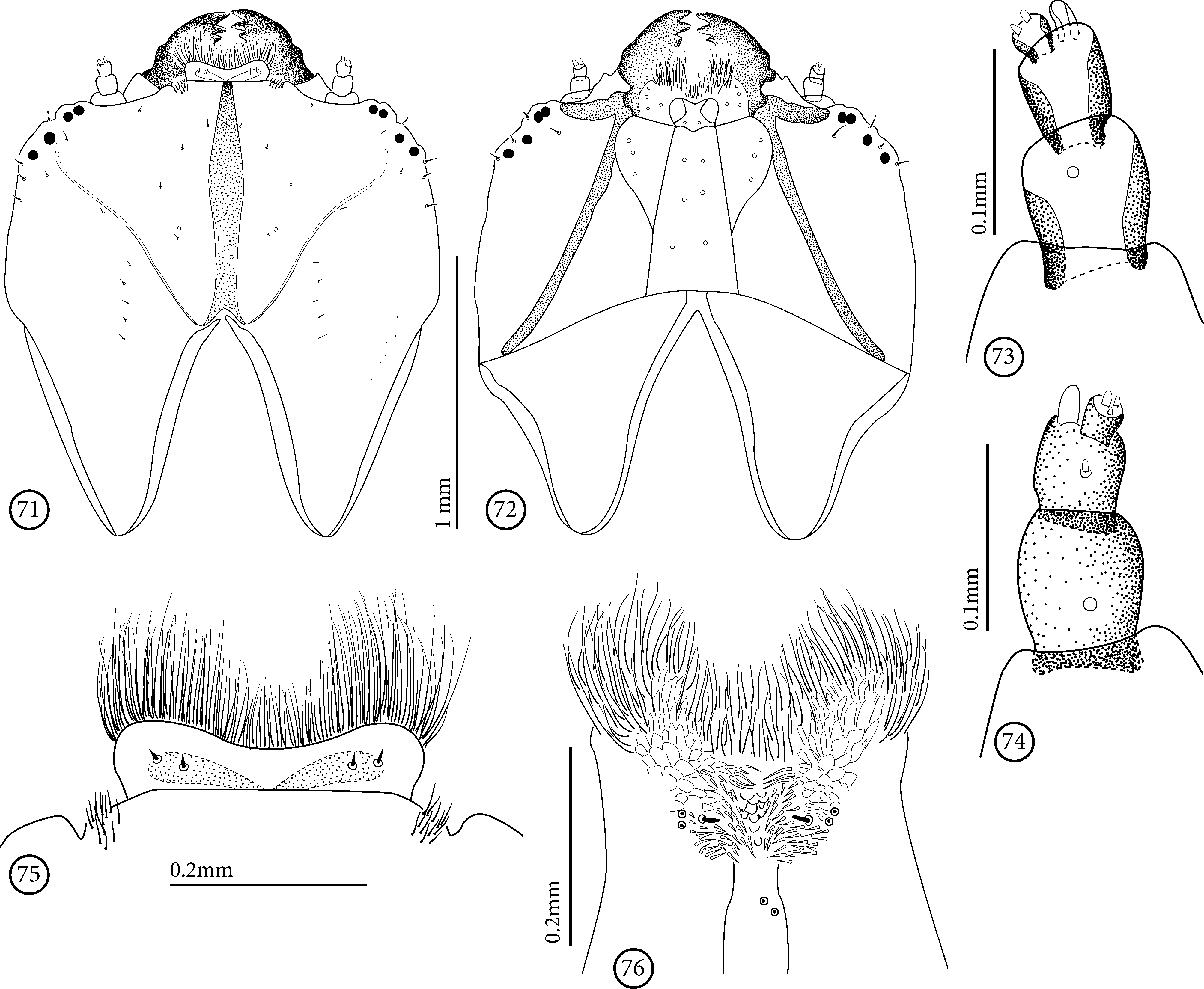
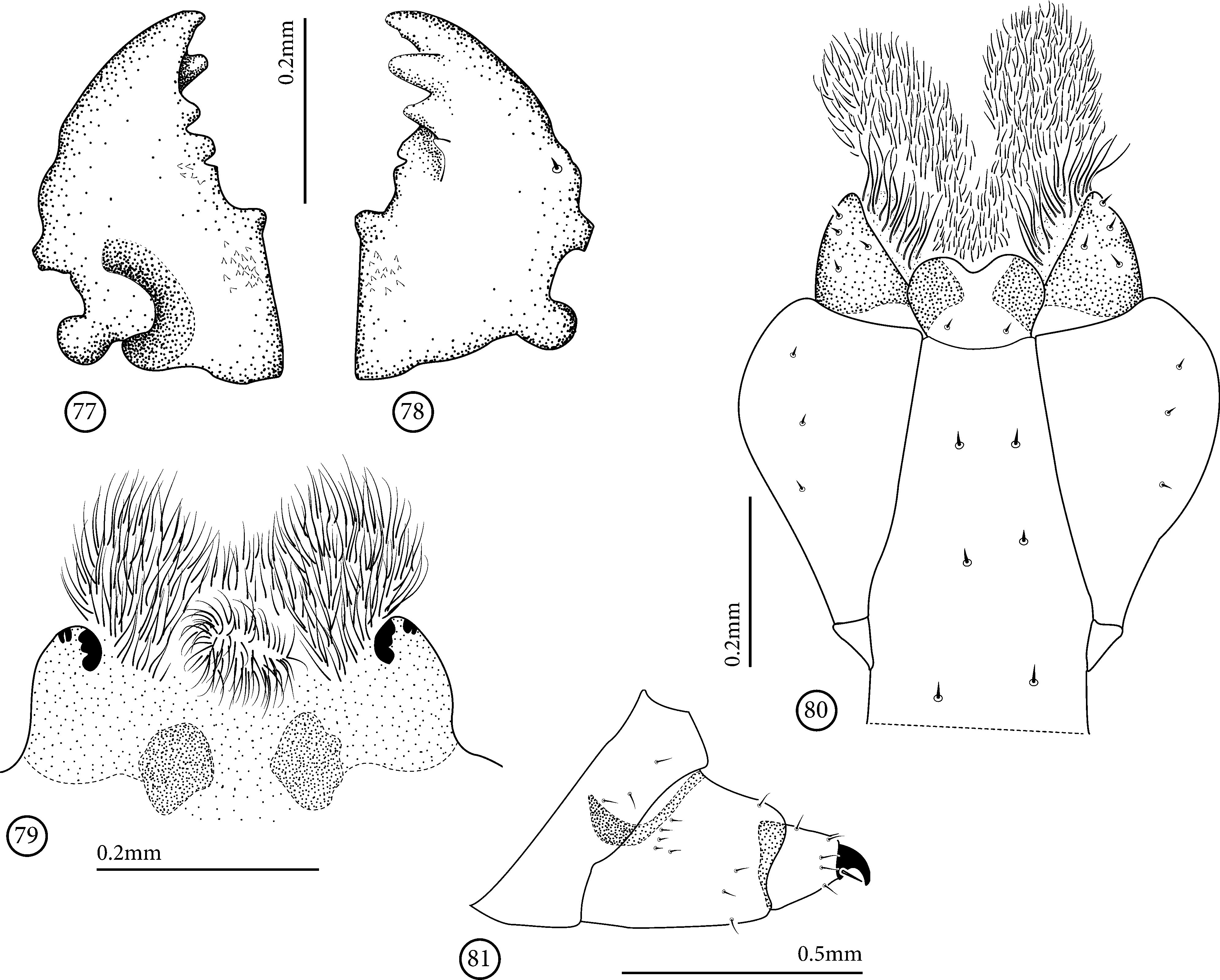
Head ( Figs 64 View FIGURES 62 – 65 , 66–72 View FIGURES 66 – 70 View FIGURES 71 – 76 ) prognathous, narrower than prothorax, strongly dorsoventrally flattened, partially retracted into prothorax; epicranial stem absent; median endocarina well-developed, extending between frontal arms; frontal arms V-shaped. Six black stemmata on each side: 2 lateroanterior, 2 dorsal and 2 ventral. Frons with 8 pairs of tiny setae: one pair on each side near anterior margin, an inclined row with 3 setae on each side of endocarina, one pair on each side near base and one pair on sagittal line; each epicranial half with 10 setae: 4 setae near lateral margin, one near frontal arm and a longitudinal row with 5 setae near middle. Hypostomal rods welldeveloped. Frontoclypeal suture indistinct. Antennae ( Figs 69, 70 View FIGURES 66 – 70 , 73, 74 View FIGURES 71 – 76 ) with 3 elongate antennomeres: basal antennomere with one campaniform sensillum dorsally and one ventrally; median antennomere bearing one ventral short and stout seta near middle, one membranous, cupuliform, sensorial appendix at apex, and the distal antennomere; sensorial appendix as long as distal antennomere; distal antennomere reduced, a half width of median antennomere, inserted latero-internally at apex, side by side with sensorial appendix, bearing at apex 3 short and stout setae. Labrum ( Figs 69 View FIGURES 66 – 70 , 75 View FIGURES 71 – 76 ) band-like, strongly emarginated medially with distal angles widely rounded; one pair of tiny setae on each side and a dense fringe of setae at anterior margin, shorter in middle. Epipharynx ( Fig. 76 View FIGURES 71 – 76 ) densely setose near anterior margin; microspined and with stout setae near middle; one short seta and one pair of campaniform sensilla on each side, near base; two basal campaniform sensilla in middle. Mandible ( Figs 77, 78 View FIGURES 77 – 81 ) with lateral margin sinuous on basal half; 3 dorsal and 3 ventral subapical, rounded teeth; one mesobasal stout rounded tooth; one lateroventral tiny seta near base. Maxilla ( Figs 70 View FIGURES 66 – 70 , 72 View FIGURES 71 – 76 , 80 View FIGURES 77 – 81 ): cardo triangular and reduced; palpus indistinguishable; stipes elongate with lateral margin rounded, narrowed at base, with 3 tiny setae laterally; mala triangular, partially sclerotized, densely setose, brush-like near apex and with 4 tiny setae near base. Labium elongate; palpi indistinguishable; prementum with sclerotized transverse band, darker in one triangular area on each side, bearing two tiny setae at base; ligula densely setose, brush-like on distal half; setae surpassing anterior margin; postmentum elongate with 3 pairs of tiny setae. Hypopharynx ( Fig. 79 View FIGURES 77 – 81 ) densely setose anteriorly; basal half slightly sclerotized with 2 small lateroanterior sclerites on each side, and 2 larger, rounded, basal sclerites near midde.
Thorax. Prothorax wider than long, longer than mesothorax; fore angles widely rounded; lateral margins almost straight; anterior margin with narrow granulated band; basal region with granulated narrow elliptical band near middle; short setae laterally. Meso- and metathorax band-like, almost of same size; each with placed laterodorsally small rounded projections, with 3–4 setae; mesothorax with laterodorsal anterior rounded spiracle located at apex of tubular projection ( Fig. 68 View FIGURES 66 – 70 ); meso- and metathorax dorsally each with two transverse and narrow granulated band with a transverse groove in middle, and ventrally with one small elliptical granulated area on each side.
Legs ( Figs 64 View FIGURES 62 – 65 , 67, 68 View FIGURES 66 – 70 , 81 View FIGURES 77 – 81 ) widely separated, located ventrolaterally; slightly sclerotized, short, robust and 4- segmented; coxa short, band-like, with 3 short setae; femur and trochanter fused, wider than long, with 4 short setae near anterior margin and 5 near base; tibia wider than long with row of sparse setae near anterior margin and one near base; tarsungulus sclerotized with lateroexternal seta at base.
Abdominal segments I–VIII ( Figs 65–68 View FIGURES 62 – 65 View FIGURES 66 – 70 ) with a lateral median elongated projection with one seta at apex; last segment shorter and more robust; segments I–VII each with one laterodorsal tubular projection with spiracular opening at apex; segments I–VII dorsally with a transverse groove with one narrow band of granulations anteriorly and 2 posteriorly; median band short and/or fused to posterior, forming an elliptical granulated area; ventrally with transverse furrow and elliptical granulated area; granulated area shorter posteriad; segment VIII shorter than previous, spiracle absent; segment IX narrower than previous, sclerotized, elongate, narrowed posterad, with distal margin emarginated, with 2 dorsal spiracular openings, innerly to distal angles. Segment X reduced, ventral, partially sclerotized with distal margin bilobed.
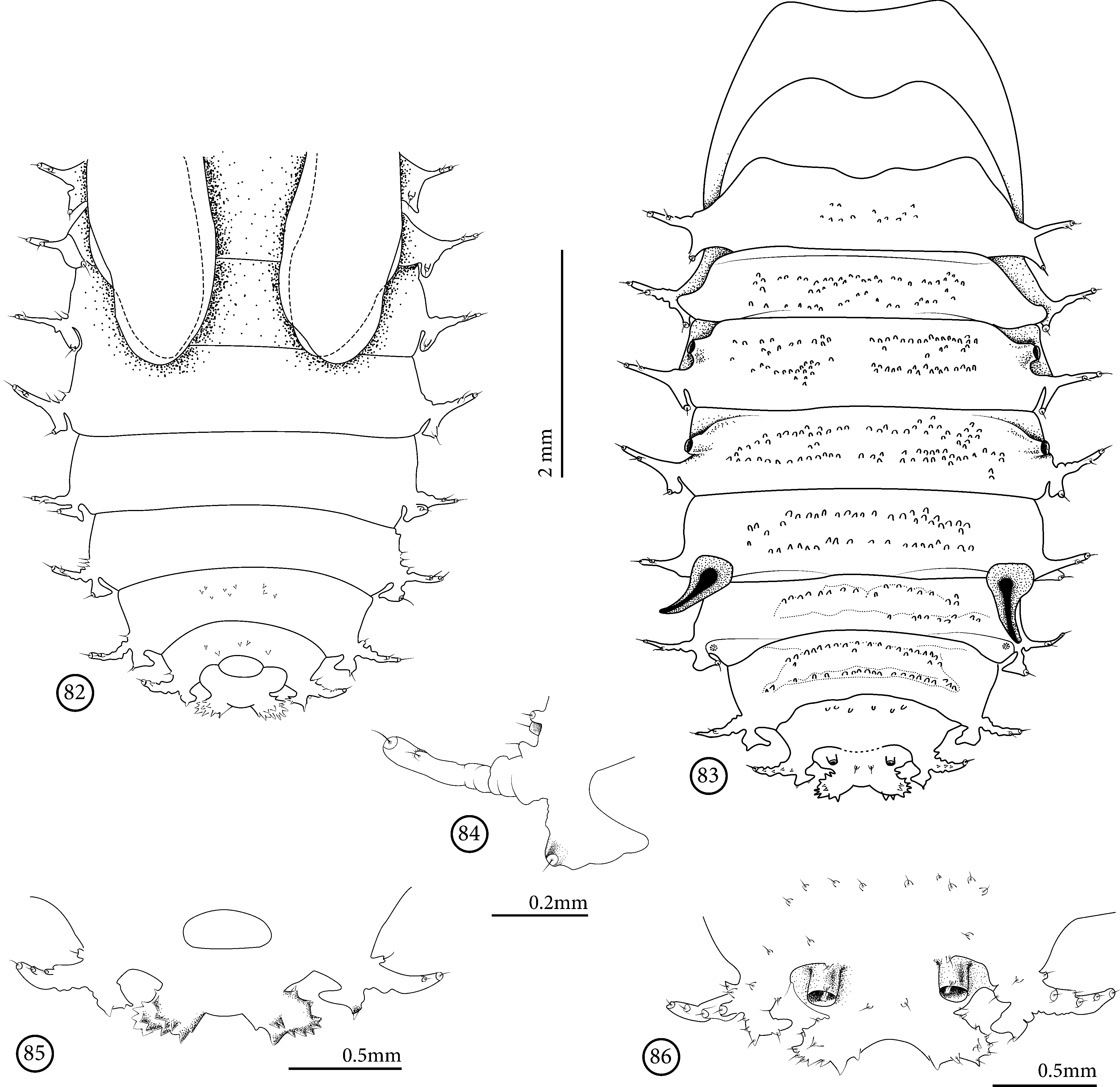
Description of pupa based on pupal exuvia ( Figs 82–86 View FIGURES 82 – 86 ).
Head and prothorax lost. Abdomen elliptical, wider on segments III–V; setae on lateral projections and on granulations. Segments I–VIII each with two lateral projections on each side: one short triangular, with sclerotized inner angle and one distal seta, and the second long with sclerotized apex, bearing one seta apically and one sclerotized projection ventrally.
Dorsally, integument with small rounded tubercles, densely granulated with one short seta at apex (setae and granules not represented on Fig. 83 View FIGURES 82 – 86 ); segment I with a few tubercles; segments II–VII with two transverse rows of tubercles; segment VIII with one transverse row and segment IX with two tubercles in middle, each with one basal seta. Spiracular openings at apex of dorsolateral tubular projection of segments III–IV; segment V with spiracular opening elongate, horn-like; a pair of rounded spiracular openings near base of segment IX. Ventrally, segments VII–VIII with some microspines near middle. Segment IX narrow, slightly sclerotized, with two apical broad lobes with strongly sclerotized teeth. Segment X reduced, placed ventrally.
Material examined. Brazil. Bahia: Serra Grande, Uruçuca (Faz. Modus Vivendi), 21–27.IX.2015, F.F. Albertoni & M. A. Ulysséa col., 1 first instar larva, 2 (1 dead) presumably mature larvae; 3 exuviae of head, 1 pupal exuvia.
Geographical distribution. Brazil (Bahia and Rio de Janeiro) (present work and Staines 2014).
Biology of Acentroptera cf. tessellata Baly, 1958 ( Figs 87–95 View FIGURES 87 – 95 )
The host plant of A. cf. tessellata is here reported for the first time, and the feeding habit of the adults of this species is similar that of A. basilica , although the host plant is Vriesea sp. Adults were searched for four consecutive nights and were not seen feeding, or on the freely open surface of leaves; they were deeply hidden among leaf bases of the rosette. During the hottest periods of the day they were also hidden among leaf bases, and were frequently seen feeding around 15:30 h. While feeding, adults stayed motionless and clinging to leaf surface when bothered, but would display tanatosis if released from leaf surface. When turned upside down on a flat surface, they efficiently turn to an upright position. Using the head and legs, the beetle managed to lean the body to one side and with the legs, hold the surface and pull the body. In this way the tibial-femur join help the movement provoking a larger body inclination, enabling the other side legs to hold the surface and definitely impulse the whole body to the upright position (this behaviour is illustrated on Figs 90–94 View FIGURES 87 – 95 ). Ants that walked on bromeliad leaves were seen interacting with adults few times including antennal perception (apparently scanning for chemical and tactile cues) of the beetle. Those were relatively quick interactions and in all cases ants seemed to have “friendship” with the beetles ( Fig. 95 View FIGURES 87 – 95 ).
Eggs of A. cf. tessellata were laid alone (three observation) or in sequence of three to four eggs (four observations) on scrapped areas of the leaf surface ( Fig. 87 View FIGURES 87 – 95 ). First instar larva hatched and immediately started mining the leaf. When uncovered by first leaf tissue layer, the ventral side of the first instar larva was turned to the thinner side of the leaf tissue corresponded to the adaxial leaf face ( Fig. 88 View FIGURES 87 – 95 ).

No known copyright restrictions apply. See Agosti, D., Egloff, W., 2009. Taxonomic information exchange and copyright: the Plazi approach. BMC Research Notes 2009, 2:53 for further explanation.