
Piezocranum, HORVATH, 1877
|
publication ID |
https://doi.org/10.1111/j.1096-3642.2011.00770.x |
|
persistent identifier |
https://treatment.plazi.org/id/03E8878D-FFAA-FFAA-5D8E-F8BBB1DFF9BB |
|
treatment provided by |
Marcus |
|
scientific name |
Piezocranum |
| status |
|
PIEZOCRANUM HORVÁTH View in CoL ( FIGS 4 View Figure 4 , 49 View Figure 49 )
Piezocranum Horváth, 1877: 92 View in CoL (gen. nov.; type species: Piezocranum simulans Horváth, 1877 View in CoL by monotypy); Atkinson, 1890: 120 (cat.); Reuter, 1891: 33, 157 (descr., key); Hueber, 1906: 3 (key); Kirkaldy, 1906: 130 (cat.); Reuter, 1910: 148 (cat.); Oshanin, 1910: 798 (cat.); Stichel, 1933: 235 (key); Kiritshenko, 1951: 127 (key); Wagner, 1952: 96, 101 (key, descr.); Carvalho, 1952: 74; Carvalho, 1955: 67 (key); Carvalho, 1958: 29 (cat.); Kerzhner, 1964a: 967 (diag., key); Wagner & Weber, 1964: 264 (syn., diag., key); Wagner, 1973: 28 (descr., key); Schuh, 1995: 68 (world cat.).
Lamprella Reuter, 1890: 253 (gen. nov.) – (syn. by Horváth, 1889: 327); Lamprella Carvalho, 1958: 29 (cat.).
Diagnosis: Recognized by the following combination of characters: macropterous individuals elongate, with transverse depression spanning vertex and frons; endosoma without spicules; DLP divided into weakly sclerotized lateral and medial plates, with paired sclerotized strips adjacent to medial margin of sclerotized rings.
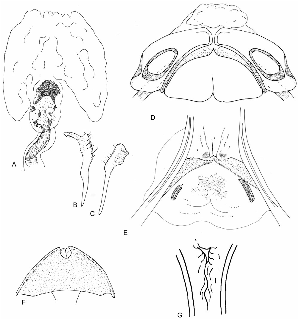
Redescription: Sexually dimorphic, with males macropterous and females macropterous or brachypterous. Coloration ( Fig. 4 View Figure 4 ): Male reddish-brown, paler on hemelytron; female glossy black. Surface and vestiture ( Fig. 4 View Figure 4 ): head smooth and shining, pronotum rugulose, especially along posterior margin, hemelytra of female sometimes glossy with deep punctures. Body of male clothed in golden simple setae; females less setaceous. Structure: head ( Fig. 4 View Figure 4 ): transverse; usually wider than tall; macropterous morphs with deep transverse depression spanning across vertex and frons, brachypterous females with at most a shallow depression; posterior margin of vertex raised and carinate; frons broadly rounded in brachypterous females; gena height two to three times eye height; labium: LI swollen, short. Antennae ( Fig. 4 View Figure 4 ): insertion below or in line with lower margin of eye; shorter than body, slender; length of AI approximately equal to eye height. Thorax ( Fig. 4 View Figure 4 ): pronotum trapezoidal in macropterous morphs, subrectangular in brachypterous females, callosite region obsolete to weakly defined in both sexes, lateral margins rounded, humeral angles rounded, posterior margin concave, more deeply in female; mesoscutum visible from above in macropterous individuals; metathoracic spiracle tear-shaped and exposed, without evaporative bodies; MTG external efferent system swollen, angled posterodorsally, ostiole ventrolateral, peritreme tongue-shaped. Hemelytra ( Fig. 4 View Figure 4 ): macropterous – parallel-sided, elongate, extending beyond apex of abdomen; cuneus elongate; membrane with two cells. Brachypterous females – undivided, posteriorly almost straight to subtriangular, sometimes extending partly over abdominal tergite VI. Legs ( Fig. 4 View Figure 4 ): short, somewhat longer in brachypterous morphs; pretarsi without fleshy pulvilli. Abdomen ( Fig. 4 View Figure 4 ): narrow in macropterous individuals, broader in females, in brachypterous females pear-shaped. Male genitalia ( Fig. 49A–C View Figure 49 ): pygophore conical; parameres roughly equal in length; left paramere L-shaped, sensory lobe broad, apophysis slender and apically hooked; right paramere slender basally, apical club elongate, weakly deflected laterally, inner margin sinuate; phallotheca unmodified; ductus seminis constricted, with flexible ribbing; secondary gonopore elongate, incompletely sclerotized, dorsoventrally compressed, apically with scale-like texturing; endosoma simple and broad, without spicules. Female genitalia ( Fig. 49D–F View Figure 49 ): sclerotized rings widely separated, ovate, weakly sclerotized; lateral margins of DLP upturned and sclerotized, closely appressed to rings, remainder of DLP and VLP membranous; posterior wall mostly membranous, with faintly sclerotized band along posterior margin; opening to vestibulum symmetrical, membranous, and unmodified.
Diversity and distribution: Piezocranum is composed of three species, all of which are found almost exclusively in the Mediterranean region, with the range of Piezocranum simulans also extending into the former Yugoslavia, Bulgaria, and Romania.
Included species: Piezocranum corvinum Puton, 1895 Syria; Turkey
Piezocranum seminulum Horváth, 1898 View in CoL Spain
Piezocranum simulans Horváth, 1877 View in CoL * southern Europe, Macedonia
Biology and host plant associations: No biological or host plant information has been recorded for this genus.
Remarks: This redescription is based on a review of the literature, particularly Wagner (1973), and examination of Pi. simulans . Our phylogeny places Piezocranum as sister to Dasyscytus , although this relationship is not well supported.

No known copyright restrictions apply. See Agosti, D., Egloff, W., 2009. Taxonomic information exchange and copyright: the Plazi approach. BMC Research Notes 2009, 2:53 for further explanation.
|
Kingdom |
|
|
Phylum |
|
|
Class |
|
|
Order |
|
|
Family |
Piezocranum
| Tatarnic, Nikolai J. & Cassis, Gerasimos 2012 |
Lamprella
| Carvalho JCM 1958: 29 |
| Reuter OM 1890: 253 |
| Horvath G 1889: 327 |
Piezocranum Horváth, 1877: 92
| Wagner E 1973: 28 |
| Kerzhner IM 1964: 967 |
| Wagner E & Weber HH 1964: 264 |
| Carvalho JCM 1958: 29 |
| Carvalho JCM 1955: 67 |
| Wagner E 1952: 96 |
| Kiritshenko AN 1951: 127 |
| Stichel W 1933: 235 |
| Reuter OM 1910: 148 |
| Oshanin B 1910: 798 |
| Hueber T 1906: 3 |
| Kirkaldy GW 1906: 130 |
| Reuter OM 1891: 33 |
| Atkinson ET 1890: 120 |
| Horvath G 1877: 92 |