
Nanniella, REUTER, 1904
|
publication ID |
https://doi.org/ 10.1111/j.1096-3642.2011.00770.x |
|
DOI |
https://doi.org/10.5281/zenodo.5479746 |
|
persistent identifier |
https://treatment.plazi.org/id/03E8878D-FFD3-FFA2-5D91-FD6FB6F3F8EC |
|
treatment provided by |
Marcus |
|
scientific name |
Nanniella |
| status |
|
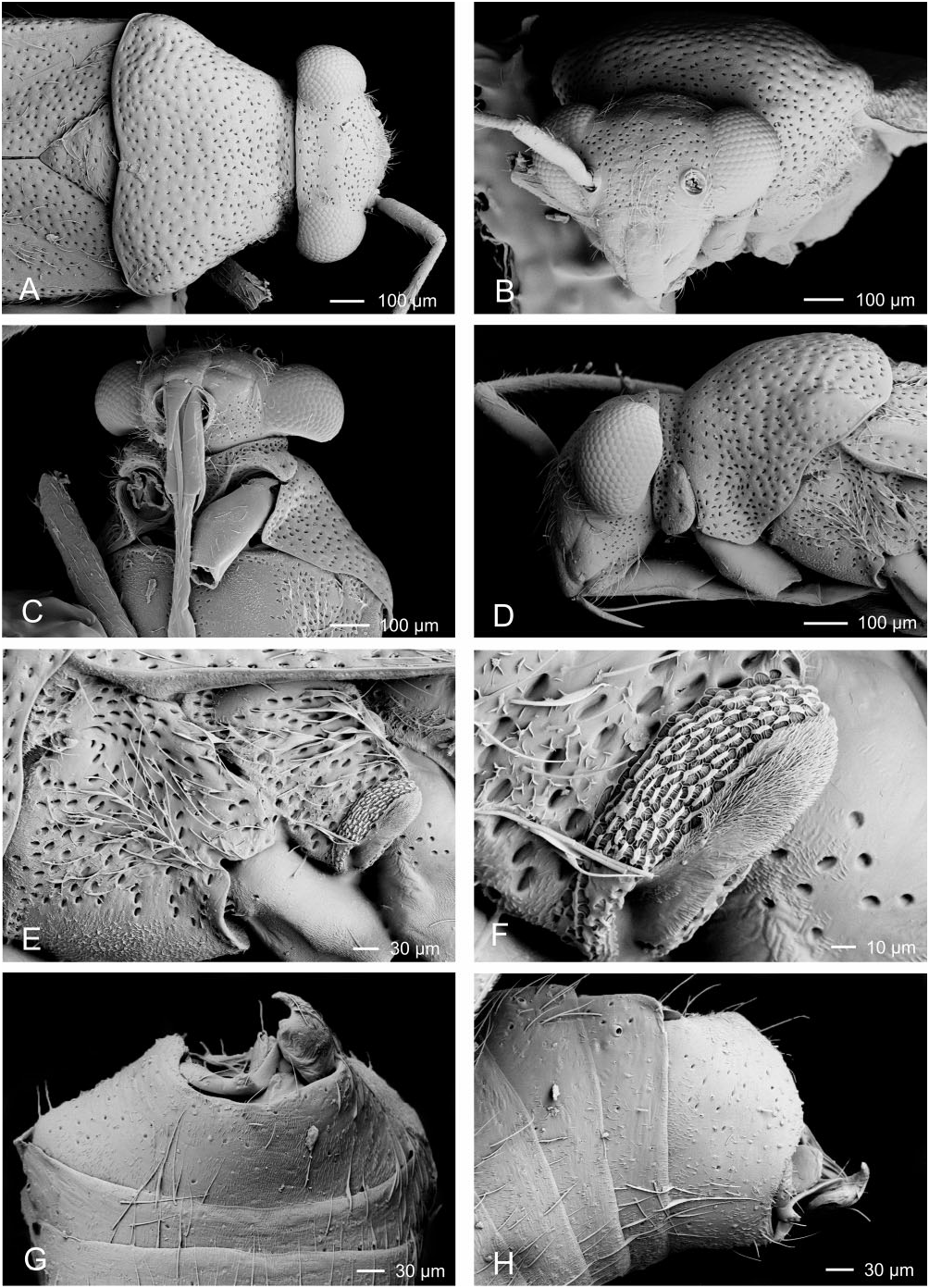
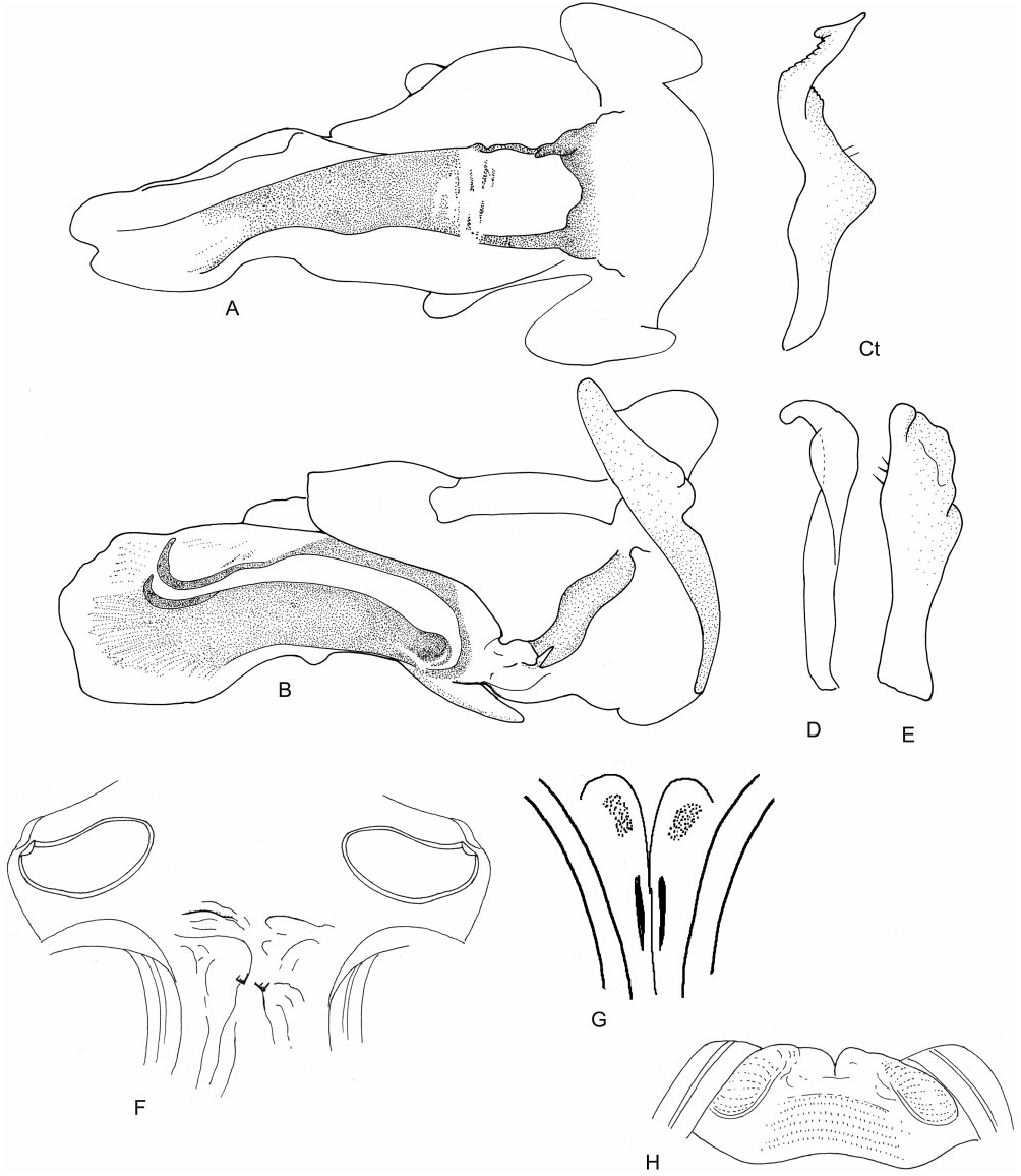
NANNIELLA REUTER View in CoL ( FIGS 4 View Figure 4 , 43–44 View Figure 43 View Figure 44 )
Nanniella Reuter, 1904: 5 View in CoL (gen. nov.; type species: Nanniella chalybea Reuter, 1904 View in CoL by monotypy); Kirkaldy, 1906: 131 (cat.); Reuter, 1910: 148 (cat.); Carvalho, 1952: 76 (syn.); Carvalho, 1958: 61 (cat.); Schuh, 1974: 28 (diag., disc. of tribal placement); Linnavuori, 1975: 48 (disc. of tribal placement, key); Linnavuori, 1994: 9 (descr., key); Schuh, 1995: 62 (world cat.).
Diagnosis: Nanniella is distinguished from other Halticini by the following combination of characters: metallic black coloration; uniformly coarsely punctate; left paramere with twisted, somewhat serrate apophysis; right paramere apicolaterally recurved with finger-like apical apophysis. Nanniella is most similar to Acratheus , but can be separated by the presence of deep punctures on the frons and vertex and the shape of the parameres.
Redescription: Length: 2.5– 4 mm. Colouration ( Fig. 4 View Figure 4 ): mostly black with yellow-brown or yellow markings. Surface and vestiture ( Figs 4 View Figure 4 , 43A–G View Figure 43 ): body with metallic lustre; densely covered with small, slit-like punctures, becoming less dense on abdomen, venter impunctate; frons with deep punctures. Body covered with long, greyish, semidecumbent, simple setae; legs with semi-erect, short spine-like setae. Structure: both sexes macropterous, body elongate and slightly ovate. Head ( Figs 4 View Figure 4 , 43A–D View Figure 43 ): transverse, slightly broader than tall, broader than anterior of pronotum, height approximately twice eye height; vertex with shallow transverse sulcus, posterior margin slightly upturned and weakly carinate, straight with eyes not touching pronotum; frons almost flat, steeply declivent; clypeus vertical, not projecting forward; buccula small and narrow. Antennae ( Figs 4 View Figure 4 , 43A–B, D View Figure 43 ): insertion in front and slightly below midpoint of eye, nearly touching eye; long and thin; AI only slightly thicker than AII, slightly longer than eye height. Thorax ( Figs 4 View Figure 4 , 43A, B, D–F View Figure 43 ): pronotum trapezoidal, elongate and bulging, collar broad and flat, callosite region poorly defined, lateral margins rounded, posterior of humeral angles weakly depressed, posterior margin wider than head, medially concave; mesoscutum not visible; scutellum flat; metathoracic spiracle elongate and exposed, without evaporative bodies; MTG efferent system narrow, angled caudally, ostiole nearly ventral, peritreme a thin strip along posteroventral margin of tergite, bordered dorsally by evaporative bodies. Hemelytra ( Fig. 4 View Figure 4 ): elongate, lateral margins subparallel, membrane extends beyond abdomen. Legs ( Fig. 4 View Figure 4 ): long and thin; metafemur not incrassate; pretarsi without fleshy pulvilli. Abdomen ( Fig. 43H View Figure 43 ): parallel-sided, slightly broader at anterior of genital capsule. Male genitalia ( Figs 43G, H View Figure 43 , 44A–E View Figure 44 ): pygophore basally broad, short and conical; both parameres long, subequal in length; left paramere elongate, sensory lobe rounded, apical apophysis angled upwards, twisted, dorsally dentate, apically bifid; right paramere projecting slightly out of pygophore, broad, laterally recurved, with curved finger-like apical apophysis; phallotheca simple and elongate; ductus seminis and secondary gonopore broad and dorsoventrally compressed; ductus seminis attenuate, weakly sclerotized, without flexible ribbing; secondary gonopore indistinct, weakly sclerotized, opening into ill-defined sclerotized channel within endosoma. Female genitalia ( Fig. 44F–G View Figure 44 ): DVP and VLP membranous; sclerotized rings small, thin, and weakly sclerotized, weakly upturned at lateral margins; posterior wall of bursa copulatrix membranous, covered with dense field of minute setae or spines, with very faint lateral swellings; margins of vestibulum weakly swollen, symmetrical, with four small sclerotized patches.
Diversity and distribution: All six species of Nanniella share an Afrotropical distribution.
Included species: Nanniella alkithoe Linnavuori, 1994 Nigeria

No known copyright restrictions apply. See Agosti, D., Egloff, W., 2009. Taxonomic information exchange and copyright: the Plazi approach. BMC Research Notes 2009, 2:53 for further explanation.
|
Kingdom |
|
|
Phylum |
|
|
Class |
|
|
Order |
|
|
Family |
Nanniella
| Tatarnic, Nikolai J. & Cassis, Gerasimos 2012 |
Nanniella
| Linnavuori RE 1994: 9 |
| Linnavuori R 1975: 48 |
| Schuh RT 1974: 28 |
| Carvalho JCM 1958: 61 |
| Reuter OM 1910: 148 |
| Kirkaldy GW 1906: 131 |
| Reuter OM 1904: 5 |