
Dasyscytus, FIEBER, 1864
|
publication ID |
https://doi.org/ 10.1111/j.1096-3642.2011.00770.x |
|
DOI |
https://doi.org/10.5281/zenodo.10544394 |
|
persistent identifier |
https://treatment.plazi.org/id/03E8878D-FFF6-FFFD-5D94-F98FB46BF91E |
|
treatment provided by |
Marcus |
|
scientific name |
Dasyscytus |
| status |
|
DASYSCYTUS FIEBER View in CoL ( FIGS 3 View Figure 3 , 19 View Figure 19 )
Dasyscytus Fieber, 1864: 84 View in CoL (gen. nov.; type species: Dasyscytus sordidus Fieber, 1864 View in CoL by monotypy); Kirkaldy, 1906: 131 (cat.); Reuter, 1910: 162 (cat.); Oshanin, 1910: (cat.); Carvalho, 1952: 73 (cat.); Carvalho, 1955: 68 (key); Carvalho, 1958: 8 (cat.); Wagner, 1973: 30 (descr.); Schuh, 1995: 47 (world cat.).
Kilicanata Seidenstücker, 1956: 66 (gen. nov., type species: Kilicanata pilifera = junior synonym of Dasyscytus View in CoL : syn. by Wagner, 1960: 99)
Diagnosis: Distinguished from all other Halticini by the presence of setae on the hemelytral membrane in macropterous males.
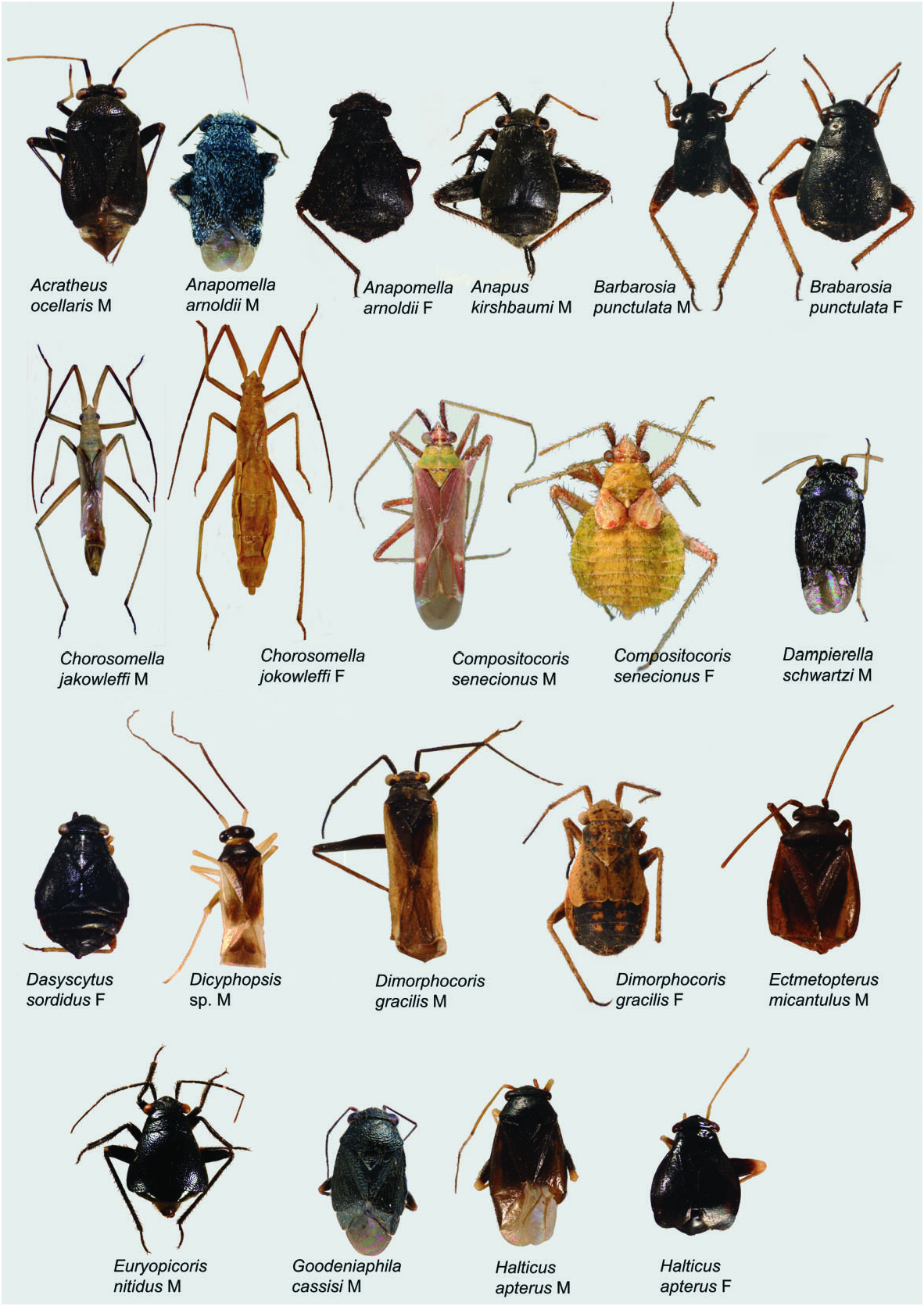
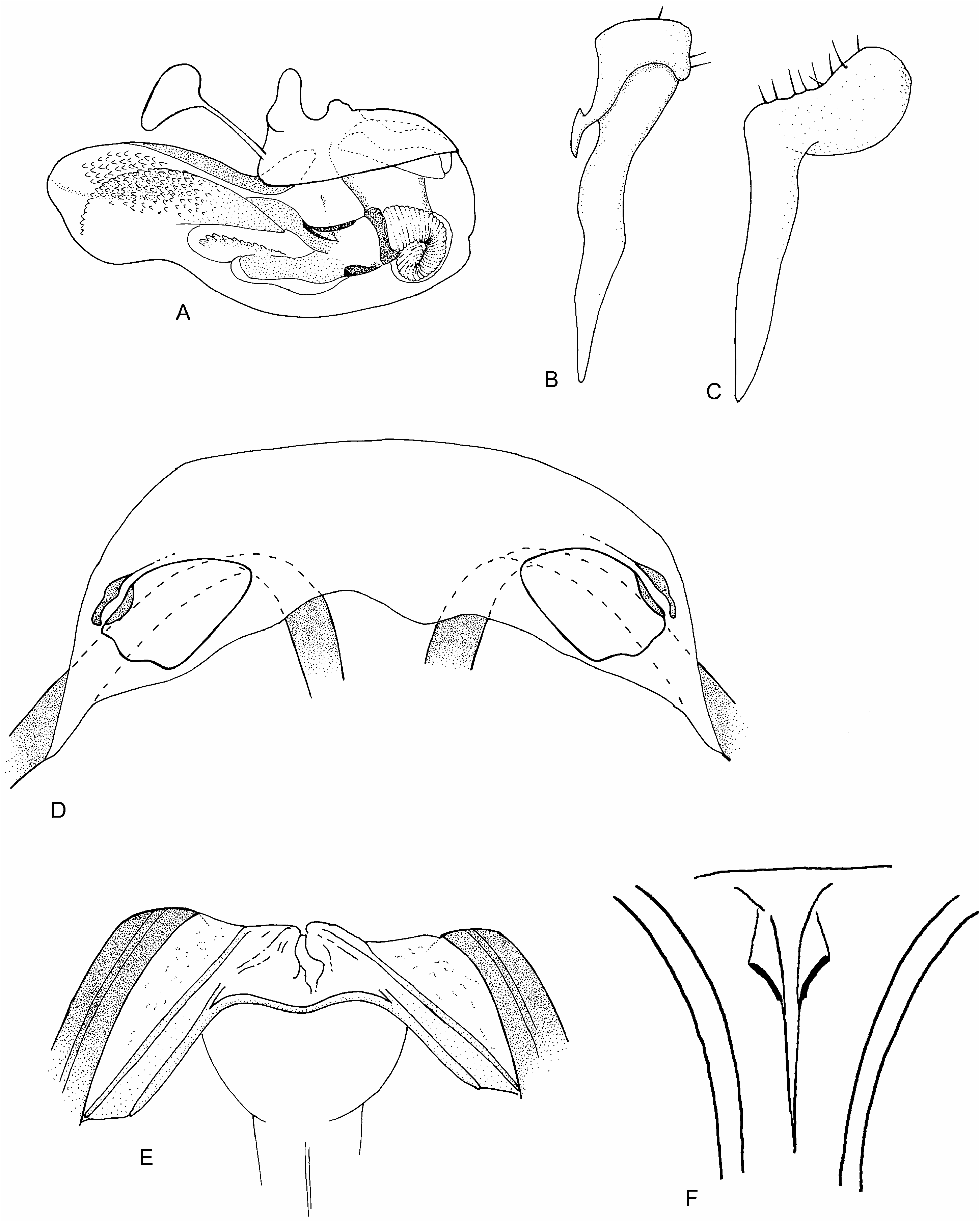
Redescription: Males macropterous, females coleopteroid or macropterous. Macropterous male 3.2–3.5 mm; macropterous female 3.3–3.5 mm; coleopteroid female 2.5– 3 mm. Coloration ( Fig. 3 View Figure 3 ): mostly dark brown to black, with some orange-brown, yellow-brown, and yellow markings. Macropterous male mostly orange-brown with yellow and tan markings. Surface and vestiture ( Fig. 3 View Figure 3 ): macropterous – body clothed in long white setae, with shorter brown setae, including on membrane of hemelytron. Coleopterous – with fewer setae, surface glossy. Antennae with white simple setae and several spinelike setae, most prominent on AI. Pronotum weakly rugulose. Structure: head ( Fig. 3 View Figure 3 ): transverse, approximately as tall as broad; width slightly wider than anterior margin of pronotum; vertex transversely sulcate, posterior margin carinate, weakly wraps around pronotum with eyes touching pronotum; genae height slightly greater than eye height; mandibular plate somewhat swollen in macropterous male; maxillary plate carinate. Antennae: insertion in line with lower margin of eye; AI weakly swollen, length approximately equal to eye height; AII weakly apically clavate, slightly more than three times AI length. Thorax ( Fig. 3 View Figure 3 ): pronotum trapezoidal, anterior weakly declivent in lateral view, collar not visible, callosite region poorly defined, posterior of humeral angles weakly depressed, posterior margin weakly concave; metathoracic spiracle large and exposed, narrowly bordered with evaporative bodies running downwards but not confluent with metathoracic evaporative area; MTG external efferent system triangular, occupies lower third of tergite, ostiole lateral, tongue-shaped, peritreme rounded, directly above ostiole, surrounded by evaporative bodies. Hemelytra ( Fig. 3 View Figure 3 ): macropterous – long and parallel-sided, extending beyond apex of abdomen, cuneus long and thin; membrane with two cells. Coleopterous – reduced to shell-like pads without division, posterior margins running diagonally from apex of claval commissure to lateral margin of abdomen, abdominal tergites IV and V partially covered, VI to apex exposed. Legs: long and slender; metafemora not swollen. Abdomen ( Fig. 3 View Figure 3 ): elongate and narrow in macropterous males, not surpassing hemelytral margins; rounded in macropterous females, laterally extending beyond lateral hemelytral margins. Coleopterous: rounded pear-shape. Male genitalia ( Fig. 19A–C View Figure 19 ): pygophore conical; left paramere apically folded, sensory lobe short and narrow, apophysis sinuate and tapering, angled downwards, apically bifid; right paramere spoon-shaped, angled laterally; phallotheca apically narrow, apicodorsally slightly compressed; ductus seminis elongate with flexible ribbing; base of secondary gonopore a sclerotized ring; apically irregularly sclerotized, transversely broadened; endosoma with weakly sclerotized dentate folds and apical lobe densely covered with fields of short teeth. Female genitalia ( Fig. 19D–F View Figure 19 ): DLP transverse, sclerotized rings indistinct, triangular-oval, lateral margin most strongly sclerotized, lateral margin and adjacent portion of DLP weakly upturned; posterior wall of bursa copulatrix membranous with thin, weakly sclerotized rods converging dorsally, ventral margin weakly sclerotized; margins of vestibulum symmetrical and very weakly sclerotized.
THE HALTICINI OF THE WORLD 589
Diversity and distribution: This monotypic genus is found throughout much of the Mediterranean region, but has not been collected in Syria, Egypt, Italy, and southern France ( Wagner, 1973).
Included species: Dasyscytus sordidus Fieber, 1864 * Mediterranean
Biology and host plant associations: Dasyscytus is found in vegetation at the edges of rivers and ditches, with adults collected in April and May ( Wagner, 1973). No clear host records exist, although specimens have been collected under Rhanterium epapposum (Asteraceae) in north-east Arabia ( Linnavuori, 1986).
Remarks: The monotypic genus Dasyscytus differs from all other halticines by the presence of setae on the hemelytral membrane in macropterous individuals (character 39-1), and the distinct folding of the left paramere ( Fig. 19B View Figure 19 : not coded in this analysis). In our phylogenetic analysis Dasyscytus is sister to Piezocranum , based on the thin hind femora of females (41-0) and the unbroken sclerotization of the apical region of the ductus seminis up to the secondary gonopore (62-1).
No known copyright restrictions apply. See Agosti, D., Egloff, W., 2009. Taxonomic information exchange and copyright: the Plazi approach. BMC Research Notes 2009, 2:53 for further explanation.
|
Kingdom |
|
|
Phylum |
|
|
Class |
|
|
Order |
|
|
Family |
Dasyscytus
| Tatarnic, Nikolai J. & Cassis, Gerasimos 2012 |
Dasyscytus
| Wagner E 1973: 30 |
| Carvalho JCM 1958: 8 |
| Carvalho JCM 1955: 68 |
| Reuter OM 1910: 162 |
| Kirkaldy GW 1906: 131 |
| Fieber FX 1864: 84 |