
Anastrepha rafaeli, Norrbom & Korytkowski, 2009
|
publication ID |
https://doi.org/ 10.11646/zootaxa.2182.1.1 |
|
DOI |
https://doi.org/10.5281/zenodo.5325601 |
|
persistent identifier |
https://treatment.plazi.org/id/03E887C3-FFB9-FFB3-FF68-0ACDFA833CD5 |
|
treatment provided by |
Felipe |
|
scientific name |
Anastrepha rafaeli |
| status |
sp. nov. |
Anastrepha rafaeli View in CoL , new species
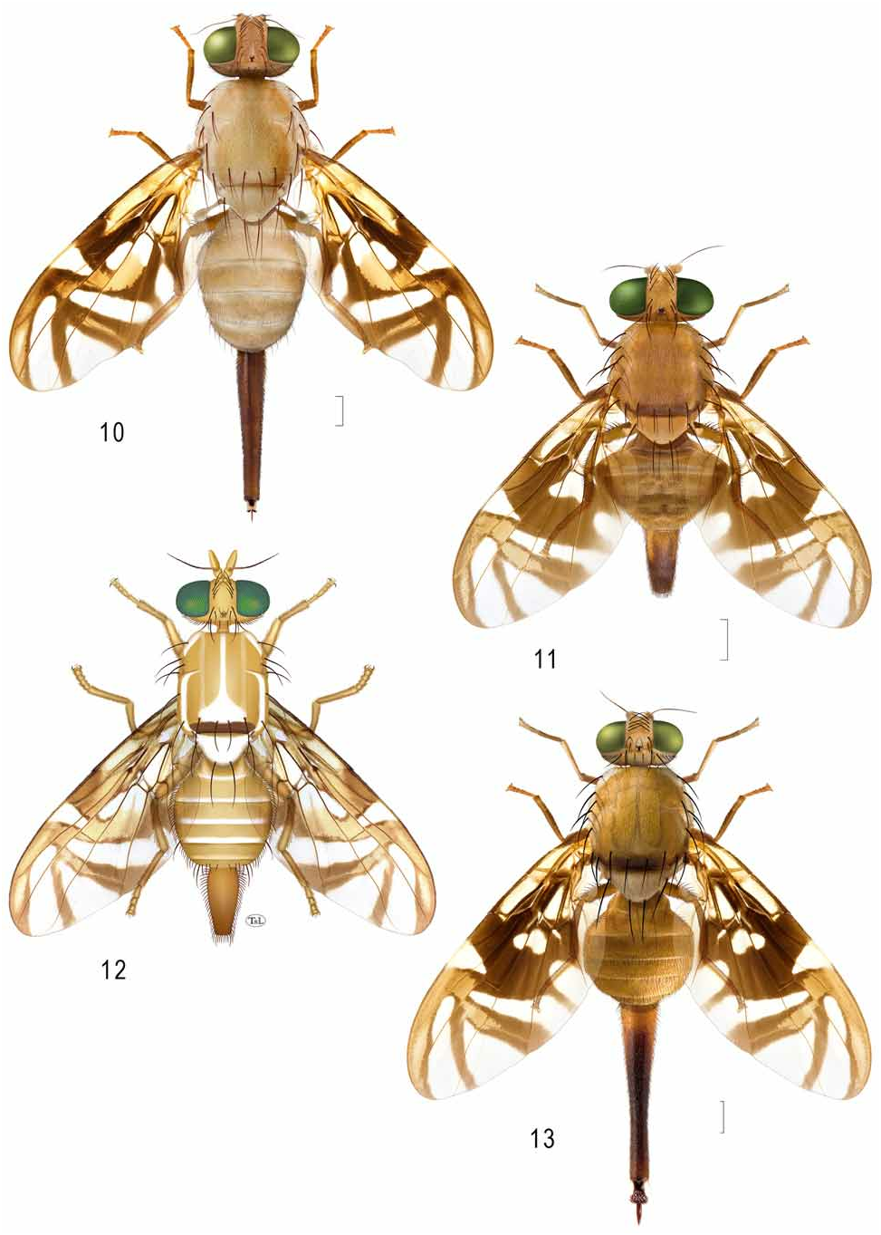
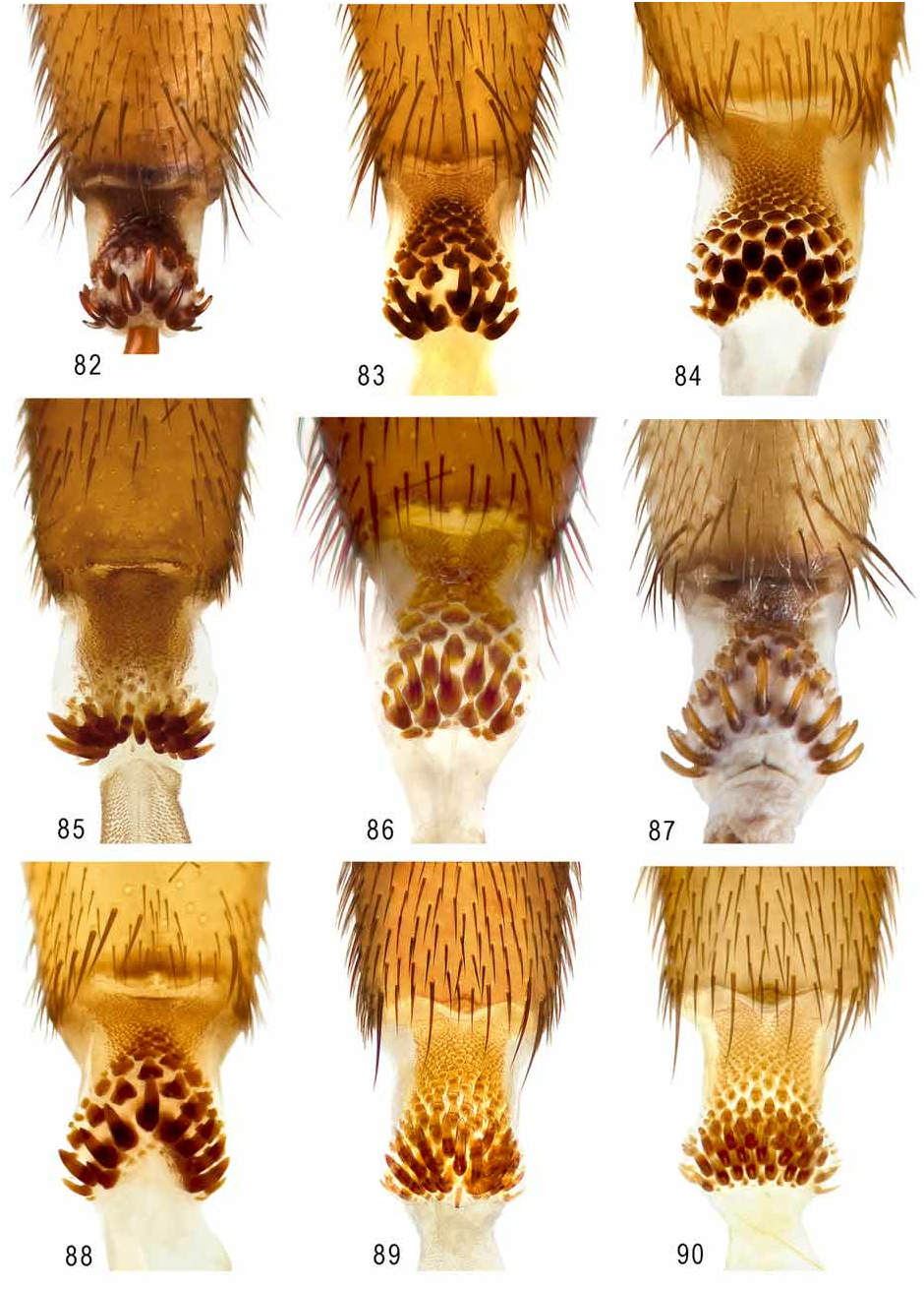
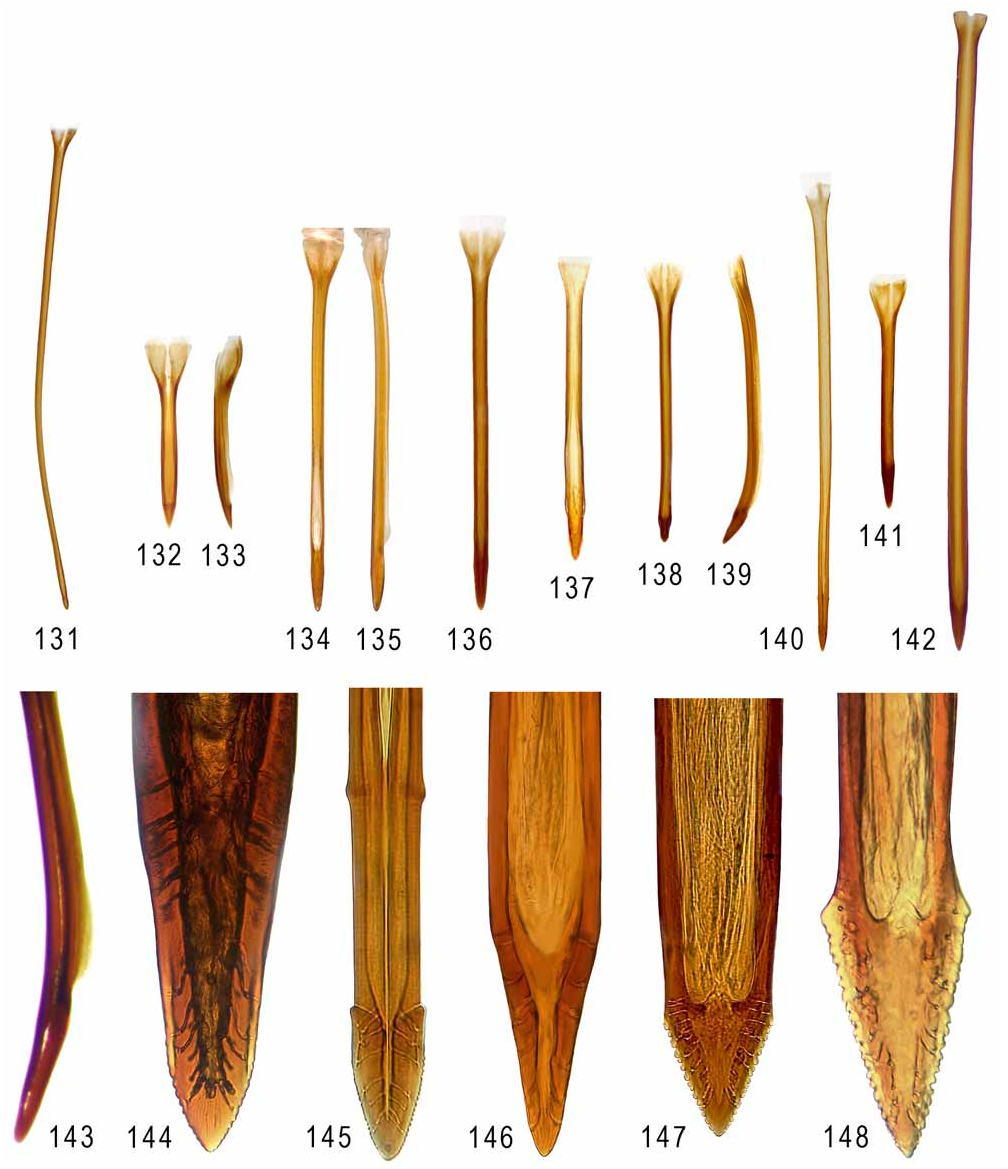
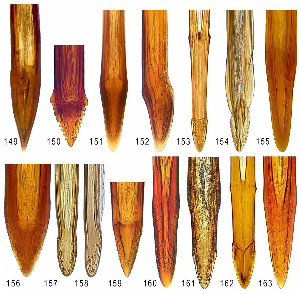
Figs. 11 View FIGURES 10–13 , 55 View FIGURES 54–62 , 89–90 View FIGURES 82–90 , 137 View FIGURES 131–148 , 161 View FIGURES 149–163
Anastrepha rafaeli Norrbom & Korytkowski View in CoL in Korytkowski 2004: 59 [nomen nudum; in key].
Diagnosis. Anastrepha rafaeli differs from most species of Anastrepha in having an extension from the basal part of the S-band to the posterior wing margin in the middle of cell cu 1 that is not connected to the posterior end of the proximal arm of the V-band. It differs from all of the other species having that wing character except A. amazonensis in having the basomarginal hyaline area in cell r 1 aligned with or slightly distal to crossvein r-m, and from all of those species except A. speciosa in usually having a brown posterior band on the scutum with a lateral extension to the intra-alar seta. It differs from A. amazonensis in having small hyaline areas in cells br and dm proximal to crossvein r-m.
Description. Mostly yellow to orange, with white to pale yellow markings. Setae orange brown to dark brown.
Head: Yellow to orange except ocellar tubercle brown and often with U-shaped brown mark on posterior half of orbital plate and vertex, touching eye, connected only to posterior side of mark on ocellar tubercle. 3 (rarely 2 or 4) frontal setae; 2 orbital setae, posterior seta well developed. Ocellar seta weak, at most 1.5 times as long as ocellar tubercle. Facial carina, in profile, straight to slightly concave dorsally and medially. Antenna extended 0.75–0.85 distance to ventral facial margin.
Thorax ( Fig. 11 View FIGURES 10–13 ): Mostly yellow to orange with following areas white or pale yellow (not always well differentiated in dried specimens): postpronotal lobe; quadrate medial area between dorsocentral setae, often diffuse or absent; paired sublateral scutal vitta from transverse suture almost to but not including intra-alar seta; entire scutellum; dorsal margin of anepisternum; katepimeron; and most of anatergite and katatergite. Posterior margin of scutum with broad brown band, broadest medially, including acrostichal seta and extended almost to dorsocentral seta, laterally with narrower extension including intra-alar seta. Subscutellum and mediotergite entirely orange. Mesonotum 2.96–3.19 mm long. Scutum entirely microtrichose; setulae mostly yellow to orange, brownish laterally. Katepisternal seta well developed, nearly as dark as and 0.67 times as large as to subequal to anepimeral seta.
Wing ( Fig. 55 View FIGURES 54–62 ): Length 6.17–6.83 mm, width 2.74–3.07 mm, ratio 2.11–2.26. Apex of vein R 1 at 0.52–0.54 wing length. Cell c 1.14–1.30 times as long as pterostigma; pterostigma 2.50–2.83 times as long as wide. Vein R 2+3 without sharp bends or undulations. Crossvein r-m at 0.60–0.65 distance from bm-cu to dm-cu on vein M. Vein M only slightly curved apically; cell r 4+5 1.06–1.22 times as wide at apex as at level of dm-cu. Cell bcu with distal lobe moderately long, length of bcu 1.62–1.70 times as long as anterior margin. Wing pattern mostly dark brown. C-band with cell bc yellowish to subhyaline; cell c yellowish to brownish basally and narrowly brown anteriorly and distally, posteriorly with elongate medial hyaline area, not extended into pterostigma or cell r 1; remainder of C-band dark brown. Cell c with elongate nonmicrotrichose posteromedial area small, 0.25–0.50 width of cell. C-band and S-band broadly connected along vein R 4+5; hyaline area in cell br small, usually not reaching vein R 4+5 and at most 0.67 times as long as distal brown area of cell; cell dm with basal hyaline area relatively small, but extending to posterior margin. Basal half of S-band entirely brown, without orange area in cell dm or bordering crossvein r-m; with lobelike projection to posterior wing margin in middle of cell cu 1; distal section of band orange with brown margins or sometimes mostly brown (at least with orange medial area between r-m and costa), moderately broad, at apex of vein R 2+3 0.64–0.87 times width of cell r 2+3, slightly to distinctly broadening in cell r 2+3, well separated from apex of vein M; hyaline area proximal to it ending at vein R 2+3. Hyaline basomarginal spot in cell r 1 short triangular, extended beyond vein R 2+3 but usually not to R 4+5, its apex aligned with or slightly distal to crossvein r-m. V-band complete, mostly brown, narrowly to broadly connected to S-band in cell r 2+3; proximal arm with medial orange area bordering anterior 0.33–0.50 of dm-cu, extending anteriorly beyond vein R 4+5; proximal arm moderately broad, gradually broadening posteriorly, without basal extension along wing margin, at level of vein M 1.5–2.8 times as wide as distal arm and 1.45–2.30 times as wide as hyaline area proximal to it in cells r 4+5 and dm; distal arm slender.
Abdomen: Mostly orange, without brown markings.
Male terminalia: Lateral surstylus short, extended beyond prensisetae by 1.0–1.5 times length of prensiseta; in lateral view broad, posteroapical corner bluntly acute; in posterior view gradually tapered to blunt, truncate apex, lateral and medial margins convex. Proctiger with ventral and lateral sclerotized areas connected but lateral areas separate dorsally. Phallus 2.45–2.75 mm long, 0.82–0.93 times as long as mesonotum; glans 0.40–0.50 mm long.
Female terminalia: Oviscape ( Fig. 11 View FIGURES 10–13 ) 2.00– 2.17 mm long, 0.65–0.68 times as long as mesonotum; distal half to two-thirds brown; spiracle at basal 0.38–0.39. Eversible membrane ( Figs. 89–90 View FIGURES 82–90 ) with 20–25 large, hooklike dorsobasal scales in triangular pattern. Aculeus ( Fig. 137 View FIGURES 131–148 ) straight to slightly ventrally curved in lateral view, 1.71–1.72 mm long; in ventral view base 0.18–0.19 mm wide; shaft 0.10 mm wide at midlength; tip ( Fig. 161 View FIGURES 149–163 ) 0.16 mm long, 0.08–0.09 mm wide, 1.78–2.39 times as long as wide, 0.04–0.05 mm wide in lateral view, 0.50–0.56 times ventral width, in ventral view basal part nearly parallel-sided with lateral margin slightly concave, apical 0.69–0.75 triangular, finely serrate, basal serrations curving slightly onto dorsal side. Spermathecae spherical.
Distribution. Anastrepha rafaeli is known from Brazil (Roraima) and possibly western Venezuela.
Biology. The host plants and other aspects of the biology of this species other than dates of capture of adults are unknown.
Type Data. Holotype female ( INPA, USNMENT00216288 View Materials ), BRAZIL: Roraima: Rio Uraricoera, Ilha de Maraca , armadilha de Malaise, 2–13 May 1987, J. A. Rafael, J. E. B. Brasil & L. S. Aquino . Paratypes: same data as holotype, 5♂ 2♀ ( INPA USNMENT00216289-90 View Materials , USNMENT00216295-99 View Materials ) 2♂ 3♀ ( USNM USNMENT00052106 About USNM , USNMENT00216287 About USNM , USNMENT00216292-94 About USNM ) ; same, 20–30 Mar 1987, L. S. Aquino, 1♂ ( USNM USNMENT00216300 About USNM ) .
Other specimens examined. VENEZUELA: Zulia: Lake Maracaibo , 1913, 1 adult ( UMSP USNMENT00054868 View Materials ); this specimen, which lacks most of its abdomen, is tentatively identified, but matches A. rafaeli in wing pattern and setal color. It is not designated as a paratype .
Etymology. This species is named for Dr. J. A. Rafael (INPA), one of the collectors of the type series.

No known copyright restrictions apply. See Agosti, D., Egloff, W., 2009. Taxonomic information exchange and copyright: the Plazi approach. BMC Research Notes 2009, 2:53 for further explanation.
|
Kingdom |
|
|
Phylum |
|
|
Class |
|
|
Order |
|
|
Family |
|
|
Genus |
Anastrepha rafaeli
| Norrbom, Allen L. & Korytkowski, Cheslavo A. 2009 |
Anastrepha rafaeli
| Korytkowski, C. A. 2004: 59 |