
Arenopontia syltensis, Sak & Karaytuğ & Huys, 2024
|
publication ID |
https://doi.org/10.11646/zootaxa.5433.1.1 |
|
publication LSID |
lsid:zoobank.org:pub:06E5A735-A276-41D7-A9EE-B09642D953B6 |
|
DOI |
https://doi.org/10.5281/zenodo.10953780 |
|
persistent identifier |
https://treatment.plazi.org/id/4B7EBE47-018E-471B-8451-22B86E4CB9B0 |
|
taxon LSID |
lsid:zoobank.org:act:4B7EBE47-018E-471B-8451-22B86E4CB9B0 |
|
treatment provided by |
Plazi |
|
scientific name |
Arenopontia syltensis |
| status |
sp. nov. |
Arenopontia syltensis sp. nov.
https://zoobank.org/ 4B7EBE47-018E-471B-8451-22B86E4CB9B0
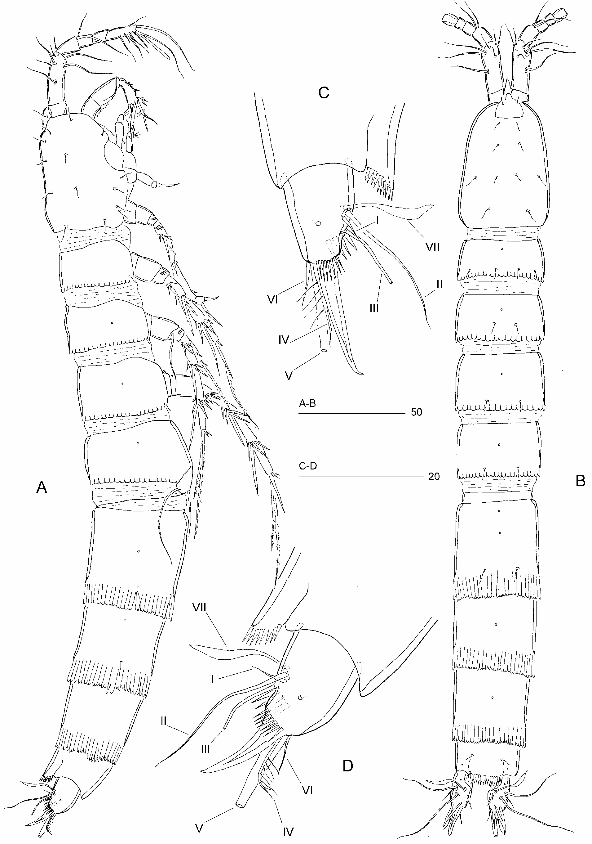
( Figs 17–20 View FIGURE 17 View FIGURE 18 View FIGURE 19 View FIGURE 20 )
Type locality. Germany, Isle of Sylt , List; sandy beach .
Material examined. Holotype ♀ (dissected on six slides) ( NHMUK reg. no 2024.1044) . Paratypes are 1 ♀ dissected on three slides ( NHMUK reg. no 2024.1045), 1 ♂ dissected on eight slides ( NHMUK reg. no 2024.1046), and 7 ♀♀ and 3 ♂♂ in ethanol ( NHMUK reg. nos 2024.1047–1049); all collected at type locality; leg. R. Huys & S. Conroy-Dalton, 25 August 1996 .
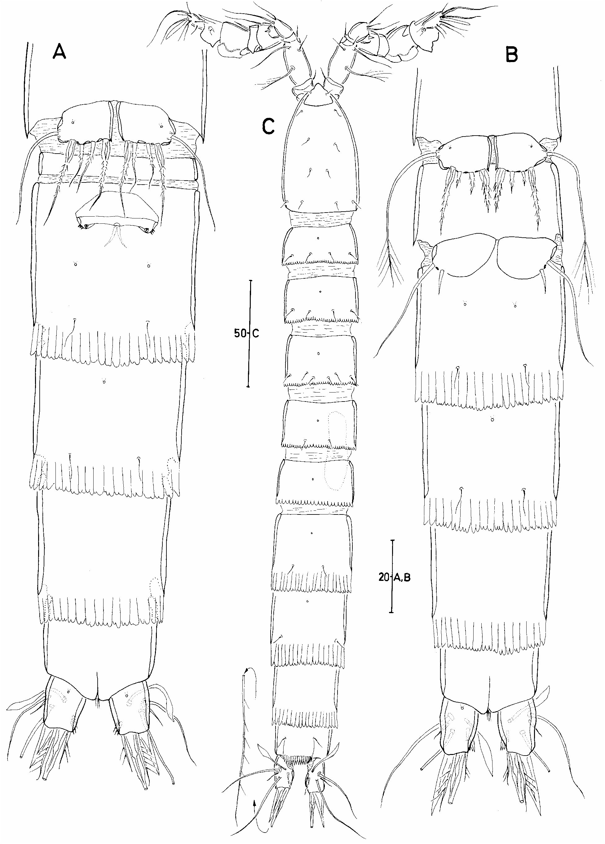
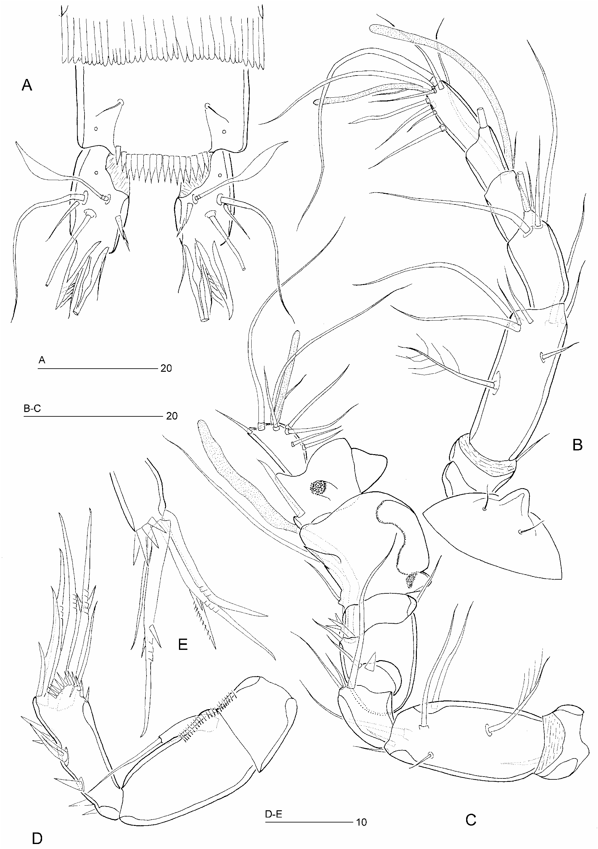
Description of female. Total body length from tip of rostrum to posterior margin of caudal rami 341–391 μm (mean = 368 μm; n = 7; holotype = 375 μm). Maximum width 44 μm measured at posterior margin of cephalothorax. Body slender and cylindrical, without clear distinction between prosome and urosome ( Fig. 17A, B View FIGURE 17 ). Hyaline frills of thoracic somites weakly developed and crenulated, those of genital double-somite and free abdominal somites strongly developed and consisting of rectangular digitate lappets ( Figs 17A, B View FIGURE 17 ; 18A View FIGURE 18 ; 19A View FIGURE 19 ). Genital double-somite ( Figs 17A, B View FIGURE 17 ; 18A View FIGURE 18 ) 1.15 times longer than wide (measured in ventral aspect); without chitinous ribs marking original segmentation; with two middorsal, two lateral and two ventral pores. Anal somite ( Figs 17C, D View FIGURE 17 ; 19A View FIGURE 19 ) with two dorsal, two lateral and two ventral pores. Anal operculum spinulose; with coarse spinules along free distal margin ( Fig. 19A View FIGURE 19 ). Anus positioned subterminally between caudal rami. Rostrum ( Fig. 19B View FIGURE 19 ) small, broadly subtriangular, tapering distally, with two delicate sensilla.
Caudal rami ( Figs 17D View FIGURE 17 ; 19A View FIGURE 19 ) approximately 2.9 times longer than maximum width (measured in dorsal view), tapering posteriorly; with single pores dorsally in anterior quarter ( Fig. 19A View FIGURE 19 ) and laterally at level of insertion of seta III ( Fig. 17D View FIGURE 17 ); outer distal corner produced into posteriorly directed, recurved spinous process, accompanied at base by outer spinular row ( Fig. 17D View FIGURE 17 ); dorsal surface without spur-like process, inner margin with few spinules ( Fig. 19A View FIGURE 19 ). Armature consisting of seven setae; seta I small; setae II and III long and naked; seta IV short, sparsely pinnate, located between seta V and posterior spinous process; seta V long and with fracture plane; seta VI small, naked and located at inner distal corner; seta VII foliaceous and tri-articulate at base.
Antennule ( Fig. 19B View FIGURE 19 ) slender, six-segmented. Segment 1 with a short seta near anterodistal margin. Segment 2 longest, about 2.6 times longer than wide. Segment 4 with long aesthetasc (L: 32 μm) fused at base with seta. Distal segment with seven naked setae (one of which spatulate) and apical acrothek consisting of short aesthetasc (L: 17 μm) and two slender setae. All setae naked except for plumose seta on dorsal surface of segment 2. Armature formula: 1-[1], 2-[7 + 1 plumose], 3-[5], 4-[1 + (1 + ae)], 5-[1], 6-[7 + acrothek].
Antenna ( Fig. 19D, E View FIGURE 19 ). Coxa small (not figured), without ornamentation. Basis and proximal endopodal segment discrete, each with spinule row along exopodal margin. Exopod one-segmented, elongate, with a naked apical seta (about 1.4 times longer than exopod). Distal endopodal segment with spinules on medial surface and at outer distal corner; medial armature consisting of two short spines; apical armature consisting of two spines and three geniculate setae, longest of which with spinules around geniculation and fused basally to naked accessory seta.
Mandible, maxillule, maxilla and maxilliped as in A. anatolica sp. nov.
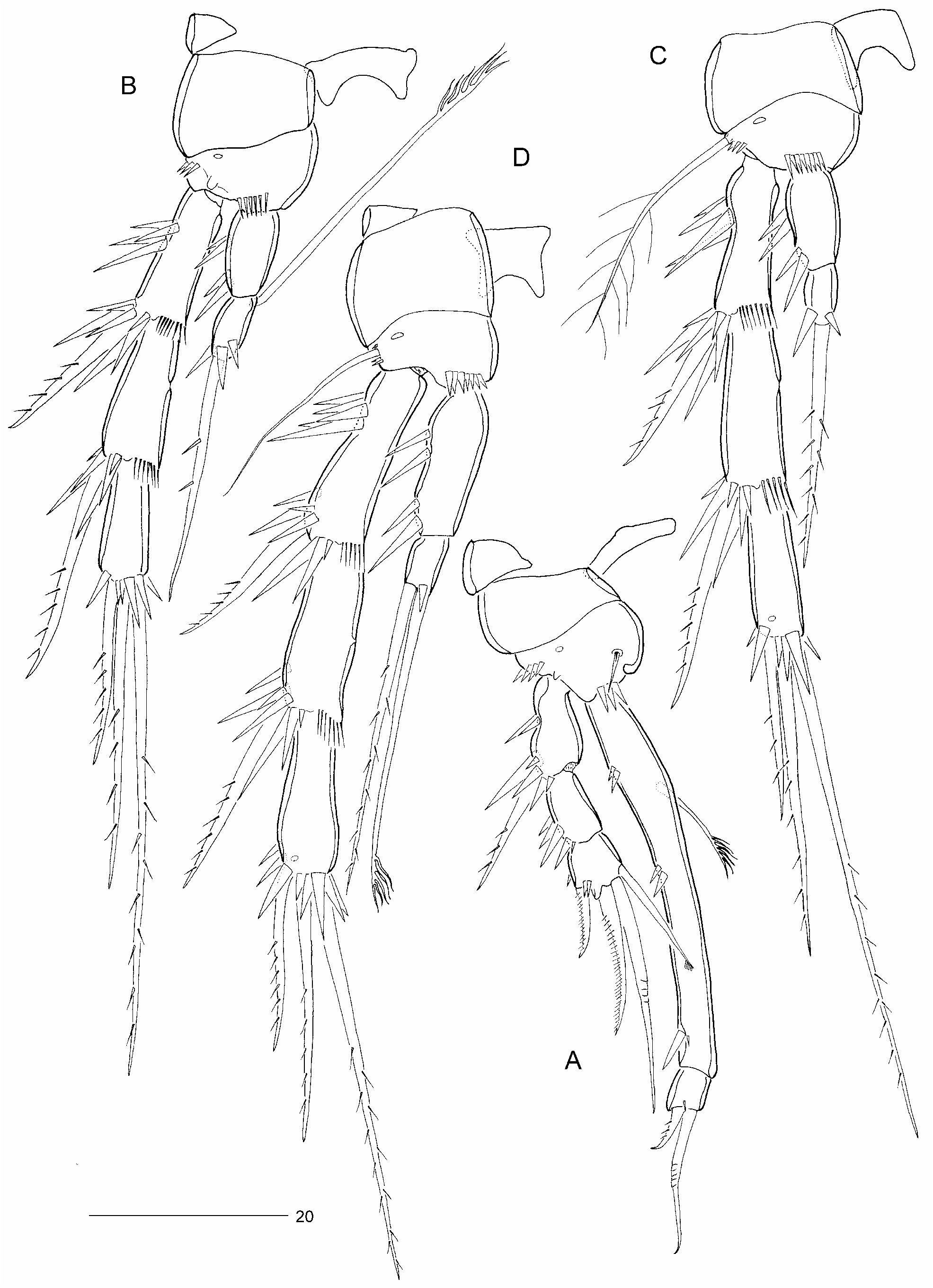
P1 ( Fig. 20A View FIGURE 20 ). Intercoxal sclerite wide and subrectangular. Praecoxa represented by triangular naked sclerite. Coxa without ornamentation. Basis with few coarse spinules near base of endopod and around outer margin; anterior surface with a pore and a small, setiform, naked spine near medial margin. Exopod three-segmented; all segments with spinules around outer margin (smaller and fewer on exp-3); exp-1 1.3 times longer than exp-2, with unipinnate outer spine; exp-2 without outer element; exp-3 with short unipinnate outer spine, a longer curved unipinnate spine and one geniculate seta distally, and one inner, apically penicillate seta subdistally. Endopod two-segmented, prehensile; enp-1 about 11 times longer than wide, and 1.85 times longer than exopod; with a serrate inner seta in proximal third, and three groups of two spinules along outer margin; enp-2 about as long as wide, with a short unipinnate outer spine and a slightly longer geniculate inner claw, in addition to one small spinule.
P2–P4 ( Fig. 20B–D View FIGURE 20 ) intercoxal sclerites naked, with concave distal margin. Praecoxae small and naked. Coxae wider than long and without ornamentation. Bases smaller than coxae, with a spinular row near base of endopod and at outer distal corner; anterior surface with a pore near coxa-basis boundary; outer basal seta absent (P2), plumose (P3) or naked (P4). Exopods three-segmented; segments with spinular ornamentation as figured; inner distal spine of exp-3 bipinnate, all other exopodal elements unipinnate; hyaline frills of exp-1 and -2 well developed; exp-2 with lateral pore halfway down inner margin length; P3–P4 exp-3 with anterior surface pore. Endopods two-segmented, with enp-1 distinctly longer than enp- 2 in P4; P2–P4 enp-1 about 1.4, 1.7 and 3 times longer than their respective distal segments, with few coarse spinules along outer margin as figured. P2 enp-2 with a long, apically serrate, posteriorly directed seta near proximal inner margin. Enp-2 with a long sparsely unipinnate (P2) or bipinnate (P3) seta terminally. P4 enp-2, with a basally fused, apically serrate seta, and a long bipinnate seta at outer distal corner. Spine and seta formula as follows:
Fifth legs ( Fig. 18A View FIGURE 18 ) closely set together but not touching medially. Baseoendopod and exopod fused forming a subrectangular plate with anterior surface pore; distal margin with four pinnate setae, middle ones shortest and about equally long, inner and outer ones swollen in proximal third; outer basal seta long and plumose.
Genital field positioned near anterior margin of genital double-somite ( Fig. 18A View FIGURE 18 ). Genital apertures ( Fig. 18A View FIGURE 18 ) fused, forming median common slit; closed off by fused P6 forming operculum, each with three rudimentary armature elements; copulatory pore small, located midventrally, close to genital slit; seminal receptacles difficult to discern.
Description of male. Total body length from tip of rostrum to posterior margin of caudal rami 328–343 μm (mean = 336 μm; n = 4). Body ornamentation ( Figs 18B, C View FIGURE 18 ) essentially as in female. Sexual dimorphism in antennule, urosomal segmentation, P5, P6 and caudal ramus. Spermatophore length approximately 37 μm.
Antennule ( Fig. 19C View FIGURE 19 ) nine-segmented, haplocer; geniculation between segments 7 and 8. Segments 7 and 8 swollen, expanded along posterior margin, each with spinulose process. Segment 2 longest and about twice longer than wide; segment 4 an incomplete sclerite with one small spiniform element and one small naked seta; segment 5 with one naked seta and one spinulose element plus long aesthetasc (L: 38 μm) fused basally to a long slender seta; segment 6 with one seta; segment 7 with one modified spine and a seta; segment 8 with a strong naked spine; distal segment with seven naked setae (none of which noticeably spatulate) and apical acrothek. Setal formula: 1-[1], 2-[6 + 1 plumose], 3-[3], 4-[2], 5-[1 + 1 spinulose + (1 + ae)], 6-[1], 7-[1 + 1 modified], 8-[1], 9-[7 + acrothek]. Acrothek consisting of short aesthetasc (L: 17 μm) fused basally to two slender setae.
P5 ( Fig. 18B View FIGURE 18 ) with anterior surface pore and with armature as in female but all elements on distal margin comparatively shorter and more spiniform; outer element longest and bipinnate, middle ones short and bipinnate, inner one bipinnate but without flagellate distal part observed in ♀. Outer basal seta plumose.
Sixth legs ( Fig. 18B View FIGURE 18 ) asymmetrical, with smallest P6 closing off functional gonopore; each with short inner and long outer seta; both elements naked.
Caudal ramus ( Fig. 17C View FIGURE 17 ) as in ♀ except for posterior spinous process being comparatively longer.
Etymology. The specific epithet (a noun in the genitive case) is derived from the name of the island where the type locality is located, the Isle of Sylt.
Remarks. Arenopontia syltensis sp. nov. differs from its congeners by the distinctive spinulose ornamentation of the anal operculum ( Fig. 19A View FIGURE 19 ) and the modified male antennules ( Fig. 19C View FIGURE 19 ) which are characterized by the enlarged segments either side of the geniculation (segments 7–8), each one of which displaying an expansion along the posterior margin and a spinulose process on the dorsal surface.The species shares with A. riedli and A. basibuyuki sp. nov. the presence of five elements on the fifth legs of both sexes (four in all other species) but differs from the former in the presence of only one apical element on P2–P3 enp-2 (instead of two) and the absence of a dorsal spur on the caudal ramus, and from the latter in the ornamentation of P1 enp-1 and the presence of two elements on the male P6 (instead of one).
It is conceivable that A. syltensis sp. nov. occurs sympatrically with A. subterranea in sandy beaches on the Isle of Sylt and possibly in other localities along the German North Sea coast. This is clearly indicated by Mielke’s (1975: 109) statement that his material of A. subterranea showed variability in the armature of the fifth legs (sometimes with five elements) and the ornamentation of the anal operculum (sometimes with coarse spinules), suggesting that it included A. syltensis sp. nov. His illustration of P1 (Abb. 73D), particularly the relative length of enp-1, also indicates that it was based on A. syltensis sp. nov. rather than A. subterranea which displays a shorter endopod ( Kunz 1937: Abb. 9–Fig. 43).
| NHMUK |
Natural History Museum, London |
| R |
Departamento de Geologia, Universidad de Chile |
No known copyright restrictions apply. See Agosti, D., Egloff, W., 2009. Taxonomic information exchange and copyright: the Plazi approach. BMC Research Notes 2009, 2:53 for further explanation.
|
Kingdom |
|
|
Phylum |
|
|
Class |
|
|
Order |
|
|
Family |
|
|
Genus |