
Phareicranaus divisor, Pinto-Da-Rocha, Ricardo & Bonaldo, Alexandre B., 2011
|
publication ID |
https://doi.org/10.5281/zenodo.207840 |
|
DOI |
https://doi.org/10.5281/zenodo.6185984 |
|
persistent identifier |
https://treatment.plazi.org/id/03EB8794-FF8F-FFD7-FF08-F990017FBEDB |
|
treatment provided by |
Plazi |
|
scientific name |
Phareicranaus divisor |
| status |
sp. nov. |
Phareicranaus divisor View in CoL sp. n.
( Figs. 8 View FIGURE 8 , 9 View FIGURE 9 A–E)
Types. Male holotype: Brasil. Acre, Parque Nacional da Serra do Divisor (between 09º24’ and 73º12’ S, 07º32’ and 73º59’W), 22.XI–12.I.2000, M.B. Souza leg. (MZSP-19238). Paratype male: Brasil. Acre, Cruzeiro do Sul, Rio Moa ( 07º24'09"S e 73º16'44"W), XI.1996, R.S. Vieira leg. (MZSP-15915).
Etymology. In reference to the type locality. Name in apposition.
Diagnosis. Phareicranaus divisor sp. n. belongs to the curvipes species-group, which has circular white spots around tubercles on the posterior part of prosoma, area I–III, and the posterior margin ( P. angelicus , P. gracilis , P. hermosa , P. ortizi and P. singularis ). It differs from them based on the combination of the following characters: male femur IV with one curved submedian spine; prosoma with 10 circular white spots, both halves of area I with 12; area II with 9, III with 11, posterior margin with 17, lateral margin without white spots.
Description. Male holotype. Measurements (mm): Dorsal scutum length 9.3; width 9.2; prosomal length 4.6; width 6.3; pedipalpal femur 5.1; femur IV 19; leg I 26; II 56; III 40; IV 56.
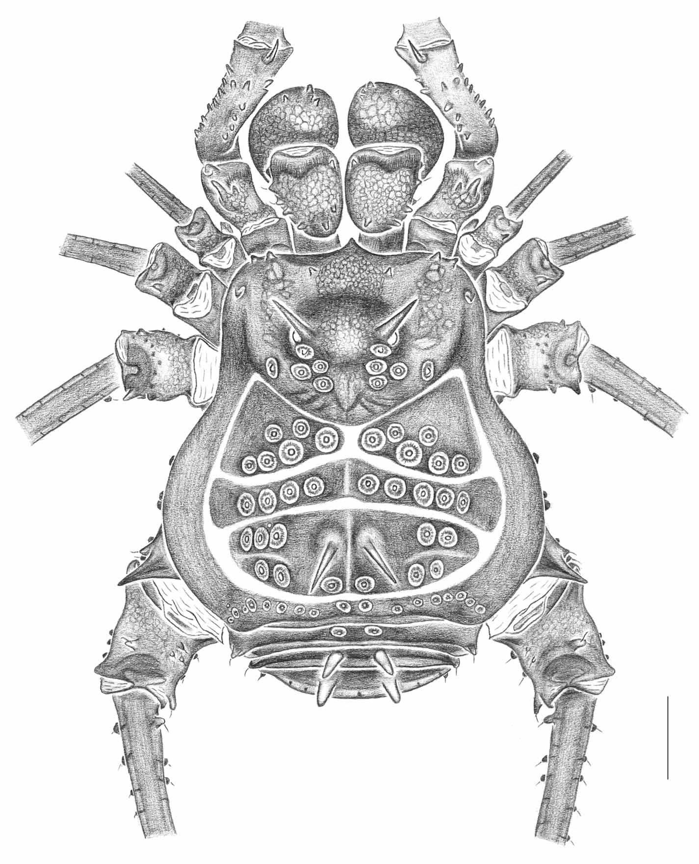
Dorsal scutum ( Figs. 8 View FIGURE 8 , 9 View FIGURE 9 A). Anterior margin with 2 tubercles each side, 2 on paramedian region. Ocularium with 2 sharp divergent long spines, 1 pair of anterior and 1 pair of posterior tubercles. Carapace with 4 pairs of tubercles behind ocularium. Lateral margin of scutum without tubercles, with small pits close to coxa IV. Area I with 6 tubercles in each half; area II with 4–5 each side; III with 2 sharp divergent spines, 5–6 each side; posterior margin with 15 tubercles. Free tergite I with 2 paramedian tubercles, 1 tubercle each corner; II–III with 2 paramedian spines, 1 tubercle each corner. Anal operculum irregularly tuberculate.
Venter. Coxa I with median row of 7 tubercles (3 larger), 3 posterior, 4 anterior, 4 apical; II with 5 anterior tubercles, median row of 5, 4 posterior, 4 apical; III with a row of 7 tubercles, 2 anterior, 4 posterior; IV with scattered tubercles, one pair of blunt apophyses near the stigmata. Stigmatic area and free sternites with one row of low setiferous tubercles.
Chelicera. Basichelicerite with 9 tubercles; hand with many frontal tubercles; fixed finger with one large median 3 small distal tubercles; movable finger with 4 large tubercles.
Pedipalp. Coxa smooth dorsally. Trochanter with two dorsal (one larger), four ventral (two large), and two retrolateral and small tubercles. Femur with ventral row of six large tubercles (basal bifid), dorsal row of five and apical very long and sharp, five mesal, and two prolateral, small tubercles. Patella with eight dorsal tubercles. Tibia with many dorsal tubercles, mesal iiIi/Ii, ectal iIi. Tarsus mesal/ectal IiIi ( Fig. 9 View FIGURE 9 B).
Legs. Coxa I dorsally with 1 anterior long and 1 posterior smaller tubercle; II with one long tubercle in front of ozopore; III with anterior tubercle directed to coxa II and posterior directed to IV; IV with some latero-ventral tubercles, 1 apical tubercle, 1 sharp dorsoapical apophysis. Trochanters I–IV small-tuberculate; II and IV with dorsomedian larger tubercle. Femora tuberculate; III–IV with two dorsoapical tubercles, posterior larger; IV with retrolateral submedian upcurved tubercle. Patellae tuberculate. Tibia IV with two retrolateral upcurved tubercles (basal larger) ( Fig. 9 View FIGURE 9 C). Tarsal formula: 8, 13–14, 8, 9–10.
Male genitalia. Ventral plate pyriform, with a slight shallow cleft in distal border, with 6 lateral setae from median part to apical of lateral margin. Stylus smooth, straight, apex bent in obtuse angle, not swollen, apical ridges bearing spiniform ventro-distal stylar apophysis ( Fig. 9 View FIGURE 9 D, E).
Color. Body, chelicerae and pedipalps dark brown with black reticulate sculpturing on median anterior region to ocularium, sulci light brown. Tubercles of dorsal scutum and free tergite I yellowish circled with white. Spines of ocularium and free tergites II–III yellowish, area III spines black with tip yellowish. Legs, trochanter brown, femur-tibia black, metatarsus greenish, tarsus yellowish.
Female. Unknown.
Distribution. Recorded in State of Acre, westernmost Brazil.

No known copyright restrictions apply. See Agosti, D., Egloff, W., 2009. Taxonomic information exchange and copyright: the Plazi approach. BMC Research Notes 2009, 2:53 for further explanation.
|
Kingdom |
|
|
Phylum |
|
|
Class |
|
|
Order |
|
|
Family |
|
|
Genus |