
Aprostocetus dehradunensis Singh, 2022
|
publication ID |
https://doi.org/10.11646/zootaxa.5129.1.1 |
|
publication LSID |
lsid:zoobank.org:pub:3770D8E0-7496-4704-8587-F302E5250EBC |
|
DOI |
https://doi.org/10.5281/zenodo.6502091 |
|
persistent identifier |
https://treatment.plazi.org/id/03EBF447-361A-D076-56E4-FE6DFDF74D67 |
|
treatment provided by |
Plazi |
|
scientific name |
Aprostocetus dehradunensis Singh |
| status |
sp. nov. |
3. Aprostocetus dehradunensis Singh View in CoL sp. nov.
( Figs 51–73 View FIGURES 51–60 View FIGURES 61–66 View FIGURES 67–73 )
Diagnosis. FEMALE: Body yellow with a longitudinal black line on the sub-lateral side of pronotum, and one black band across the middle of gaster. Head with toruli placed just below the middle of face, their lower margins slightly below a line joining the lower margins of the eyes; subantennal grooves short, reaching midway between toruli and clypeus. Antenna with scape not extending beyond vertex; with one anellus; funicle 3-segmented, all funicle segments elongated and subequal; club 3-segmented, appearing 2-segmented, apical segment small and indistinctly separated from second. Forewing: SMV with 2 dorsal setae; marginal vein 4.2× as long as stigmal vein. Rim of propodeal spiracle entirely visible; paraspiracular carina present; median carina not sharp; area of propodeum between paraspiracular carinae with weak longitudinal striae converging posteriorly. Ovipositor sheaths slightly exserted, about 0.5× as long as postcercale.
MALE: Body color lighter than in female, black spot rather than line on lateral side of pronotum, black band on gaster wider and placed at the middle of posterior half. Toruli above a line joining the lower margins of the eyes, their lower margins touching the line; subantennal grooves long, reaching two-thirds the distance between toruli and clypeus. Antennal scape with a pale brown ventral plaque at distal half covering about 0.3× of ventral margin; funicle 4-segmented, without whorls of longer setae at base; F1 shortest; club clearly 3-segmented. Forewing with marginal vein 3× as long as stigmal vein. Digitus of genitalia with one long digital spine, paramere with 2 setae.
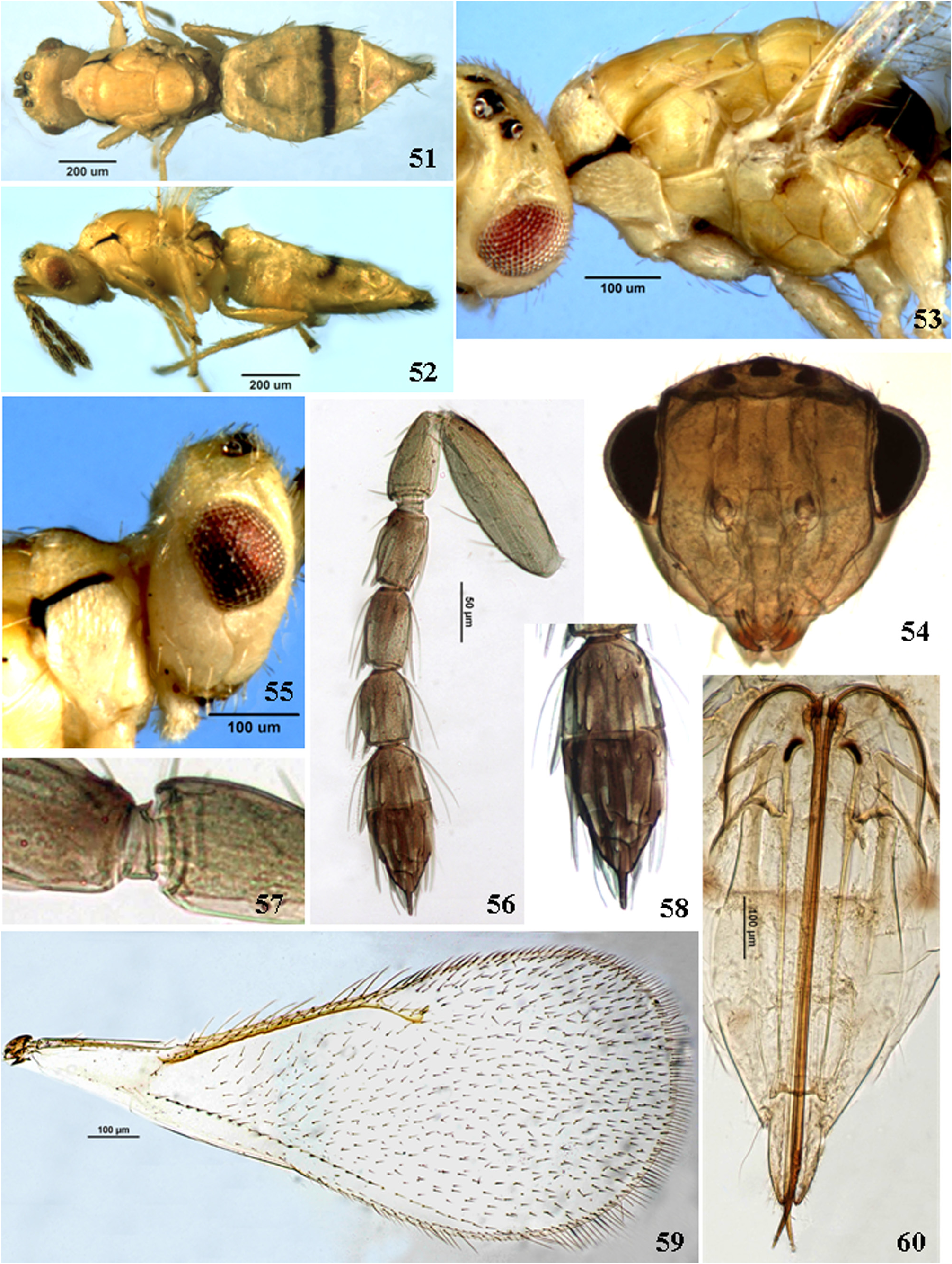
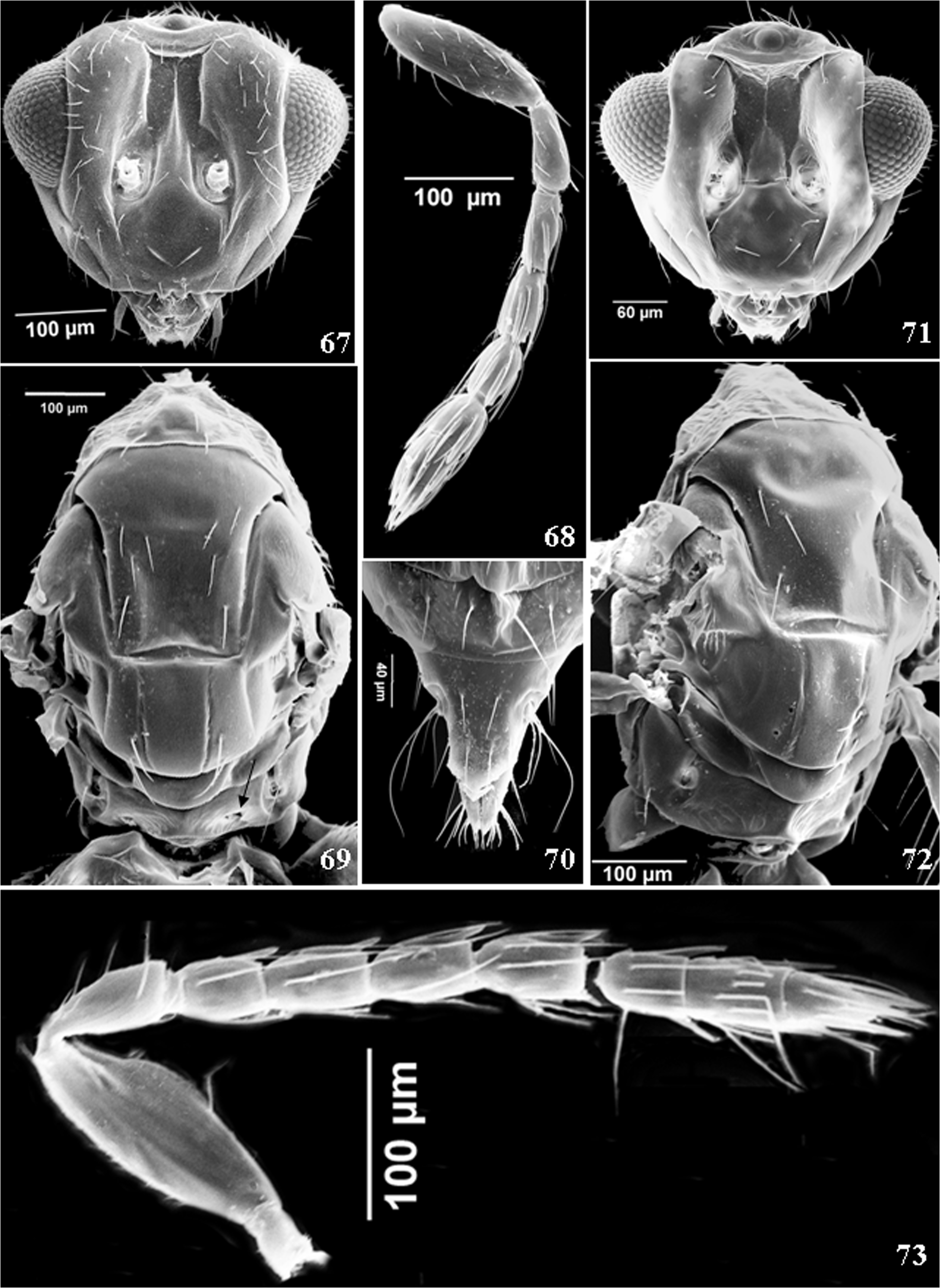
Description. FEMALE ( Figs 51–60 View FIGURES 51–60 , 67–70 View FIGURES 67–73 ). Body size 1.37 ± 0.04mm (freshly killed in alcohol, n = 3, range 1.32 to 1.40 mm). Holotype 1.4 mm (Card-mounted).
Color/Sculpture. Body ( Figs 51, 52 View FIGURES 51–60 ) completely yellow with dark brown stripe on sub-lateral side of pronotum, originating from pronotal spiracle and running anteriorly. Gaster with sharp black band on Gt4 and Gt5. Lateral sides of metanotum and propodeum with brownish hue. Antenna yellowish, ventrally lighter; dorsal margin of scape, basal third of each funicle and basal club segments light brown; distal 2 club segments brown. Pretarsi dark brown. Head ( Fig. 67 View FIGURES 67–73 ) triangular, sparsely setose and with very faint longitudinal reticulate sculpture, more prominent on lower face; parascrobal area dorsally with 4 rows of setae; vertex with 40-50 setae; scrobes deep; interantennal area with a pair of setae at the base; subantennal grooves short, almost reaching one-third the distance between toruli and cylpeal margin; malar sulcus distinctly curved ( Fig. 55 View FIGURES 51–60 ); lower face between malar sulci with 7 pairs of setae; eyes with small setae. Mesosoma ( Fig. 69 View FIGURES 67–73 ), sculpture of pronotum longitudinal reticulate. Mesoscutum and scutellum silky with fine and delicately engraved longitudinal reticulations. Midlobe of mesoscutum without median line, with single row of 4 adnotaular setae (second seta from anterior margin is out of alignment with other 3 setae); lateral lobe of mesoscutum with faint longitudinal reticulations and with 3–4 setae on the lateral and posterior margins. Scutellum with finer reticulation than mid lobe of mesoscutum, with 2 pairs of equal setae outside submedian groove, anterior pair placed at the distal two-thirds near submedian groove, posterior pair at the distal scutellum margin, nearer to submedian groove; dorsellum sculptured as scutellum. Propodeal median carina not prominent; paraspiracular carina developed; area between median and paraspiracular carinae with longitudinal striae converging posteriorly, at the middle; slightly posterior to the middle with a pit on either side ( Fig. 69 View FIGURES 67–73 , arrow). Callus with 2 setae, anterior seta stronger and twice as long as posterior one. Legs yellow; wings hyaline, veins light brown; SMV of forewing with 2 dorsal setae.
Structure. Head clearly wider than mesosoma (370: 339) ( Fig. 51 View FIGURES 51–60 ); in frontal view triangular ( Fig. 54 View FIGURES 51–60 ) 1.2× as wide as high (370: 300), 1.6× as wide as frontovertex (370: 231); toruli placed just below middle of face, their ventral margins slightly below a line joining the lower margins of the eyes; malar space about 0.9× as long as eye length (118: 134), mouth slightly wider than malar space length (125: 118); in dorsal view 2.2× as wide as long (370: 170); 1.3× as wide as eye length (370: 128); POL 1.3× OOL (95: 74). In lateral view ( Fig. 55 View FIGURES 51–60 ) eye as long as malar sulcus. Antenna ( Figs 56 View FIGURES 51–60 , 68 View FIGURES 67–73 ) with scape 3.1× as long as wide (159: 51) not extending beyond vertex dorsally; pedicel about 1.8× as long as wide (73: 40); with one anellus ( Fig. 57 View FIGURES 51–60 ); funicle segments subequal in length, longer than wide; F1 0.8× as long as pedicel length (74: 62) and 1.6× as long as wide (62: 40), F2 1.7× as long as wide (63: 36), F3 1.4× as long as wide (61: 44). Club 3-segmented; C1 and C2 both slightly wider than long (54: 50, 54: 51, C3 ( Fig. 58 View FIGURES 51–60 ) indistinctly separated from C2, cone-like, 1.1× as long as wide (length includes terminal spine) (43: 39), suture between C2 and C3 faint and oblique; terminal spine 0.5× as long as C3 width (19: 39), without apical seta.
Mesosoma 1.6× as long as wide. Pronotum 0.4× as long as mesoscutum (106: 244); mesoscutum 1.9× as long as scutellum (244: 128); scutellum about 1.7× as wide as long, 2.7× dorsellum length; submedian lines further from each other than from sublateral line, enclosing a space about 2× as long as wide. Dorsellum 2.7× as wide as long. Propodeum medially 0.9× as long as dorsellum. Forewing ( Fig. 59 View FIGURES 51–60 ) 2.2× as long as wide (1247: 559); marginal vein 1.2× as long as costal cell (377: 305), 4.3× as long as stigmal vein (377: 88); postmarginal vein about two-thirds of stigmal vein (59: 88). Mesotibial spur 0.5× as long as basitarsus.
Gaster ( Figs 51, 52 View FIGURES 51–60 ) slightly wider than mesosoma; 1.3× as long as mesosoma; petiole indistinct; Gt7 4× as long as exserted part of ovipositor, postcercale 0.6× length of Gt7. Hypopygium reaching about middle of gaster. Exserted part of ovipositor sheaths half as long as postcercale ( Fig. 70 View FIGURES 67–73 ).
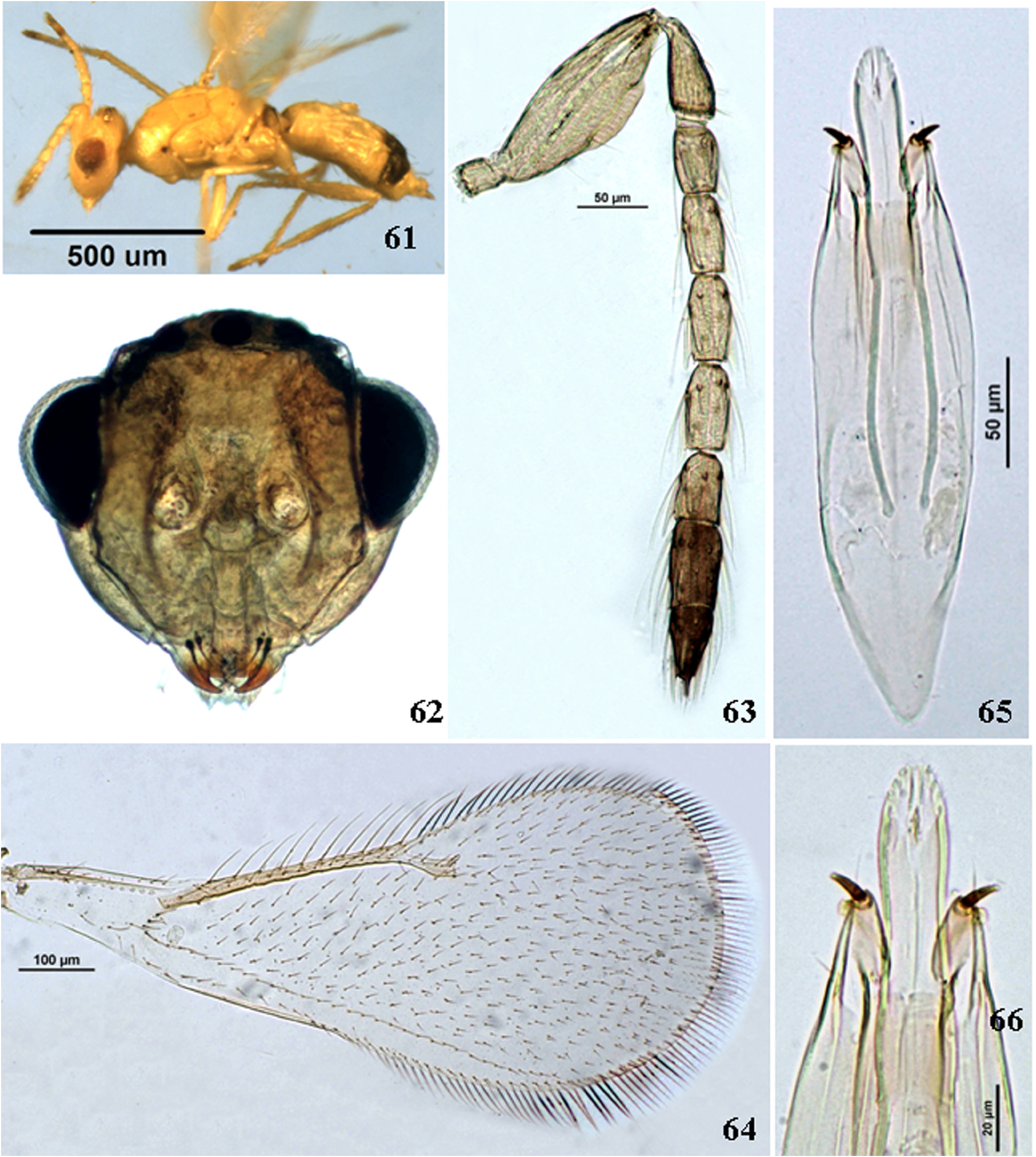
MALE ( Figs 61–66 View FIGURES 61–66 , 71–73 View FIGURES 67–73 ). Body size 1.08 ± 0.01mm (freshly killed in alcohol, n = 5, range 0.95 to 1.27 mm).
Color/Sculpture. Similar to female except the color is whitish yellow ( Fig. 61 View FIGURES 61–66 ) and band on gaster wider. Antenna ( Fig. 63 View FIGURES 61–66 ) pale yellow; basal club segment light brown, distal 2 segments dark brown; whorls of long setae on flagellar segments absent.
Structure. Head clearly wider than mesosoma (360: 327); in frontal view ( Fig. 62 View FIGURES 61–66 ) 1.2× as wide as high (360: 300), 1.7× as wide as frontovertex (360: 202); toruli placed at middle of face, their lower margins touching a line joining the lower margins of the eyes; malar space about 0.8× as long as eye length (106: 134), malar sulcus curved, mouth as wide as malar space length (130: 128); in dorsal view POL 1.3× OOL (94: 72). Antenna ( Fig. 63 View FIGURES 61–66 ) with one anellus; scape 2.5× as long as wide (157: 62), ventral plaque at distal half covering about 0.4× of ventral margin; pedicel about 2.4× as long as wide (74: 31); F1 shortest 1.4× (44: 31), F2 2.1× (56: 26), F3 and F4 each 2× (57: 28, 60: 29) as long as wide. Club 3-segmented, as long as preceding 3 funicle segments; C1 1.5× (46: 31), C2 1.8× (63: 35) and C3 2× (63: 30) as long as wide. Terminal spine 0.3× as long as C3 width (10: 30), without apical seta.
Mesosoma 1.4× as long as wide (478: 329), mesoscutum 1.5× as long as scutellum (202: 138); scutellum 1.5× as wide as long (202: 138), about 4.3× dorsellum length (138: 32); dorsellum 3.3× as wide as long. Propodeum 1.3× as long as dorsellum (43: 32), Forewing ( Fig. 64 View FIGURES 61–66 ) 2.4× as long as wide (941: 398); marginal vein narrowly shorter than costal cell (260: 262), 3.8× as long as stigmal vein (260: 69).
Gaster ( Fig. 61 View FIGURES 61–66 ) as long as mesosoma; genitalia ( Figs 65, 66 View FIGURES 61–66 ), aedeagus 0.8× as long as phallobase length (213: 270), phallobase 3.7× as long as wide (270: 73), digitus with one long digital spine.
Variation. There is variation in size for both sexes; slight variations are also found in intensity of dark markings on the body.
Material examined. Holotype: ♀, card mounted; INDIA: Uttarakhand, Dehradun, New Forest ; 31.i to 10.ii.2017; Sudhir Singh ; ex. galls of Madhuca longifolia (Koenig) (Sapotaceae) . Deposited in NFIC-FRI, Accession No. H-22039.
Paratypes: 3 ♀, 10 ♂, same data as holotype ; 1 ♀ (antennae and wings removed and mounted on a slide), 6 ♂ on 5 cards; 2 ♀ and 4 ♂ dissected and mounted on 2 slides,; NFIC-FRI, Accession No. P-22039 .
Host. Associate of galls on Madhuca longifolia , perhaps parasitoid of Selitrichodes madhucae sp. nov.
Distribution. India: Uttarakhand.
Etymology. Named after the collection locality, Dehradun.
Comments. In the key to Aprostocetus species of Indian sub-continent by Narendran (2007) A. dehradunensis runs to couplet 24, where A. coimbatorensis keys out, but does not match any of its character except SMV with 2 dorsal setae. It differs from coimbatorensis on the basis of following characters (characters given in brackets are those of A. coimbatorensis ):
Female: Body yellow, see description (body mainly black with blue reflections except following lemon yellow: lower frons, lateral margins of pronotum, sides of mesoscutum, posterior part of scutellum, metanotum, posterior margins of tegulae and dorsal margin of mesepimeron; legs white with dusky tarsi). Antenna with one anellus (4); F1 1.5×, F2 1.7× and F3 1.4× as long as wide (F1 and F2 each 2× and F3 1.2×); club 3-segmented (unsegmented), as long as F2 and F3 combined (distinctly longer).
No known copyright restrictions apply. See Agosti, D., Egloff, W., 2009. Taxonomic information exchange and copyright: the Plazi approach. BMC Research Notes 2009, 2:53 for further explanation.
|
Kingdom |
|
|
Phylum |
|
|
Class |
|
|
Order |
|
|
Family |
|
|
Genus |