
Cobanocythere ikeyai, Higashi, Ryouichi & Tsukagoshi, Akira, 2011
|
publication ID |
https://doi.org/ 10.5281/zenodo.207881 |
|
DOI |
https://doi.org/10.5281/zenodo.6183261 |
|
persistent identifier |
https://treatment.plazi.org/id/03ED87E7-FFC5-590E-FF13-829CFB2FFC7B |
|
treatment provided by |
Plazi |
|
scientific name |
Cobanocythere ikeyai |
| status |
sp. nov. |
Cobanocythere ikeyai View in CoL sp. nov.
( Figs 2–5 View FIGURE 2 View FIGURE 3 View FIGURE 5 , 6 View FIGURE 6 A)
Type series. Holotype: adult male (SUM-CO-1942), right valve length 219 µm, height 73 µm, left valve length 226 µm, height 73 µm, appendages mounted on slide and valves preserved in a cardboard cell slide, Paratypes: 10 adult males (SUM-CO- 1943–1952) and 6 adult females (SUM-CO- 1953–1958). All illustrated specimens were collected from interstitial pore water at the type locality on October 25, 2008.
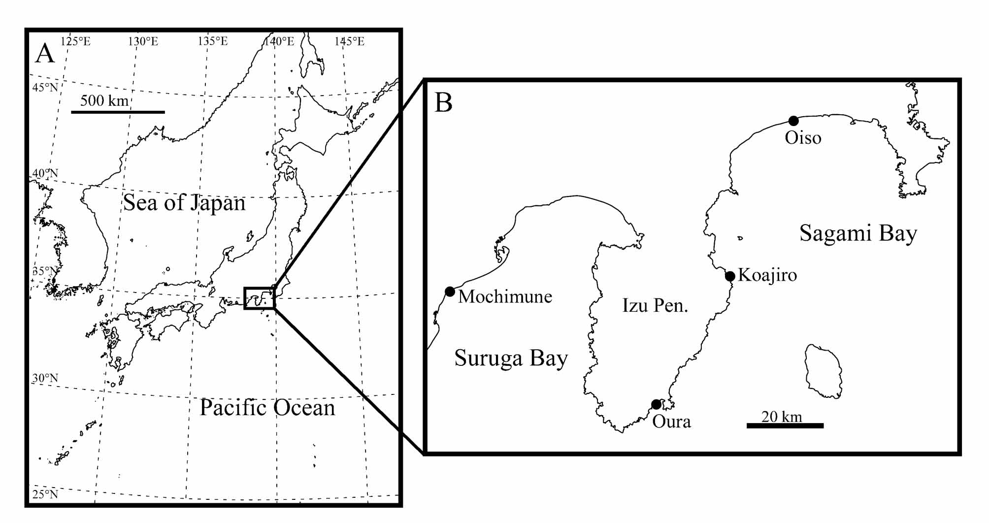
Type locality. Koajiro, Ito City, Shizuoka Prefecture, Pacific coast of central Japan ( Fig. 1 View FIGURE 1 B), 34°57'15"N, 139°08'31"E. Marine littoral beach; sediment composed of shell and clastic coarse sand. Sampling depth approximately 5 cm.
Etymology. Named in honour of the late Emeritus Professor Noriyuki Ikeya of Shizuoka University for his outstanding contributions to Japanese research on Ostracoda.
Diagnosis. Female larger than male. Carapace rounded triangular in anterior and posterior views. Carapace surface covered with fine puncta in mid- and postero-ventral areas in lateral view. Anterior part of carapace rounded and shallowly concaved along ventral margin in lateral view. Sixty one pore systems per valve. Ten marginal pores along anterior margin and no marginal pore along posterior margin. Three vein-like structures running forward on inner surface of anterior marginal infold. Hingement of lophodont type. No rib behind adductor muscle scars. Right and left male copulatory organs consisting of thin and round-tipped distal lobe, right-angled tip of capsule and curved copulatory duct with two swollen parts.
Description. Carapace ( Figs 2 View FIGURE 2 , 3 View FIGURE 3 ). Depressed dorso-ventrally and elongated. Rounded triangular-shaped outline in anterior and posterior views. External surface generally smooth and ornamented with fine puncta in mid- to postero-ventral area in lateral view. Anterior part protruded in dorsal view and rounded in lateral view. Ventral area flat and shallowly concaved in antero-ventral margin. All pore systems of simple type and 61 per valve. Marginal infold broad in anterior area and narrow in posterior areas. Three vein-like structures running forward on inner surface of centre of anterior marginal infold. Vestibula developed in both anterior and posterior marginal infolds. Left valve slightly overlapping right valve along anterior and posterior margins. Hingement of lophodont type. Three adductor muscle scars in oblique row.
Antennula (Fig. 4A). Five articulated podomeres. First podomere bare. Second podomere as long as first podomere, with one long seta on outside of distal end and long setulae along anterior margin. Third podomere as long as second podomere, with one medium seta on antero-distal end. Fourth podomere as long as third podomere, with one long seta on antero-medial suture, two long and one medium setae on antero-distal end, one medium seta on middle of posterior margin and one very long seta on postero-distal end. Fifth podomere three-fourths as long as fourth podomere, with one short seta on inside of distal end, one very long seta, and one pair of basally fused setae composed of slender and round-tipped setae on distal end.
Antenna (Fig. 4B). Four articulated podomeres. First podomere with two segmented spinneret (exopodite) on outside distal end. Second podomere half as long as first podomere, with one short seta on postero-distal end and one short and thin seta on centre of outside. Third podomere three-halves as long as second podomere, with one long seta on antero-medial suture, one short seta on middle of posterior margin and one medium seta on posterodistal end. Fourth podomere one-third as long as third podomere, with claw on antero-distal end and one short and one thick setae on postero-distal end.
Mandibula (Fig. 4C1, C2). Coxa (Fig. 4C1) long, with one thick seta on antero-ventral part, two setae between first and second anterior coxal endites, one seta between second and third anterior coxal endites and two setae on postero-ventral part. Five coxal endites. Palp (Fig. 4C2) consisting of four articulated podomeres. First podomere (basis) with short process (exopodite) on middle outside of dorsal margin and one long seta on ventro-distal end. Second podomere four-thirds as long as first podomere, with one long seta on inside ventro-distal end. Third podomere half as long as second podomere, with two long and one medium setae on middle of dorsal margin, one medium seta on ventro-distal end and one medium seta on outside distal end. Fourth podomere half as long as third podomere, with two medium and one long setae on distal end.
Maxillula (Fig. 4D). Thin branchial plate (exopodite) with eight plumose setae. Basal podomere with one palp and three endites. Palp consisting of two indistinct podomeres: first podomere with three long setae on dorso-distal end; second podomere as long as first podomere, with two long and one medium setae on distal end. Endites with five setae, respectively.
Fifth limb (Fig. 4E). Four articulated podomeres. First podomere with one very short antero-proximal seta, one short antero-distal seta and very short postero-proximal seta. Second podomere five-thirds as long as first podomere, with one short seta on antero-distal end. Bare third podomere half as long as second podomere. Fourth podomere six-fifths as long as second podomere, with stout distal claw.
Sixth limb (Fig. 4F, G). Remarkably reduced leg in male (Fig. 4F): three articulated podomeres; first podomere with one medium seta on antero-distal end; second podomere as long as first podomere, with one very short seta on antero-distal end; third podomere one-third as long as second podomere, with very short seta on distal end. Normal walking leg in female (Fig. 4G): four articulated podomeres; first podomere with one medium setulous seta on antero-distal end and one short seta on postero-proximal part; second podomere seven-fifths as long as first podomere, with medium seta on antero-distal end; third podomere half as long as second podomere; fourth podomere fivefourths as long as third podomere, with stout distal claw.
Seventh limb (Fig. 4H). Four articulated podomeres. First podomere with three short setae on antero-proximal, antero-distal and postero-proximal parts, respectively. Second podomere three-halves as long as first podomere, with one medium seta on antero-distal end and setulae distally along two-thirds of anterior margin. Third podomere half as long as second podomere, with setulae distally along half of anterior margin. Fourth podomere five-fourths as long as third podomere, with one stout distal claw and setulae distally along half of anterior margin.
Male brush-shaped organ (Fig. 4I). Consisting of two branches. Each branch with about 12 setae on distal margin.
Male copulatory organ ( Fig. 5 View FIGURE 5 A, B). Capsule square. Tip of capsule (Tc) right-angled. Distal lobe (Dl) thin and round-tipped triangular: almost same shape between right and left but left Dl slightly larger than right. Copulatory duct (Cd) about half as long as capsule, curved on distally one-third and formed two arches along posterior margin.
Caudal part of female ( Fig. 6 View FIGURE 6 A). Genitalia consisting of strongly sclerotised structure and rounded frame-work connected to ovum. Caudal rami near genitalia, with two setulous setae on anterior and posterior parts, respectively. Caudal process with one long spine on distal end, surrounded by thin and long setulae.
FIGURE 4. Appendages of Cobanocythere ikeyai sp. nov. A–E, H and I, Holotype (SUM-CO-1942); F, Paratype (SUM-CO- 1943); G, Paratype (SUM-CO-1953). A, antennula; B, antenna; C1, coxa of mandibula; C2, palp of mandibula; D, maxillula; E, fifth limb; F, sixth limb of male; G, sixth limb of female; H, seventh limb; I, brush-shaped organ. Scale bar indicates 50 µm.
Eye. Present.
Dimensions. See Table 1.
Occurrence. Type locality and Oura, Shimoda City, Shizuoka Prefecture, Pacific coast of central Japan (34°40'06"N, 138°56'28"E).
Length (µm) Height (µm)
Mean Observed range N Mean Observed range N Male Right valve 222 218–227 4 72 68–73 4
Left valve 228 224–235 3 75 73–78 3 Female Right valve 259 251–264 5 87 84–90 5
Left valve 265 254–272 5 88 83–93 5
Remarks. This new species resembles Cobanocythere lanceolata Gottwald, 1983 , C. psammophila Gottwald, 1983 , C. pacifica Gottwald, 1983 , C. fernandinensis Gottwald, 1983 , C. hoodensis Gottwald, 1983 and C. sublitoralis Gottwald, 1983 , which were referred to as the lanceolata group in Gottwald (1983), based on the shape of carapace and most features of the appendages. For the carapace morphology, the elongated shape and the shallow concavity on the antero-ventral margin are features shared with C. ikeyai sp. nov. and the species of the lanceolata group. The shape and proportion of the antennular podomeres are also similar between these species. Therefore C. ikeyai sp. nov., belongs to the lanceolata group. On the other hand, this new species does not have a rib behind the adductor muscle scars, which distinguishes it from other species of the lanceolata group. Moreover, C. ikeyai sp. nov., differs from other species of lanceolata group in the following features of the male copulatory organ: rightangled Tc; round-tipped and triangular Dl; and Cd relatively longer than the other species ( Fig. 5 View FIGURE 5 ).


No known copyright restrictions apply. See Agosti, D., Egloff, W., 2009. Taxonomic information exchange and copyright: the Plazi approach. BMC Research Notes 2009, 2:53 for further explanation.
|
Kingdom |
|
|
Phylum |
|
|
Class |
|
|
Order |
|
|
Family |
|
|
Genus |