
Tazoudasaurus (Cooper, 1984)
|
publication ID |
https://doi.org/ 10.5281/zenodo.5371775 |
|
persistent identifier |
https://treatment.plazi.org/id/03EF8786-326A-7415-FD0E-0827FBD361E9 |
|
treatment provided by |
Marcus |
|
scientific name |
Tazoudasaurus |
| status |
|
EARLY JUVENILE TAZOUDASAURUS BONES
Among the several hundred bones collected in Toundoute, some very small elements are perfectly preserved and can be confidently assigned to Tazoudasaurus . Because juvenile material of sauropods is relatively rare in the fossile record ( Norman et al. 2004b), some bones of these early juvenile sauropods are described below. A detailed ontogenetic and histological study is beyond the scope of this paper.
ANTERIOR DORSAL NEURAL ARCH
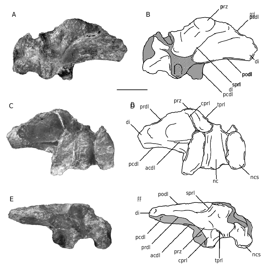
The incomplete neural arch of an anterior dorsal vertebra of a juvenile individual (MHNM To1- 383) has been recovered in Toundoute ( Fig. 38 View FIG ).
The left transverse process, the neural spine and the postzygapophyses are missing. The interdigitating neurocentral suture is clearly visible in ventral view ( Fig. 38C, D View FIG ), confirming the immaturity of the specimen. The parapophyses are not present on the neural arch and thus were located on the centrum, suggesting this was probably an anterior dorsal neural arch. The neural arch lamination is as well developed as in the adult specimen To1-69. Four diapophyseal laminae (acdl, pcdl, prdl, podl), and four zygapophyseal laminae (cprl, sprl, tprl, tpol) have been identified on the centrum ( Table 2), and the cpol is absent. The spinodiapophyseal lamina, which is very reduced in the anterior dorsal vertebra of the adult (see above), is absent in this juvenile vertebra. The articular facets of the prezygapophyses face dorsally ( Fig. 38A, B View FIG ). The hyposphenehypantrum system is not developed in To1-383, and the tprl’s do not fuse to each other above the neural canal ( Fig. 38E, F View FIG ). Each tprl defines with the cprl and the dorsolateral margin of the neural canal a deep fossa on the anterior surface of the neural arch as in To1-69, but without additional lamina inside the fossa ( Fig. 38E, F View FIG ). The transverse process is directed laterally. The tpol’s meet above the neural canal but extend laterally to the neural canal, and thus form an X, in posterior view.
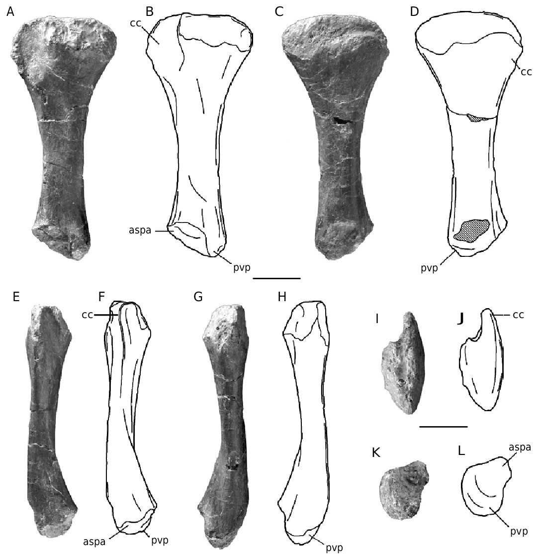
LEFT HUMERUS
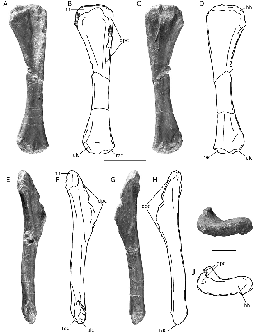
The left juvenile humerus (MHNM To1-93) is only 18% the length of the largest known adult humerus (Pt-1) ( Fig. 39 View FIG A-J; Table 3). Its circumference/length ratio (0.35) is slightly less than that of the adult (0.39). The proximal end is less expanded transversely and is 13% of the adult proximal width. Similarly, the distal end is less expanded anteroposteriorly with a length which is only 8% of the length of To1-193 while it is 14% in Pt1. In medial view, the humerus is slightly sigmoidal ( Fig. 39E, F View FIG ). Its lateral margin is nearly straight on its entire length in anterior and posterior views ( Fig. 39 View FIG A-D). The proximal end is compressed anteroposteriorly. Its anterior surface is more concave than in the adult specimen ( Fig. 39A, B, I, J View FIG ). The deltopectoral crest extends down the anterolateral margin of the humerus over 82 mm, which represents 44% of the total length of the bone (49% in Pt-1), and is slightly more prominent than in the adult humerus. The crest running over the anterolateral margin of the distal end of the humerus is absent in the juvenile specimen ( Fig. 39G, H View FIG ). As in Pt-1, the distal end is rotated about 15° anticlockwise with respect to the proximal end.
LEFT ULNA
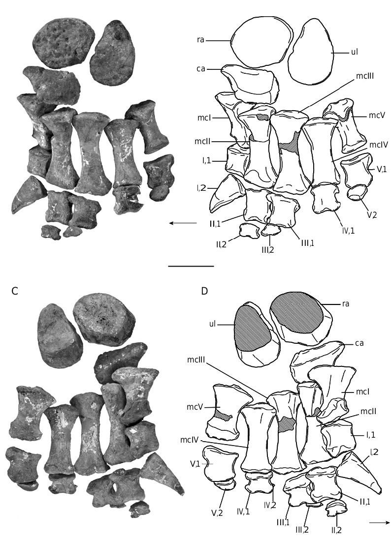
The juvenile left ulna MHNM To1-374 is better preserved than the adult ulna (Pt-24) described above. It is a slender bone, which is 30% the length of the largest known adult ulna (Pt-24) ( Fig. 40A F View FIG ; Table 3). Its circumference/length ratio (0.37) is not substantially different than that of the adult (0.34). The proximal end is triradiate, but one of the processes is broken distally. This bone was first thought to be a right ulna, but according to the complete left manus found articulated with a distal radius and ulna ( Fig. 23 View FIG ), it would appear to be the left ulna, the anteromedial process of which is broken ( Fig. 40A, C, E View FIG ). The anterolateral process is more developed than in the adult specimen and in Vulcanodon and must be only slightly shorter than the anteromedial process. The radial fossa is deeper than in the adult specimen. The proximal articular surface lacks an olecranon ( Fig. 40 View FIG A-D). The medial surface of the anteromedial process is slightly concave, whereas the lateral surface of the anterolateral process is slightly convex ( Fig. 40E View FIG ). Below the proximal end, the shaft of the ulna is subtriangular in cross section, with the apex directed anteriorly. The shaft of the ulna is concave laterally and medially and straight posteriorly. The distal end is elliptical with an almost laterally directed long axis. A small bump and roughened ridges mark the contact of the radius on the lateral side of the anterior surface of the ulna, but these are not as prominent as in the adult specimen ( Fig. 40A View FIG ). The rugose distal articular surface of the ulna is expanded transversely.
RIGHT ISCHIUM
The maximum width across the proximal end of the right juvenile ischium MHNM To1-379 is 24% that of the adult specimen ( Table 5). Only the distalmost part of the ischium is missing (Fig. 41). The mildly convex proximal end of the iliac peduncle is elliptical in outline, with the long axis of this ellipse directed anteroposteriorly. The pubic peduncle is as long as the iliac peduncle and is also elliptical in outline. The acetabular surface slopes medially and has an upstanding medial rim (Fig. 41C, D). As in the adult specimen, the blade of the ischium is thicker posterodorsally than anteroventrally where it is articulated with the left ischium. The ischial blade is thus triangular in cross section. A prominent groove extends along the laterodorsal margin of the proximal part of the blade (Fig. 41A, B). Unlike the adult specimen, the angle between the blade and the pubic peduncle is greater than that between the blade and the iliac peduncle, implying that the ischium was more ventrally directed in juvenile individuals. The distal blade is slightly twisted laterally relative to the plane of the proximal plate, but both ischia would meet on an angle, forming a triangle in distal view, unlike macronarians ( Wilson & Sereno 1998). The distal end of the ischium is expanded ventromedially.
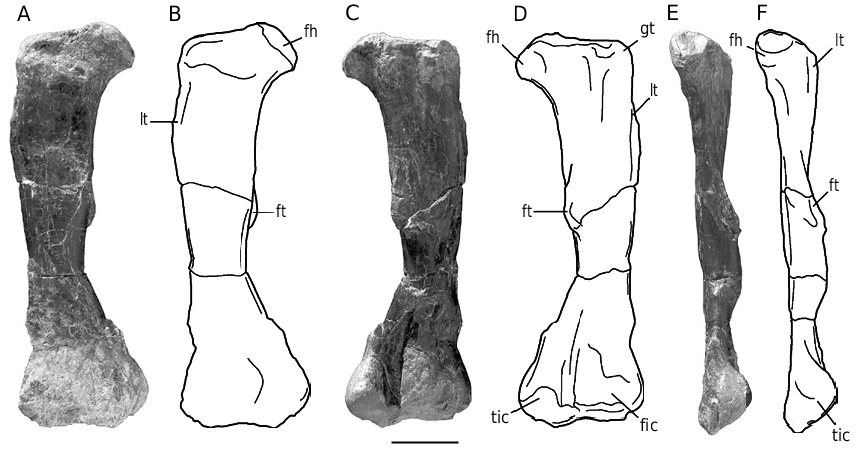
FEMUR
The right juvenile femur (MHNM To1-256) is 24% the length of the longest femur To1’-381. Although it is complete, this femur is severely crushed anteroposteriorly, and thus lost its shape in its distal half ( Fig. 42 View FIG ). The juvenile femur is straight and anteroposteriorly compressed.The head of the femur is direct- ed dorsomedially ( Fig. 42A, B View FIG ). The lesser trochanter is clearly visible and extends as a developed ridge on the anterolateral margin of the femur, terminating about 104 mm below the femoral head ( Fig. 42 View FIG A-E). The fourth trochanter, which lies at the posteromedial margin of the femur, is more prominent than in the adult specimen ( Fig. 42 View FIG C-F). A deep intercondylar groove separates the fibular and rounded tibial condyles ( Fig. 42C, D View FIG ). Poor preservation prevents any assessment of the extent of the distal condyles.
LEFT TIBIA
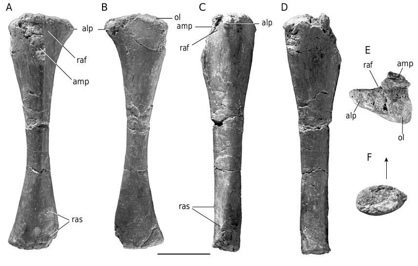
The juvenile left tibia (MHNM To1-76) is 35% the length of the largest known tibia of Tazoudasaurus (To1’-380) ( Fig. 43 View FIG ). The circumference/length ratio of the juvenile tibia (0.48) is equivalent to that of the adult (0.49). The shaft of the tibia is less compressed transeversely, its mediolateral width at midlength ilpd ilpd
FIG. 41. — Tazoudasaurus naimi , right ischium (To1-379): A, B, lateral view; C, D, proximal view. Abbreviations: act, acetabulum; gr, groove; ilpd, iliac peduncle; pupd, pubic peduncle; sy, symphisis. Scale bar: 5 cm.
being 52% its anteroposterior length, otherwise the morphology of the juvenile tibia is in all respects identical to that of the adult specimen ( Fig. 43 View FIG ).
Astragalus
The mediolateral width of the juvenile astragalus (MHNM To1-135) is 39% that of the adult specimen described above.Most of the outer surface of the bone is shaved away, which suggests that the ossification of the astragalus was not complete (Fig. 44). The shape of the juvenile astragalus is similar to that of the adult, with a subrectangular outline in proximal view (Fig. 44C, D), and a wedge-shaped in anterior and posterior views (Fig. 44A, B). The smooth
pf cas asp cas cas
FIG. 44. — Tazoudasaurus naimi , right astragalus (To1-135): A, B, posterior view; C, D, proximal view; E, F, lateral view.Abbreviations: asp, ascending process of the astragalus; cas, calcaneal articular surface; cr, crest; pf, posterior fossa; tas, tibial articular surface. Scale bar: 5 cm.
and concave lateral fibular articular surface is more elliptical than in the adult astragalus (Fig. 44E, F). The ascending process is directed dorsoposteriorly, but its posterior extension is less than in the adult specimen and the ascending process fails to reach the posterolateral margin of astragalus (Fig. 44C, D). The main difference between the juvenile and adult specimens is in the shape of the proximal articular surface. The posterior fossa of the astragalus is undivided in the juvenile specimen and it faces more laterally. It is separated from the tibial articular surface by a sharp crest which extends from the posteromedial corner of the ascending process to the posterior tongue of the astragalus (Fig. 44C, D). The tibial articular surface is larger than in To1-31 and occupies more than a half of the transverse length of the astragalus.
No known copyright restrictions apply. See Agosti, D., Egloff, W., 2009. Taxonomic information exchange and copyright: the Plazi approach. BMC Research Notes 2009, 2:53 for further explanation.