
Dentalion Hansson
|
publication ID |
https://doi.org/ 10.5281/zenodo.207919 |
|
DOI |
https://doi.org/10.5281/zenodo.6184926 |
|
persistent identifier |
https://treatment.plazi.org/id/03EF87C4-FF88-FFD4-FF67-FC6CFD87875A |
|
treatment provided by |
Plazi |
|
scientific name |
Dentalion Hansson |
| status |
|
Dentalion Hansson gen. nov.
Type species: Dentalion pinguicornis sp. nov., designated here.
Etymology. Named for the tooth on the medioventral part of the hind femora, from the Latin dentis = tooth. The gender is neuter.
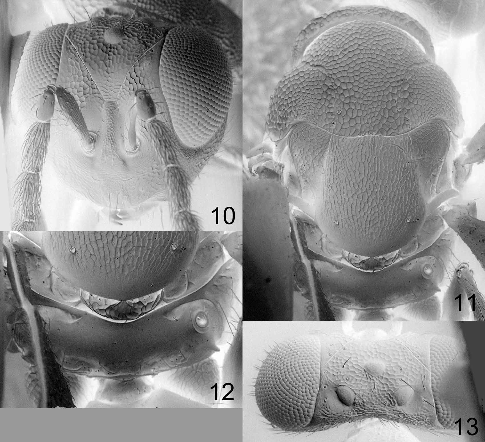
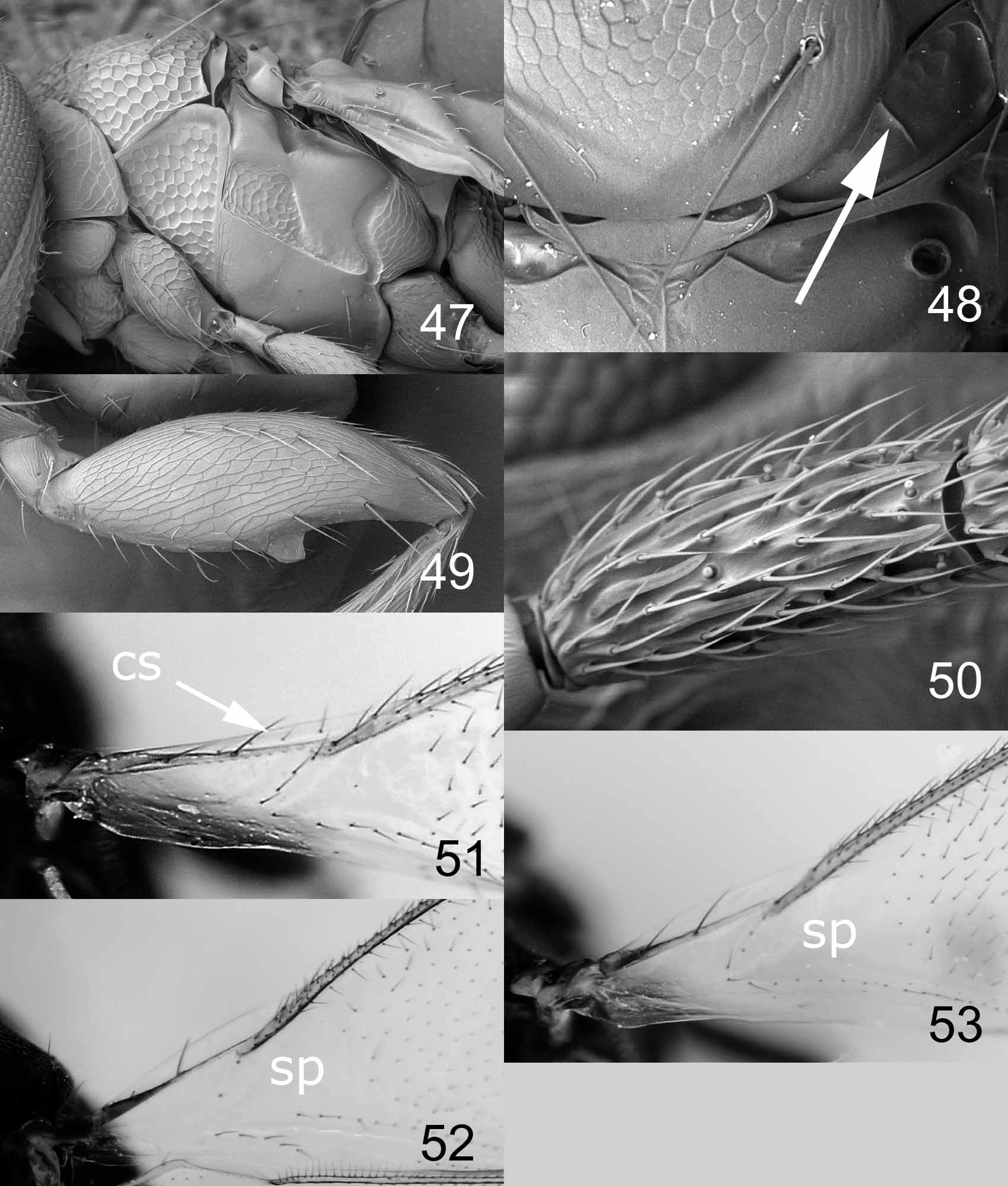
Diagnosis. Frontal suture V-shaped in a narrow angle (usually 35–60°), and reaching eyes high up on frons (e.g. Figs 6 View FIGURES 6 – 9 , 10 View FIGURES 10 – 13 , 14 View FIGURES 14 – 18 ); antennal scrobes wide and indistinctly delimited (e.g. Figs 2 View FIGURES 2 – 5 , 6 View FIGURES 6 – 9 , 10 View FIGURES 10 – 13 ); lateral panels of metanotum divided into two about equal-sized parts by a longitudinal carina ( Fig. 48 View FIGURES 47 – 53 ); hind femur with a tooth medioventrally ( Fig. 49 View FIGURES 47 – 53 ); postmarginal vein always longer (1.3–3.0X) than stigmal vein.
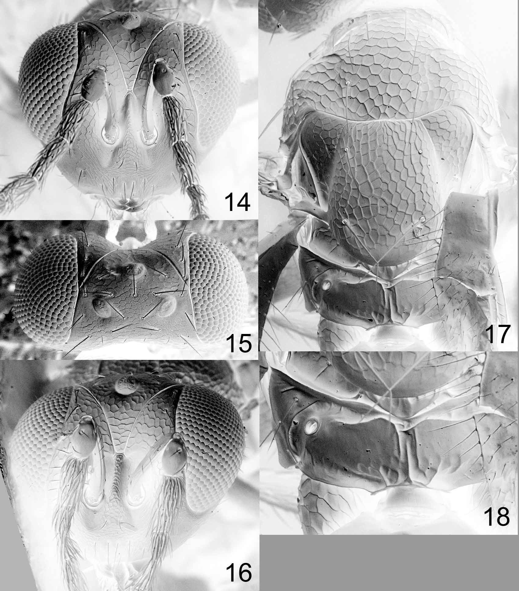
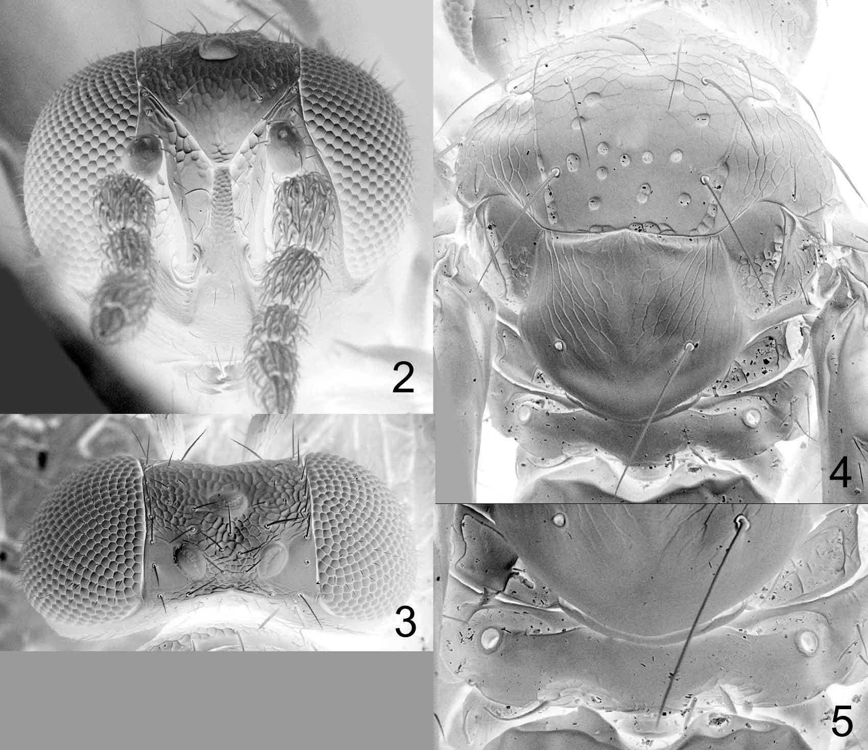
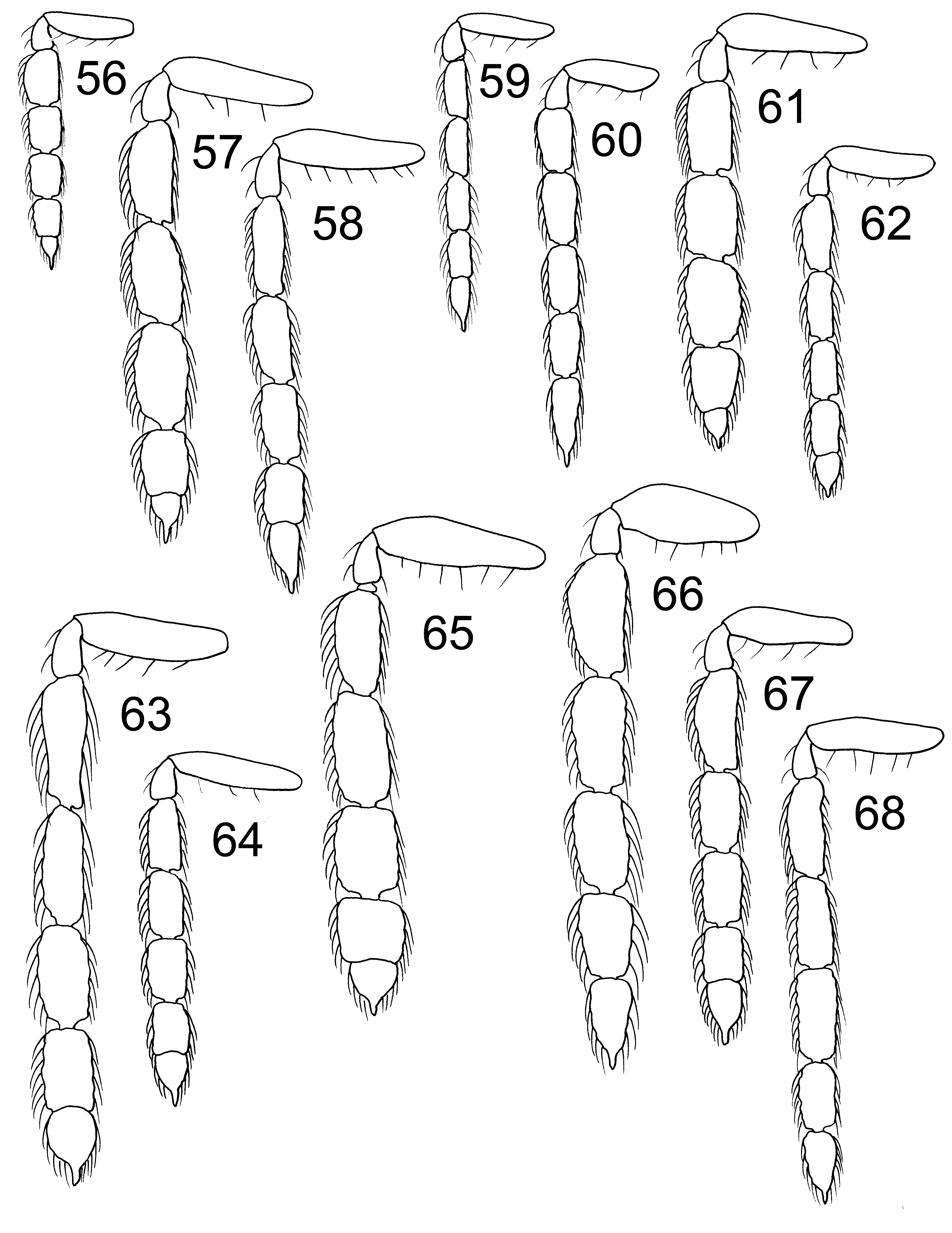
Description. Female flagellum with a 2-segmented clava (e.g. Figs 56–58 View FIGURES 56 – 68 ), in male with all 5 flagellomeres distinctly separated ( Figs 60, 64, 67 View FIGURES 56 – 68 ); male flagellomeres with scattered setae; male scape slightly wider than in female (Figs, 60, 64, 67), with ventral sense area present along entire scape; sensilla ampullacea globular, symmetric (type I sensu Hansson (1990)), present on all flagellomeres. Antenna with discoid anelli. Mandibles with three large teeth at apex. Clypeus not delimited. Malar sulcus missing, sometimes indicated by fine small meshed reticulation. Frontal suture V-shaped in a narrow angle, usually 35–60° (e.g. Figs 6 View FIGURES 6 – 9 , 10 View FIGURES 10 – 13 , 14 View FIGURES 14 – 18 ) but in one species 85° ( Fig. 2 View FIGURES 2 – 5 ). Antennal scrobes joining frontal suture separately (e.g. Figs 6 View FIGURES 6 – 9 , 10 View FIGURES 10 – 13 , 14 View FIGURES 14 – 18 ). Occipital margin rounded; occiput without a median fold or groove between occipital margin and occipital foramen.
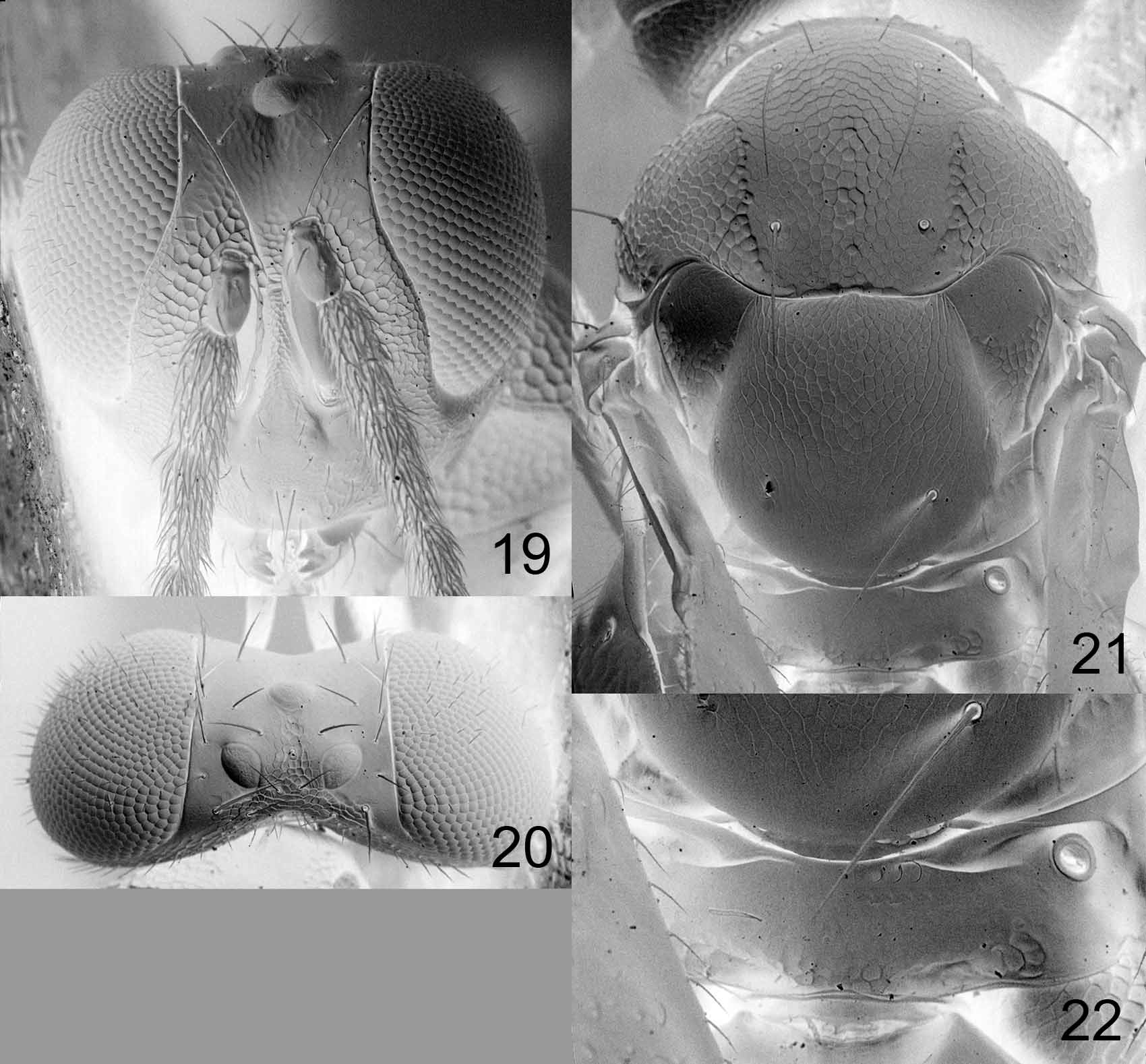
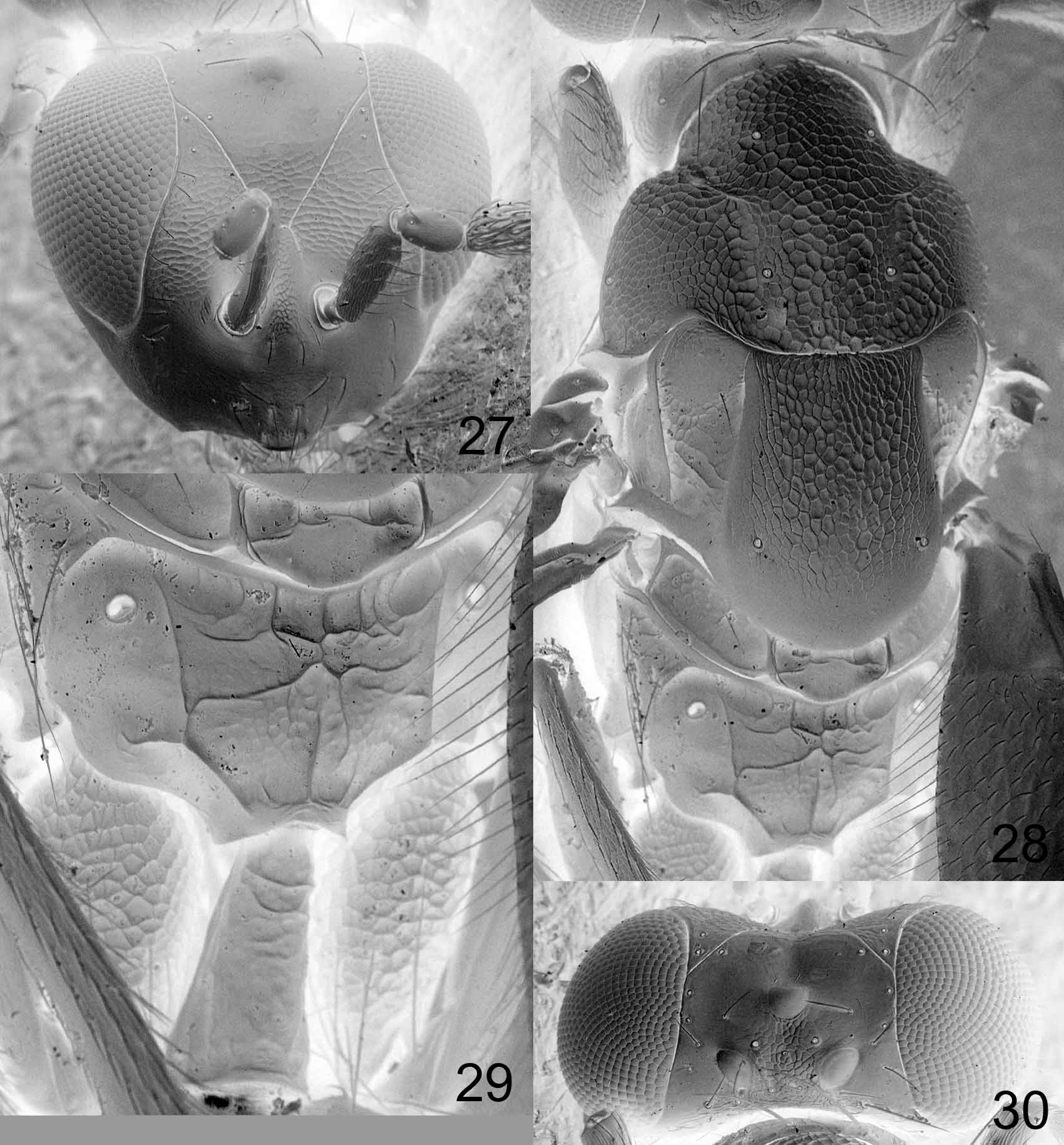
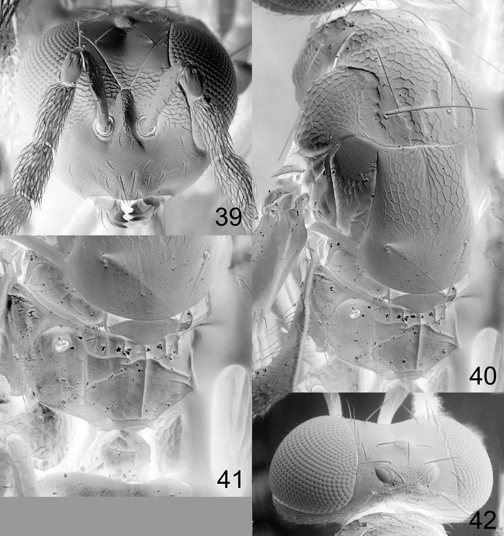
Pronotum usually without transverse carina close to posterior margin (e.g. Figs 21 View FIGURES 19 – 22 , 24 View FIGURES 23 – 26 , 28 View FIGURES 27 – 30 ), but in one species with a weak carina ( Fig. 7 View FIGURES 6 – 9 ). Midlobe of mesoscutum with 2 pairs of setae; notauli distinct and narrow in anterior 1/ 3, in posterior 2/3 either not visible ( Fig. 44 View FIGURES 43 – 46 ), or as indistinct impressions ( Fig. 28 View FIGURES 27 – 30 ), or as distinct wide grooves ( Fig. 7 View FIGURES 6 – 9 ), or as rows of foveae ( Fig. 4 View FIGURES 2 – 5 ). Scutellum with 1 or 2 pairs of setae (e.g. Figs 7 View FIGURES 6 – 9 , 11 View FIGURES 10 – 13 ). Transepimeral sulcus wide and curved in a 90° angle ( Fig. 47 View FIGURES 47 – 53 ). Dorsellum visible in dorsal view, with strong sculpture. Lateral panels of metanotum divided into two about equal-sized parts by a longitudinal carina ( Fig. 48 View FIGURES 47 – 53 ). Forewing with postmarginal vein 1.3–3.0X as long as stigmal vein; speculum usually closed below ( Fig. 53 View FIGURES 47 – 53 ), but open in one species ( Fig. 52 View FIGURES 47 – 53 ). Propodeum usually smooth or with irregular carinae in median part (e.g. Figs 5 View FIGURES 2 – 5 , 8 View FIGURES 6 – 9 , 18 View FIGURES 14 – 18 ), but in one species with a complete median carina, plicae and costulae ( Fig. 41 View FIGURES 39 – 42 ); propodeal callus with 2–7 setae.
Petiole 0.2–1.8X as long as wide, but usually distinctly transverse, smooth and shiny. Male genitalia as in most other entedonine genera ( Hansson 1996), i.e. phallobase with two equally large digital spines, volsellar setae as “normal” thin setae, parameres not protruding.
Distribution. Brazil, Costa Rica, Ecuador, Trinidad & Tobago.
Biology. Unknown.
Identification. Dentalion is a member of the Entedoninae , as indicated by the following combination of characters: submarginal vein with 2 setae on dorsal surface; forewing with a distinct break between submarginal and marginal veins; scutellum without sublateral grooves; flagellum with five large flagellomeres.
In the key to Nearctic genera of Eulophidae by Schauff et al. (1997), Dentalion can be separated from all other genera by the presence of a ventral dent on the hind femur ( Fig. 49 View FIGURES 47 – 53 ). Another option for identification is to use the matrix key to the Neotropical genera of Entedoninae on the website http://www.neotropicaleulophidae.com/, where also colour illustrations for each species are included.
Putative autapomorphies. Hind femur with a ventral dent ( Fig. 49 View FIGURES 47 – 53 ). Some species of Pleurotroppopsis Girault (e.g. P. podagrica (Waterston)) have a row of teeth along the ventral margin of hind femur, which is a different character state from the single tooth in Dentalion .
Frontal suture V-shaped in a narrow angle, 35–60°, and reaching eyes high up on frons, in level with a distance of 1.5X the diameter of one ocellus in front of anterior ocellus ( Fig. 2 View FIGURES 2 – 5 ) or behind this level ( Figs 6 View FIGURES 6 – 9 , 10 View FIGURES 10 – 13 , 14 View FIGURES 14 – 18 , 19 View FIGURES 19 – 22 , 23 View FIGURES 23 – 26 , 27 View FIGURES 27 – 30 , 31 View FIGURES 31 – 34 , 35 View FIGURES 35 – 38 , 39 View FIGURES 39 – 42 , 43 View FIGURES 43 – 46 ).
Other apomorphies, present also in other genera of Entedoninae . L ateral panels of metanotum divided into two about equal-sized parts by a longitudinal carina ( Fig. 48 View FIGURES 47 – 53 ); also present in Emersonella ( Hansson 2002) . Antennal multiporous plate sensilla with upper surface concave ( Fig. 50 View FIGURES 47 – 53 ); also present in Microdonophagus Schauff ( Hansson 2009b) and Inti Hansson (Hansson 2010) .
Discussion. Dentalion is defined by two autapomorphies, and possess two additional apomorphies that also occur in other genera of Entedoninae . The longitudinal carina on the lateral panels of the metanotum occurs only in one other entedonine genus, Emersonella , which indicates a sister-group relationship for these two genera. Dentalion and Emersonella do not share other apomorphies, but Emersonella is characterized by six additional apomorphies ( Hansson 2002) and Dentalion by two autapomorphies. The derived state of the antennal sensilla having a concave upper surface is a putative homoplasy. Apart from occurring in Dentalion , this sensillar state also occurs in two other entedonine genera, Microdonophagus and Inti , and these two genera are not closely related ( Hansson 2010), clearly indicating that this character state has evolved at least twice. Dentalion does not share other apomorphies with either Microdonophagus or Inti .


No known copyright restrictions apply. See Agosti, D., Egloff, W., 2009. Taxonomic information exchange and copyright: the Plazi approach. BMC Research Notes 2009, 2:53 for further explanation.