
Micropsephodes bahamaensis Shockley, 2010
|
publication ID |
https://doi.org/10.5281/zenodo.4645498 |
|
persistent identifier |
https://treatment.plazi.org/id/03F0C738-FFBC-FFEF-7F9F-E2CFFB6A3CB0 |
|
treatment provided by |
Felipe |
|
scientific name |
Micropsephodes bahamaensis Shockley |
| status |
sp. nov. |
Micropsephodes bahamaensis Shockley , new species
( Fig. 2C View Figure 2 , 5-9 View Figure 3-5 View Figure 6 View Figure 7 View Figure 9 )
Etymology. The specific epithet is based on the fact that this is the first species of Micropsephodes to be described from the Bahamas.
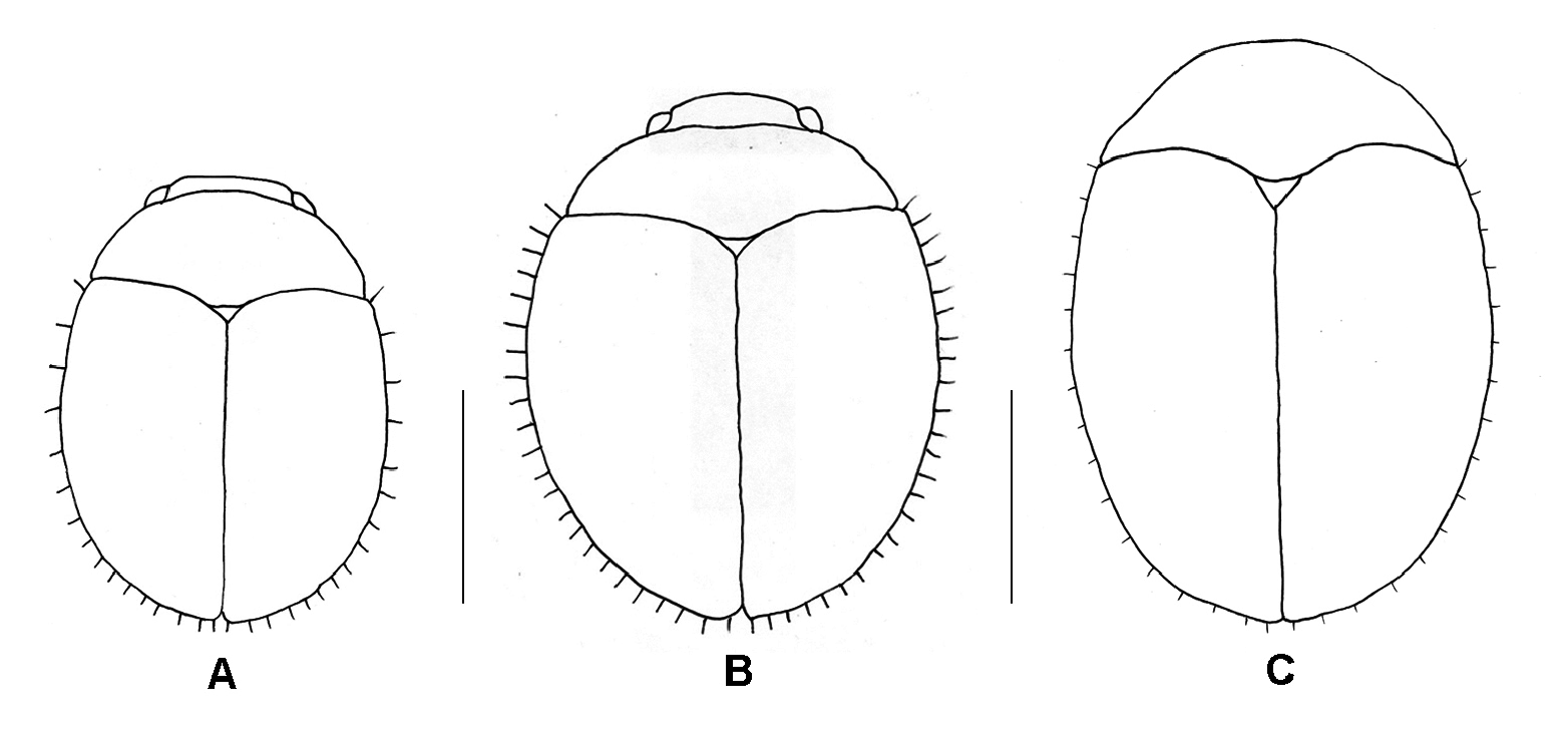
Diagnosis. Adults of M. bahamaensis may be distinguished from those of M. lundgreni and M. serraticornis by the combination of the following characters: larger size and elongate habitus; much shorter setae along the lateral margins of the elytra ( Fig. 2 View Figure 2 ); ovoid pedicel and elongate antennomere III ( Fig. 6C View Figure 6 ); lacinia unisetose apically ( Fig. 6F View Figure 6 ); galea with 10 setae apically, the two most laterad much narrower and less stout compared to the medial eight ( Fig. 6F View Figure 6 ).
Description. Length 1.35-1.41 mm (mean = 1.38); width 1.00- 1.02 mm (mean = 1.01); depth 0.70 mm (n = 3). Body ( Fig. 2C View Figure 2 , 5A, B View Figure 3-5 ) elongate oval, moderately convex; dorsum shining, dark brown-black, glabrous; venter, antennae, mouthparts, and legs reddish-brown, covered with vestiture of short, fine, palecolored setae.
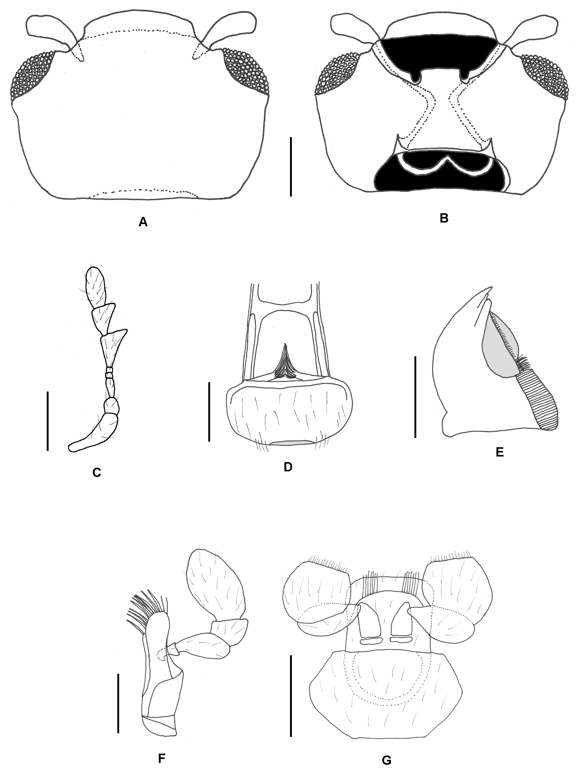
Head. Head ( Fig. 6A, B View Figure 6 ) deeply retracted into prothorax, highly deflexed and hypognathous, completely obscured when viewed dorsally; frontoclypeal suture present; gular sutures relatively short; frons finely and sparsely punctate; clypeus transverse and rectangular; tentorium with anterior arms convergent, but not fused medially, corpotentorium lyriform. Eyes large, round, and finely-faceted. Labrum ( Fig. 6D View Figure 6 ) transverse and ovoid, truncate and submembranous apically. Mandible ( Fig. 6E View Figure 6 ) bifid apically with prominent subapical tooth; prostheca divided longitudinally with a basal tuft of setae just distad of the mola; mola relatively large and finely, transversely ridged. Maxilla ( Fig. 6F View Figure 6 ) with small triangular shaped cardo, well-developed and large basistipes, and narrow, elongate dististipes; galea with 10 large apical spines, the two lateral most spines narrow, the remaining medial eight spines thickened; lacinia narrow, bearing a single prominent spine apically; maxillary palp 4-segmented, palpomere I small and narrow, palpomere II long and expanded apically, palpomere III subquadrate, palpomere IV as long as preceding 3 segments combined and expanded medially with greatest width at 2/3 its length and apex narrowly rounded. Labium ( Fig. 6G View Figure 6 ) with transverse mentum and quadrate prementum; labial palp 3- segmented, palpi narrowly separated basally, palpomere I large, elongate and subcylindrical, palpomere II transverse, palpomere III large ovoid to subquadrate and truncate apically. Antenna ( Fig. 6C View Figure 6 ) 8-segmented; scape prominent, long, widening apically; pedicel subglobose; antennomere III narrow and elongate, distinctly longer than pedicel; antennomeres IV and V very small bead-like; antennomeres VI-VIII greatly enlarged to form a large, loosely articulated club, antennomeres VI and VII internally serrate.
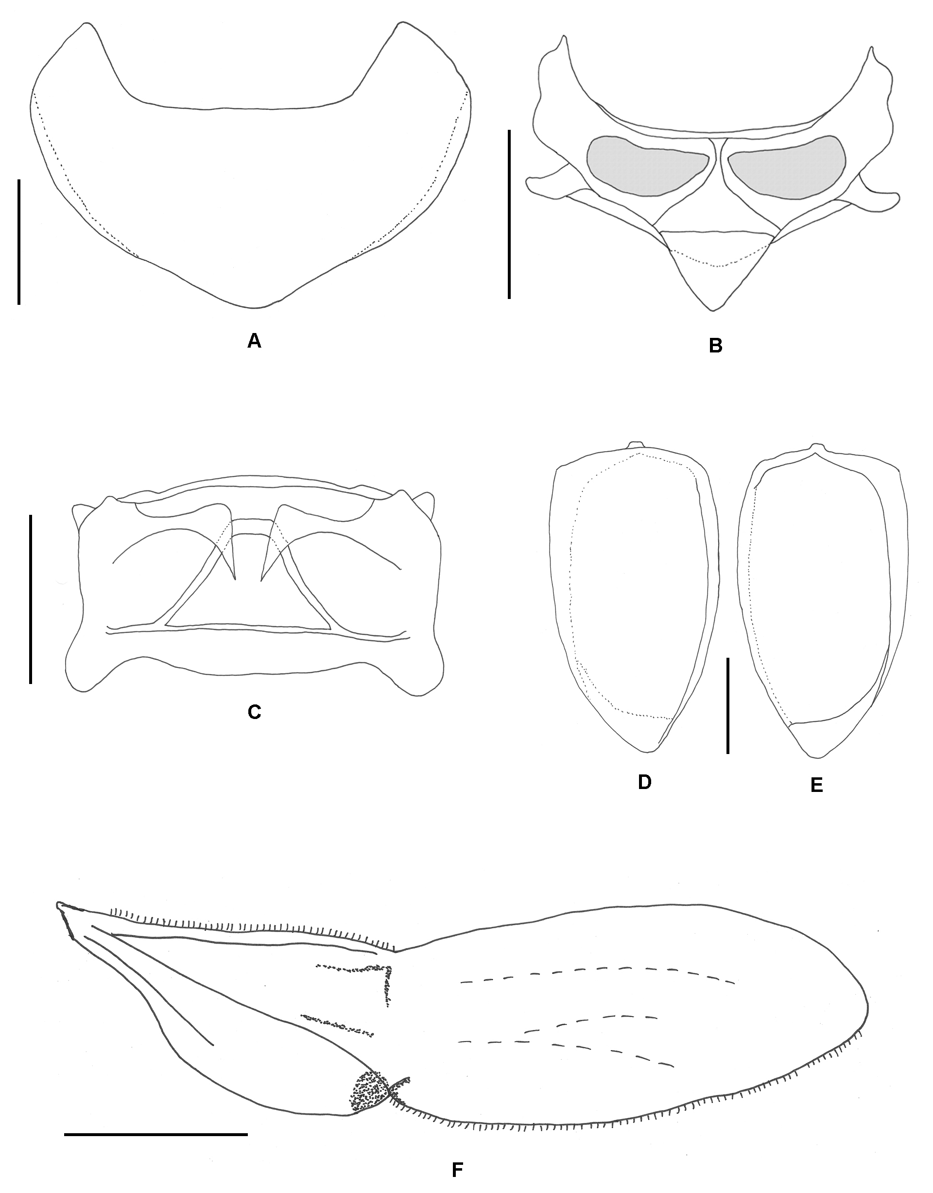
Thorax. Pronotum ( Fig. 7A View Figure 7 ) strongly transverse (length 0.40 mm; width 0.86 mm), highly convex, widest at base and narrowing anteriorly; anterior angles weakly produced and rounded apically; posterior angles roundly obtuse; lateral margins narrow; lacking lateral sulci; basal margin strongly lobed medially, opposite scutellum. Mesonotum ( Fig. 7B View Figure 7 ) with small, triangular mesoscutellum. Elytra ( Fig. 7D, E View Figure 7 ) elongate oval, convex, finely and sparsely punctate, apex slightly and narrowly reflexed; epipleuron narrowing at 2/3 length, broad basally and apically; disc and lateral margin glabrous. Metanotum ( Fig. 7C View Figure 7 ) transverse with prominent metascutellar ridge. Metathoracic wing ( Fig. 7F View Figure 7 ) with a single cubitoanal vein posteriorly and a long sclerotized media posterior; medial and subcubital flecks small, conspicuous and undivided; anal lobe lacking. Prosternum (Fig. 8A) with short, narrow intercoxal process, acute apically with two apical setae, process extending to posterior margin of the procoxal cavity but not beyond. Mesosternum (Fig. 8B) flat, intercoxal process transverse, weakly narrowing towards apex, and truncate (nearly concave) apically and extending to posterior margin of mesocoxal cavity but not beyond it. Metasternum (Fig. 8B) highly transverse; intercoxal process broad straight, extending to middle of metacoxal cavity where it broadly meets the rounded intercoxal process of abdominal ventrite I; metendosternite (Fig. 8C) with widely separated anterior arms, short anterolaterally-directed tendons and long, posterolaterally-directed struts.
Legs. Procoxae ( Fig. 9A View Figure 9 ) triangularly transverse, narrowly separated by prosternal process; mesocoxae ( Fig. 9B View Figure 9 ) globose to ovoid, widely separated by mesosternal process; metacoxae ( Fig. 9C View Figure 9 ) highly transverse, narrowing laterally, widely separated by metasternal process and intercoxal process of abdomen. Pro-, meso-, and metathoracic legs similar in structure for femora, tibiae and tarsi; femora short, stout; tibiae long, narrow slightly expanded apically. Tarsi ( Fig. 9D View Figure 9 ) 3-3-3 with long, lobed tarsomere I bearing spines ventrally and apically; tarsomere II relatively short with a narrow ventral lobe; tarsomere III long and narrow; pretarsus with claws sharp and ventrally bearing a flat, rectangular expansion basally, the apex of which is not acute or produced into a tooth. Figure 8. Thoracic and abdominal venter of
Abdomen. Abdomen (Fig. 8D) with six visible, Micropsephodes bahamaensis Shockley , new species. A) transverse ventrites, widest near the midpoint of Prosternum, ventral. B) Mesoventrite and Metaventrite, ventrite I and gradually narrowing apically, ventral. C) Metendosternite, ventral. D) Abdomen, ventrite VI approximately half as wide as ventrite ventral. Scale bar = 0.50 mm. I. Ventrite I with postcoxal femoral lines recurved and nearly complete, shallow posteriorly reaching less than half length of ventrite I, merging anteromedially with the lateral margins of the heavily sclerotized intercoxal process; ventrites II-V similar in length and narrowing apically; ventrite VI with posterior margin notched medially in female.
Note. The female genitalia were damaged during clearing and dissection, but generally appeared similar to that described and illustrated for Micropsephodes by Tomaszewska (2000). Given the small size of the type series, no additional specimens were dissected.
Material Examined. Holotype and paratype (both female) bear the following collection data: BAHAMAS: Andros ; Forfar Field Station ; mv + bl, 4 June 2001; R. Turnbow . Holotype deposited at FSCA ; Paratype
deposited in RHTC. An additional paratype (disarticulated) is deposited in FWSC and bears the following collection data: BAHAMAS: Andros ; Forfar Field Station ; 2 June 2001; R. Turnbow .


| R |
Departamento de Geologia, Universidad de Chile |
| FSCA |
Florida State Collection of Arthropods, The Museum of Entomology |
No known copyright restrictions apply. See Agosti, D., Egloff, W., 2009. Taxonomic information exchange and copyright: the Plazi approach. BMC Research Notes 2009, 2:53 for further explanation.