
Calesynthemis jeanlegrandi, Fleck, 2024
|
publication ID |
https://doi.org/ 10.11646/zootaxa.5403.3.2 |
|
publication LSID |
lsid:zoobank.org:pub:127A062D-8597-4DD8-B3EE-1B94A3F0B878 |
|
DOI |
https://doi.org/10.5281/zenodo.10568705 |
|
persistent identifier |
https://treatment.plazi.org/id/03F387EE-FFC7-FF85-FF3D-FE01FE127AD7 |
|
treatment provided by |
Plazi |
|
scientific name |
Calesynthemis jeanlegrandi |
| status |
sp. nov. |
Calesynthemis jeanlegrandi sp. nov.
( Figures 1–21 View FIGURE 1 View FIGURES 2–5. 2 View FIGURES 6–10 View FIGURES 11–14. 11 View FIGURE 15 View FIGURES 16–21. 16 )
Material. Holotype ♂ and Paratype ♀: New Caledonia, Province Nord, Mont Panié , ca 900 m asl, 27-1-1988; collector J. Legrand; specimens stored dry in envelopes .
Specimens will be deposited at the nonprofit organisation Earth-Safe ( Lagorce , France), Insect collection, collection G. Fleck, number GF2312-01 (holotype) and GF2312-02 (paratype).
Etymology. I am proud to name this species in honour to Jean Legrand, last Odonatologist of the MNHM. The species epithet is a noun in the genitive case.
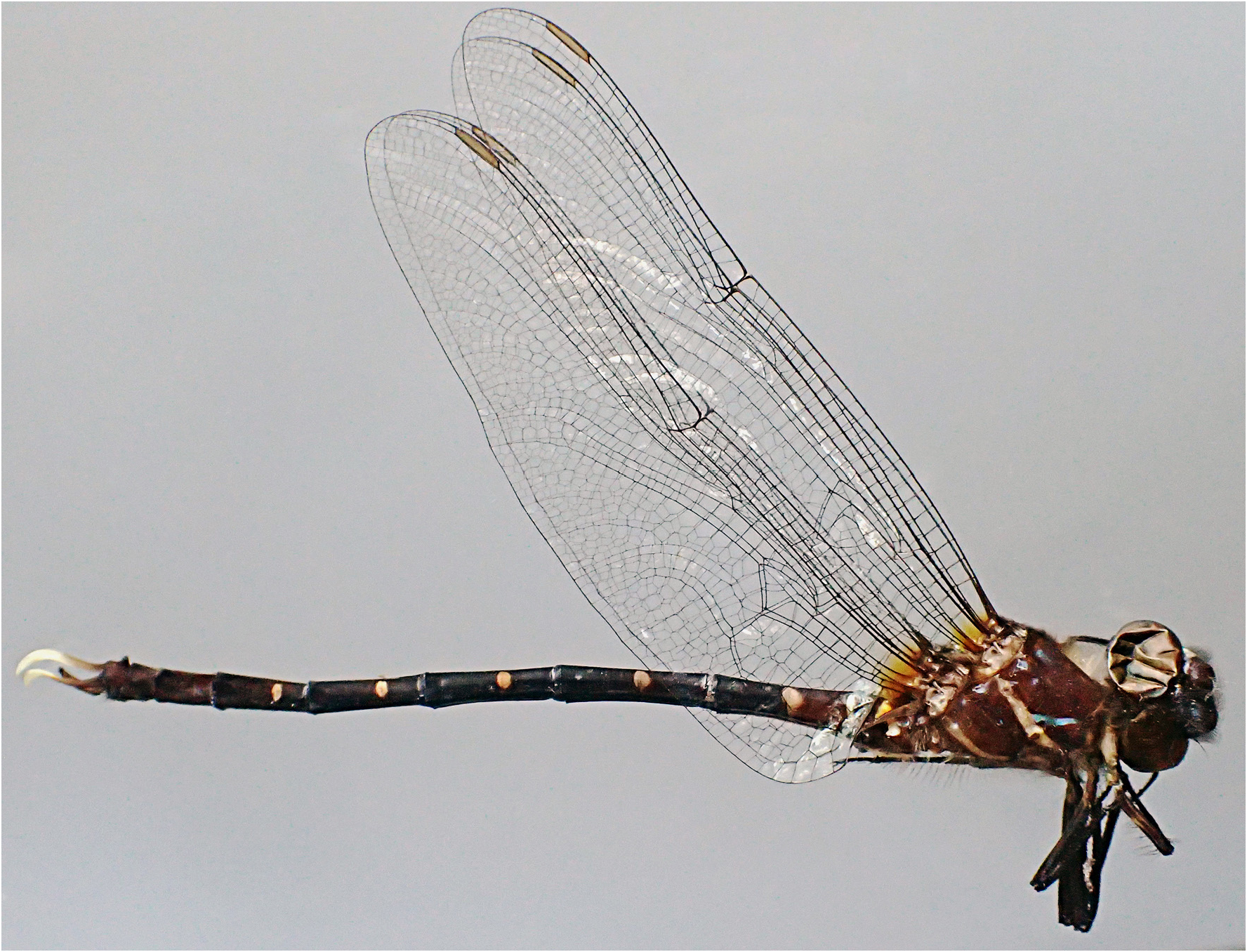
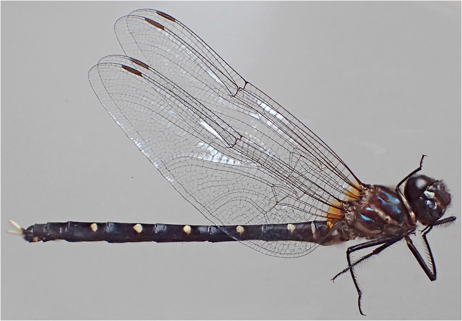
Description of male holotype. A large synthemistid with dark brown to black body showing green metallic reflections on the thorax, exhibiting white to whitish markings, and with strikingly long sinuous cerci ( Fig. 1 View FIGURE 1 ).
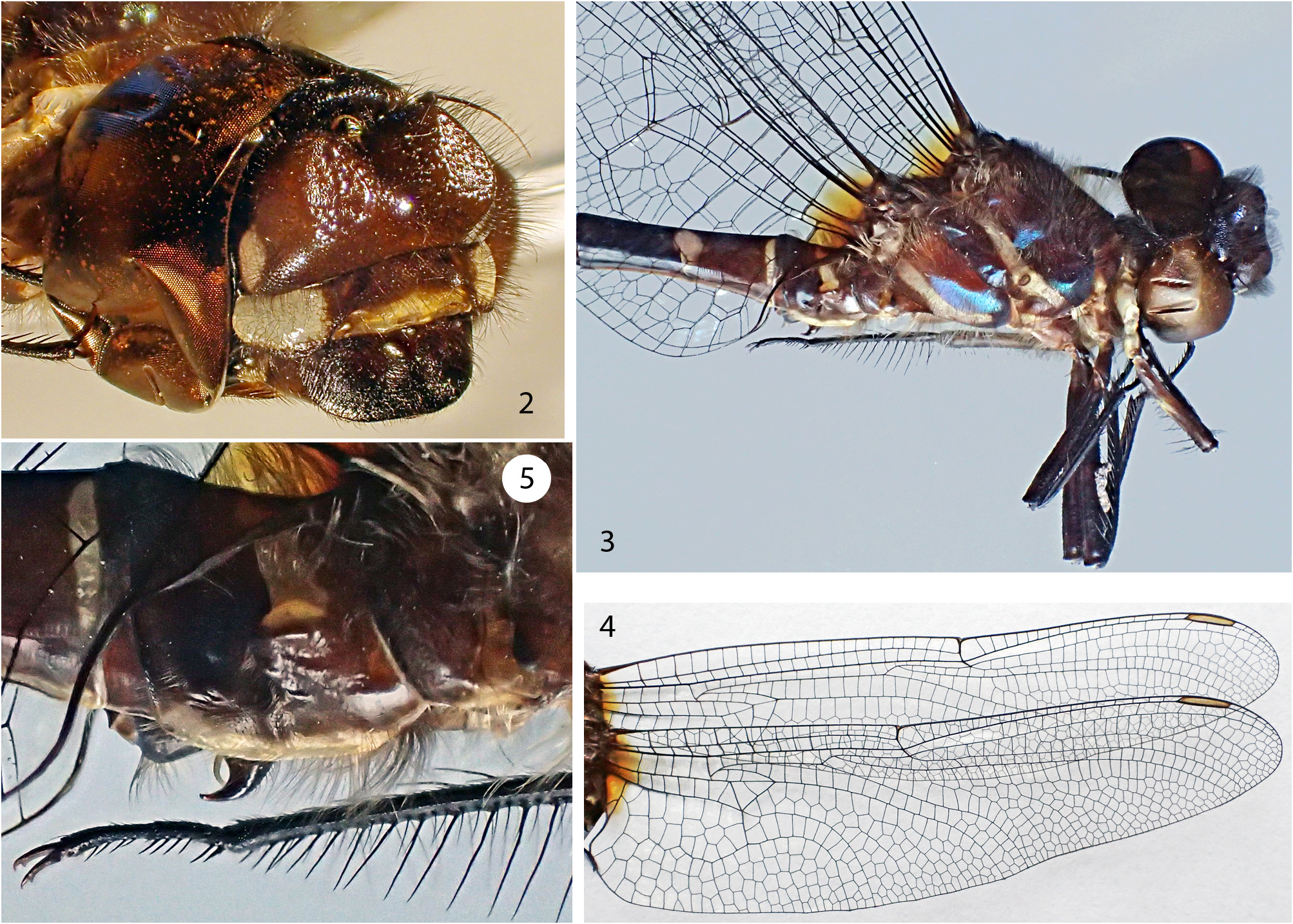
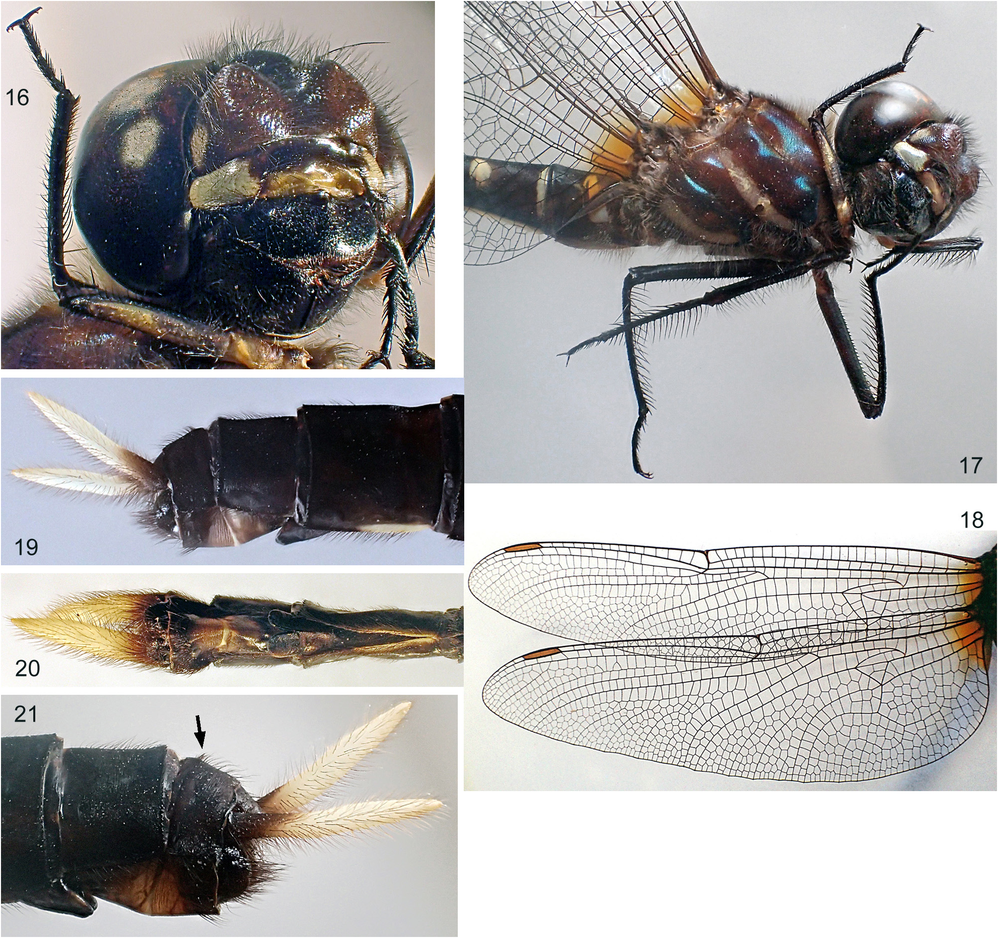
Head ( Fig. 2 View FIGURES 2–5. 2 ). Face, frons and vertex covered by long bristle-like dark brown setae except on ventral margin of labrum and anteclypeus. Labium and labrum dark brown. Anteclypeus yellowish orange, postclypeus brown with two large lateral white spots reaching eye margins. Frons glossy brown with two small lateral white spots reaching eye margins; prominent and strongly bilobed, each lobe bearing a frontal flat ovate area. Vertex dark brown uniformly rounded dorsally. Antennae brown at base and with long flagellum turning progressively light brown distally. Eyes in contact dorsally for a very short distance (ca 0.5 mm) in their anterior part. Posterolateral margin of eyes with small indentation (remnant of larval eye, often encountered in corduliids). Occiput dark brown, in dorsal view with slightly rounded posterior margin and rather long triangular anterior intrusion.
Thorax ( Figs 1 View FIGURE 1 , 3 View FIGURES 2–5. 2 ). covered by pale hair-like setae. Legs rather short compared to other body dimensions, hind leg (ca 18 mm) being distinctly shorter than half of HW length and slightly shorter than third of abdominal length including anal appendages. Legs with tibiae and tarsi dark brown to blackish; profemora brown with irregular pale yellowish spot at midlength; protrochanter light brown with yellowish streak ending at distal margin; mesofemora and metafemora brown at base becoming gradually dark brown toward apex; mesotrochanter and metatrochanter with brown outer side and paler inner side. Tibial keels present on all legs, occupying 50–55% on the protibiae, 60% on the mesotibiae and 75–80% on the metatibiae. Ventral tooth of tarsal claws well developed and situated at about 2/3 to 3/4 of claw length (see Fig. 5 View FIGURES 2–5. 2 ). Prothorax light brown to brown; hind lobe with rounded margin and regularly covered by rather short setae. Pterothorax brown with bluish to greenish metallic reflections (depending on light incidence) and with whitish and thin (ca 1 mm width) metepisternal and metepimeral stripes; pterothorax additional distinct and contrasting whitish markings on mesepisternal ante-alar ridge, on infraepisterna with small spot close to ventral margin, on mesonotum, and on meso-metanotal suture.
Wings ( Figs 1 View FIGURE 1 , 4 View FIGURES 2–5. 2 ): hyaline with saffron tinge at base. This saffron spot better developed in HW, reaching first crossvein of median and submedian spaces and occupying anal triangle. Veins dark brown to brown except extreme base of all costae (humeral plate, or DCP sensu Ninomiya & Yoshizawa 2009) showing a clearly delimited white dorsal spot. Membranulae well developed, brown, nearly as long as anal triangle in HW. Pt yellowish — light brown, rather long (ca 5% of wing length) and with proximal and distal margins roughly parallel; pterostigmal brace present, rather weak and slightly distal to Pt proximal margin. FW nodal ratio of ca 1.17 (see Fleck & Haber 2022); HW nodus shifted basally with nodal ratio ca 0.78. Basal antenodal of second rank (between ScP and R) anterior to primary Ax1 presents in both pair of wings; all primary Ax1 and Ax2 (crossvein of first rank between C and ScP and crossvein of second rank between ScP and R aligned, reinforced and bracketlike) separated by a secondary crossvein (crossvein of first rank and crossvein of second rank not aligned and not reinforced); FW Ax 23 (left wing)–21 (right wing) of the first rank (including primaries) and 23 of the second rank (including primaries and basal crossvein); several FW Ax of the first rank aligned or sub-aligned with those of the second rank but only Ax1 and Ax2 reinforced and bracketlike; HW Ax5, 7, 9, 11 and 13 (left wing) and Ax5, 7, 9 and 12 (right wing) of first rank of primary type (aligned with those of second rank, reinforced and bracketlike), reinforcement of these Ax diminishing distally (thus the HW presents a succession of primary type crossveins separated by secondary crossveins).
HW Ax 15 (left wing)–14 (right wing) of the first rank (including primaries) and 17–15 of the second rank (including primaries and basal crossvein). FW Px 16–15; HW Px 19–18. Crossveins distal to Pt 4 or 5 (left FW). Arculus distinctly distal from the level of Ax 2 in all wings; sectors fused on a rather long distance (about as long of the basal closure in FW and twice the basal closure in HW); basal closure short, about 1/4 of common stem of RP+MA. Bridges with 7 or 8 crossveins. Median spaces with 4 crossveins; submedian spaces with 7 or 8 (left FW) crossveins. Hypertriangles with 2 or 3 (left FW) crossveins. All discoidal triangles 2-celled. FW subtriangles 3- celled; no distinct HW subtriangles; position of anteroproximal angle of HW discoidal triangle distinctly distal to posterior crossvein of arculus (separated by 2 cells of submedian space). No distinct Mspl nor Rspl. FW MA and MP separated at triangle by 3 cells, then separated by 2 cells to the level of RP3/4 (distinctly convergent near triangle then parallel), and separated by 3 or more cells distal to the level of RP3/4 (distinctly divergent). Anal loop as long as broad with 9–10 cells; posterior margin of anal loop and posterior wing margin separated by 3 (left) or 3–4 (right) rows of cells; presence of a smaller secondary anal loop made by 4 cells. Anal triangle well defined, twice as long as broad and 2-celled with crossvein posteriorly shifted, oblique and strongly curved. Anal angle well defined, tornus distinct and made by slight and short enlargement of the wing margin, apparently covered by microstructures.
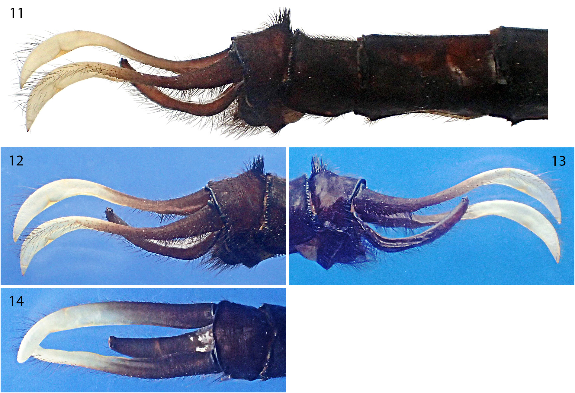
Abdomen. Slender, slightly depressed laterally, slightly longer than HW, and in lateral view moderately swollen at S2 ( Figs 1 View FIGURE 1 , 3, 5 View FIGURES 2–5. 2 ). Brown on S1–2, on anterior part of S3, on dorso-lateral part of S7 and on S8–10; remainder of S3 and S7, and S4–6 dark brown to black (passage from brown to dark brown and dark brown to black progressive) ( Figs 1 View FIGURE 1 , 3 View FIGURES 2–5. 2 ). S2 with a pair of transverse whitish lateral strips occupying dorsal surface of oreillets and ending as slender tip close to middorsal carina (thus strips not meeting dorsally) and with a pair of longitudinal whitish strips bordering ventral margins of tergite ( Figs 3, 5 View FIGURES 2–5. 2 ); S3 with a pair of longitudinal thin and distally tapering whitish strips bordering 1/4 of ventral margins of tergite; S3–4 with pair of transverse lateral stripes bordering anterior margin of segment and not meeting dorsally, those of S3 particularly distinct, developed on ca 2/3 of lateral part and whitish, those of S4 much less distinct occupying about 1/3 of lateral part and yellowish-brown ( Figs 1 View FIGURE 1 , 3 View FIGURES 2–5. 2 ); S3–7 with pair of pale yellowish lateral spots centred on transverse median carina ( Figs 1 View FIGURE 1 , 3 View FIGURES 2–5. 2 ); S8 extreme ventral base and basal 2/3 of tergal ventral longitudinal carina whitish, and S9 ventral distal half white (partly visible on Figs 11, 13 View FIGURES 11–14. 11 ); epiproct brown, cerci brown at base turning progressively white, with whitish tinge occupying ca distal 1/2 and bright white occupying ca distal 1/3 of the cerci ( Figs 1 View FIGURE 1 , 11–14 View FIGURES 11–14. 11 ). Posterior margin of oreillets covered by minute protuberances and denticles. S10 with dorso-basal protuberance bearing clump of long and strong setae mimicking a dorsal horn ( Figs 1 View FIGURE 1 , 11–13 View FIGURES 11–14. 11 ).
Accessory genitalia: lamina anteriore rather long, occupying approximately anterior 1/3 of genital fossa and presenting a posterior margin strongly excavated flanked laterally by a pair of apically strongly chitinized subrectangular flaps (hamuli anteriores likely merged with lamina sensu stricto); hamuli posteriores bipartite, with a basal part -mostly hidden in lateral view by ventral margin of tergite- slightly swollen and bearing a smooth divergent indentation (probably made to secure anterior part of the penis V 1 in copulae), and with an apical part strongly curved and back directed, narrowing distally, and ending as a pointed process slightly curved laterally ( Fig. 5 View FIGURES 2–5. 2 ).
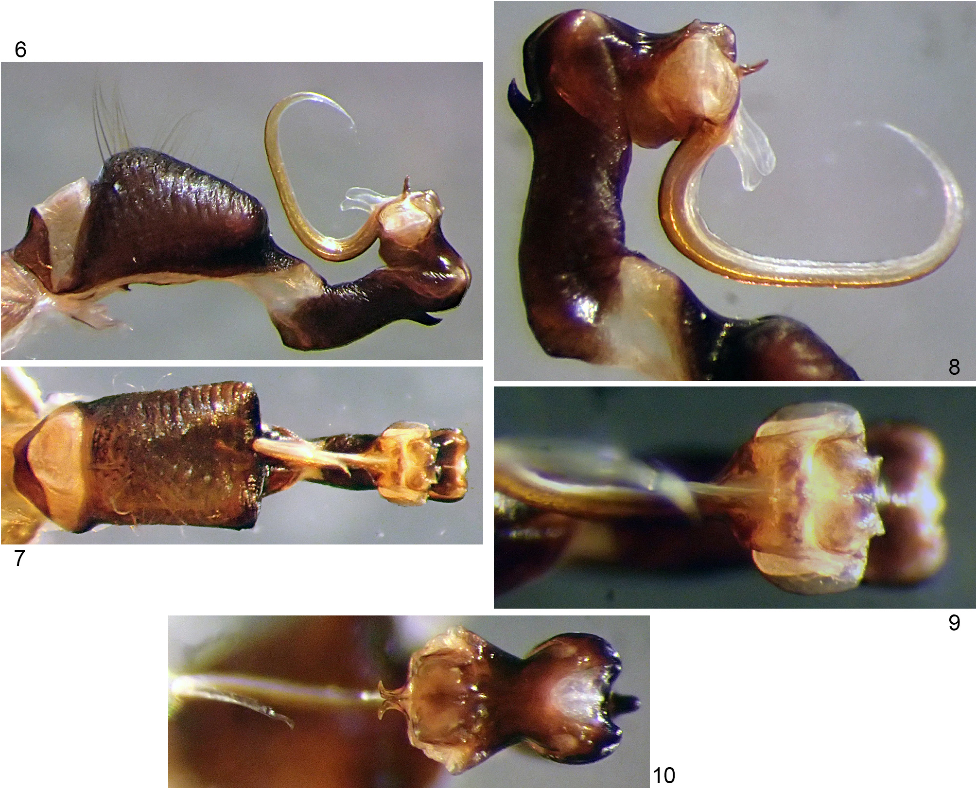
Vesica spermalis (penis): as in Figs 6–10 View FIGURES 6–10 , with noteworthy features: V1 distinctly ventrally inflated and appearing sub-rectangular in ventral view ( Figs. 6–7 View FIGURES 6–10 ); V2 with strong hump ( Figs 6, 8 View FIGURES 6–10 ); V3 rather short with low proximal horns exhibiting posterior minute rounded protuberance ( Figs 6–9 View FIGURES 6–10 ); V4 with short first distal horn (dh1) strongly chitinized ( Figs 6, 8 View FIGURES 6–10 ) exhibiting branches strongly divergent at base ( Fig. 10 View FIGURES 6–10 ), with second distal horn (dh2) appearing as two semitranslucent slightly asymmetrical lamellae ( Figs 6, 8 View FIGURES 6–10 ), and with flagellum-like very long V4- furrow apically recurved ( Fig. 6 View FIGURES 6–10 ) showing overlap of margins on its right side ( Fig. 8 View FIGURES 6–10 ).
Caudal appendages ( Figs 11–14 View FIGURES 11–14. 11 ). Cerci: exceptionally long, much longer -in dorsal view- than S9+10 (ratio of cerci length / length of S9+10 = 1.71) and longer than S8+9 [note: due to strong dorso-ventral undulation, true developed length (7.2 mm) distinctly greater than dorsal projected length (6.0 mm)]; in lateral view sinuous, strongly down curved distally with apex perpendicular to body axis; in dorsal view sub-parallel/slightly divergent to the level of epiproctal apex then convergent and distally overlapping (natural or due to preservation of specimen?); base with marked dorsal transverse bead close to S10 reinforced distal margin (acting probably as dorsal/lateral blocking system); base strong, slightly higher than broad, thinning rapidly distally and giving to 2/3 distal of cerci a flattened appearance; strong inner carina vanishing at distal 1/3; inner surface remarkably glabrous, except for basal 1/4 covered by scarce and weak setae; dense field of long and thin setae (thinner and slightly shorter than, or as long as setae forming S10 clump) covering basal 1/4 of ventral side; lateral side covering with thin setae of moderate to very important length, the longest (length twice or more that of S10 clump setae) between distal 7/10 and distal 9/10. All setae brown to blackish. Epiproct: in lateral view strongly curved with robust latero-ventral carina extending from base to basal 6/10 of length and a weaker latero-dorsal carina extending from basal 1/10 to basal 4/10; in dorsal view, thin, triangular and truncated at apex; apical region bearing (1) a sub-apical pair of strongly chitinized small tubercles made each by jointed proximal minute rounded molar tooth and minute acute distal tooth, and bearing (2) an apical pair of small spiny protuberances.
Measurements (mm). Total length (including caudal appendages) 66.0, FW length 43.0 (wingspan ca 87), HW length 42.0, abdomen length (including caudal appendages) 52.0, cerci 6.0 (dorsal projection from base to apex) (7.2 true developed dorsal length), epiproct 3.3 (ventral projection from base to apex).
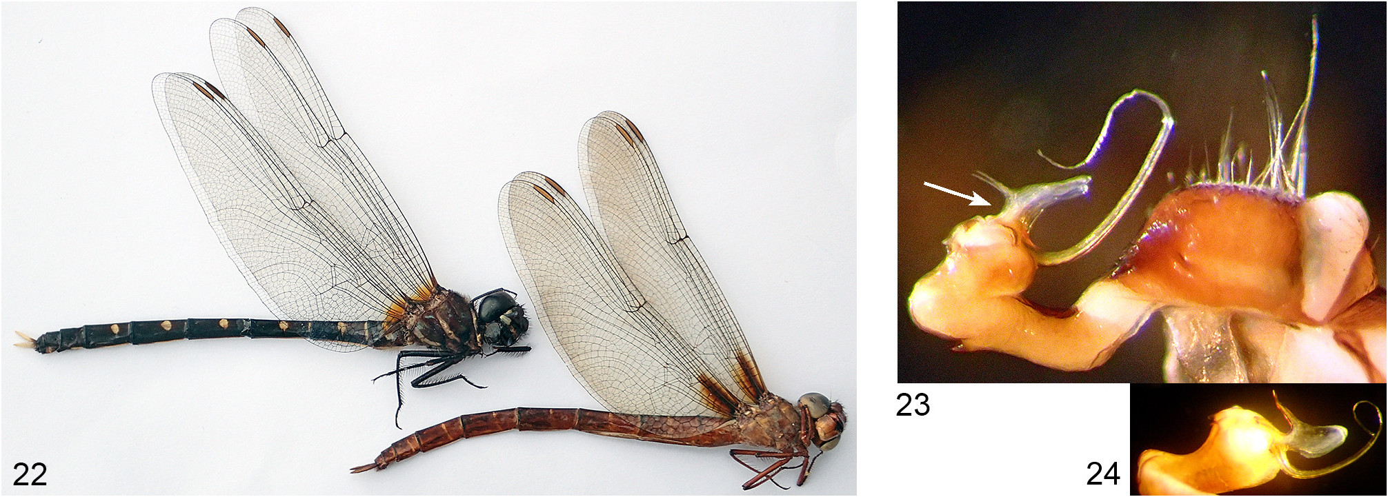
Description of female paratype. Distinctly larger and stouter than male but with similar appearance and almost identical in colour pattern ( Figs 15–17 View FIGURE 15 View FIGURES 16–21. 16 , compare with Figs 1–3 View FIGURE 1 View FIGURES 2–5. 2 ). Thus, only distinct differences with holotype mentioned.
Wings ( Figs 15 View FIGURE 15 , 18 View FIGURES 16–21. 16 ): Broader, with HW ratio length/width = 3.13 (male HW ratio = 3.7). Basal saffron tinge slightly more expanded (clearly reaching first crossvein of median and submedian spaces in both pair of wings). Pt reddish brown. HW Ax5, 7, 9, 11, 13 and 14 (left wing) and Ax5, 7, 9, 11 and 13 (right wing) of first rank of primary type. Bridges with 5 to 8 crossveins. Median spaces with 4 or 5 crossveins; submedian spaces with 8 or 9 crossveins. All discoidal triangles 3-celled. FW MA and MP separated at triangle by 4 cells. Anal loop very large with 27–31 cells; posterior margin of anal loop and posterior wing margin separated by 5 rows of cells; smaller secondary anal loop with 8 cells.
Abdomen. Well developed dorso-ventrally, strongly depressed laterally, and in lateral view very slightly swollen at S2 ( Figs 1 View FIGURE 1 , 17 View FIGURES 16–21. 16 ). Pair of thin distally tapering whitish strips bordering ventral margins of tergites slightly better developed and occupying basal ventral 6/10 to 7/10 of S3, 6, 7, 8; those of S6 the thinner and somewhat inconspicuous, those of S8 well marked and forming distinct subrectangular markings (see Fig. 19 View FIGURES 16–21. 16 ). Pair of S4 transverse lateral stripes bordering anterior margin of segment distinct and whitish. S9 ventral distal half and S10 proximal half greyish brown, intersegmental membrane whitish ( Fig. 19 View FIGURES 16–21. 16 ). S10 with a dorsal small but dense field of setae of moderate length close to posterior margin (probably homologous to remarkably developed male structure) ( Fig. 21 View FIGURES 16–21. 16 , indicated by arrow). Cerci long, longer than S9+10; slightly flattened dorso-ventrally tapering distally with obtusely pointed apex; covered by long brown to dark brown setae; brown at base turning rapidly white with clear white occupying ca distal 2/3 to distal 3/4 depending on view-point, i.e. less extended dorsally. Epiproct well developed grossly tile-shaped, down tilted, and nearly as long as S10 ( Fig. 21 View FIGURES 16–21. 16 ).
Ovipositor: as in Figs 19–21 View FIGURES 16–21. 16 , with remnant of V1 (vulvar lamina) reaching about middle of S9, sub-triangular in lateral view, showing rounded apex in ventral view, and with medial notch well marked, deep and V-shaped ( Fig. 20 View FIGURES 16–21. 16 ). Remnant of V2 (median process) hidden by V1 and hardly visible, subcylindrical, slightly curved with apex in contact with V1.
Measurements (mm). Total length (including caudal appendages) 74.0, FW length 50.0 (wingspan ca 102), HW length 49.0, abdomen length (including caudal appendages) 57.5, cerci 4.0.
Differential diagnosis
Calesynthemis jeanlegrandi is a large and well differentiated species. The female belongs among the fullest bodied representatives of the family and is comparable in dimension to Calesynthemis serendipita Winstanley considered to be the largest species ( Fig. 22 View FIGURES 22–24. 22 ); C. serendipita has a broader abdomen, but the length of the abdomen and wings is about the same, and C. jeanlegrandi shows larger head, stouter thorax and broader HW base. This new species is probably closely related to C. miranda Selys. The male can easily be separated from allied species but also from all other synthemistids by the unique shape, length, and colour of the caudal appendages, and by the remarkable S10 dorsal brush mimicking a horn. The female can be separated from congeners by the following combination of characters: (1) pterothorax with green metallic reflexions and with two distinct whitish lateral thin stripes, (2) wingspan more than 90 mm, costae brown with distinct basal white spot, and basal saffron tinge not overpassing distinctly first costal, subcostal, median and submedian spaces crossveins, (3) cerci largely white, longer than S9+10, and not up turned.
No known copyright restrictions apply. See Agosti, D., Egloff, W., 2009. Taxonomic information exchange and copyright: the Plazi approach. BMC Research Notes 2009, 2:53 for further explanation.