
Adosomus
|
publication ID |
https://doi.org/10.11646/zootaxa.4021.3.3 |
|
publication LSID |
lsid:zoobank.org:pub:39804549-C2A5-4534-973B-4312AC140EDD |
|
DOI |
https://doi.org/10.5281/zenodo.6098559 |
|
persistent identifier |
https://treatment.plazi.org/id/03F487E9-FFC3-FFC9-FF48-FE0A5A4AFE41 |
|
treatment provided by |
Plazi |
|
scientific name |
Adosomus |
| status |
|
Adosomus View in CoL (s.str.) roridus (Pallas, 1781)
Material examined. SLOVAKIA: Bratislava region: Pezinok-Grinava env., ( 48°16'9.66"N, 17°13'52.95"E). 27.v.2012 ( 1 pupa (♂)), 15.v.2014 ( 4 larvae, 1 pupa (♀)), 13.vii.2014 ( 1 pupa (♂)), all leg. F. Trnka.
Description of mature larva. COLOURATION. Brown or dark brown head with a distinct pale pattern around the frontal suture and lateral sides ( Fig. 4 View FIGURE 4 C). All thoracic and abdominal segments white; only dorsum of pronotum with elongated light brown stripe ( Figs. 4 View FIGURE 4 C–D).
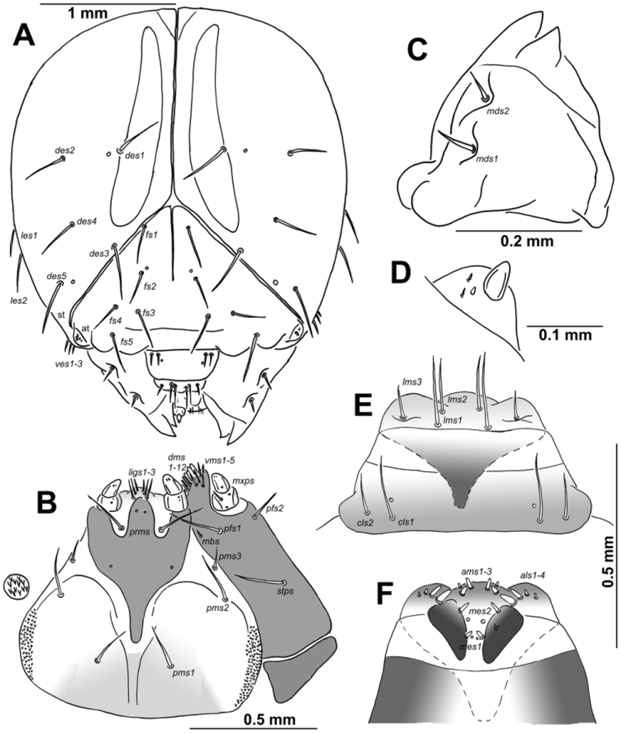
HEAD CAPSULE AND MOUTH PARTS. Head width: 2.5–3.0 mm (mean 2.8 mm), suboval, flattened laterally, endocarinal line long exceeded almost to the half of frons. Frontal sutures on head distinct, extended to antennae. Single stemma (st), in the form of a dark pigmented spot, located on each side anterolaterally. Des1 and des2 located in upper part of the central part of epicranium, des1 near to the middle part of epicranium, and des2 near to side of epicranium, des3 located anterially near to frontal suture, des4 located in the central part of epicranium, des5 located anterolaterally; all des long, almost all equal in length ( Fig. 1 View FIGURE 1 A). Fs1 and fs2 placed medially, fs3 located anteriomedially, fs4 located anteriolaterally, and fs5 located laterally, close to the epistoma; all setae relatively long, fs5 slightly longer than the remaining four setae ( Fig. 1 View FIGURE 1 A). Les1–2 as long as des1; and ves1–3 short. Epicranial area without pores. Antennae located at the end of the frontal suture on each side, membranous and slightly convex basal article bearing conical triangular sensorium, relatively long; basal membranous article with 3 sensillae different in both shape and length ( Fig. 1 View FIGURE 1 D).
Clypeus ( Fig. 1 View FIGURE 1 E) approx. 2.2 times as wide as long with 2 relatively long cls, almost equal in length, localized posteriolaterally and 1 sensillum; anterior margin rounded to the inside; median part covered by thorn-shaped cuticular processes. Labrum ( Fig. 1 View FIGURE 1 E) approximately 4 times as wide as long, with 3 pairs of hairform lms, of different length; lms3 distinctly shorter than very long lms1 and lms2; lms1 placed close to the margin with clypeus, lms2 located anteriomedially and lms3 located anteriolaterally; anterior margin double sinuate. Epipharynx ( Fig. 1 View FIGURE 1 F) with 4 blunt, finger-like als, unequal in length, 2 laterally als distinctly shorter than 2 medially als; 3 very short. blunt, finger-like ams, ams1 and ams2 distinctly larger than ams3; 2 pairs of short, blunt mes, unequal in length; labral rods (lr) rather broad, sub-triangular, approximating towards base. Mandibles ( Fig. 1 View FIGURE 1 C) distinctly broad, bifid, tooth of unequal height; slightly truncate; both mds relatively long, hairform, located in distinct holes. Maxilla ( Fig. 1 View FIGURE 1 B) stipes with 1 stps, 2 pfs and 1 mbs, stps and pfs1–2 very long, equal in length, mbs very short; mala with 12 bacilliform dms in two different lengths (6 very long and 6 relatively long); 5 short vms, almost equal in length; vms distinctly shorter than dms. Maxillary palpi with two palpomeres; basal palpomere with 1 very short mxps and two sensilla; length ratio of basal and distal palpomeres: 1:0.9; distal palpomere with one sensillum and a group of conical, cuticular apical processes. Praelabium ( Fig. 1 View FIGURE 1 B) heart-shaped and distinctly elongated, with 1 very long prms; ligula with sinuate margin and 3 hairform short ligs, unequal in length; premental sclerite well visible. Labial palpi with two palpomeres; length ratio of basal and distal palpomeres: 1:0.8; distal palpomere with one sensillum and short, cuticular apical processes; basal palpomere with 1 dorsal sensillum. Postlabium ( Fig. 1 View FIGURE 1 B) with 3 pms, pms1 located anterially, remaining two pairs laterally; pms1 and pms2 very long, pms3 distinctly shorter; surface of postlabium partly covered by distinct cuticular processes.
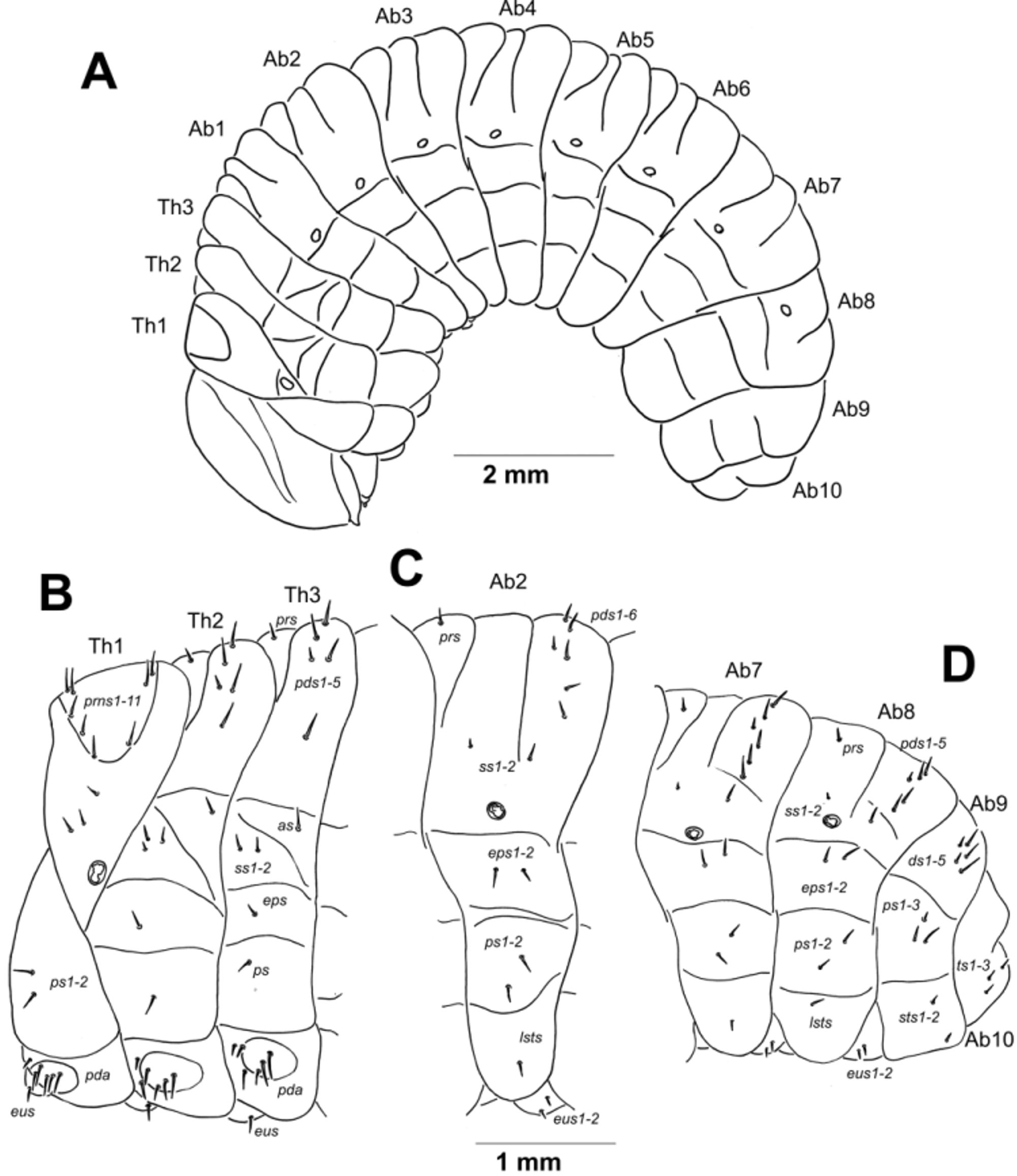
THORAX AND ABDOMEN. Body length: 12.0– 16.4 mm (mean 14.5 mm) stocky, slightly curved, rounded in cross section ( Fig. 2 View FIGURE 2 A). The widest place in the body (metathorax and abdominal segments I–IV) measuring up to 6.0 mm. Prothorax distinctly smaller than meso- and metathorax. Metathorax and abdominal segments I–IV of almost equal length, next abdominal segments decreasing gradually to the terminal parts of the body. Abdominal segment X reduced to four anal lobes of unequal size, the dorsal being distinctly the largest, the lateral pair equal in size, and the ventral lobe very small. Anus located terminally. Spiracles (9 pairs) unicameral, the first placed between the pro- and mesothorax (see Material and methods), the abdominal spiracles located laterally, close to the anterior margin of abdominal segments I–VIII.
Chaetotaxy of mature larva. Setae thin, short to relatively long, light yellow or orange. Thorax. Prothorax ( Fig. 2 View FIGURE 2 B) with 11 prns unequal in length, 8 of them on weakly pigmented dorsal sclerite, this sclerite subdivided in two triangular plates medially, 3 of them closely to spiracle; 2 relatively long ps and 1 eus. Mesothorax ( Fig. 2 View FIGURE 2 B) with 1 prs; 5 pds unequal in length, pds1–3 and pds5 long, pds4 very short; 1 relatively long as; 3 ss unequal in length, ss1 and ss2 short, ss3 very short; 1 relatively long eps; 1 relatively long ps and 1 short eus. Chaetotaxy of metathorax ( Fig. 2 View FIGURE 2 B) almost identical to mesothoracal, metathorax with only 2 ss almost equal in length, both short. Each pedal area of thoracic segments well separated, with 6 relatively long pda, 3 of them on pigmented pedal area, unequal in length. Abdomen. Abdominal segments I–VII ( Figs 2 View FIGURE 2 C–D) with 1 short prs; 6 relatively long pds, pds5–6 the longest one, pds3 the shortest; 2 ss of unequal length, ss1 very short, ss2 long as pds6; 2 relatively long eps of almost equal length; 2 relatively long ps of equal length; 1 short lsts and 2 short eus. Abdominal segment VIII ( Fig. 2 View FIGURE 2 D) with 1 short prs; 5 pds, pds6 lacking, pds3 less than half of length of the four relatively long remaining setae; 2 ss of unequal length, ss1 very short, ss2 long as pds5; 2 relatively long eps of almost equal length, 2 relatively long ps of equal length, 1 short lsts and 2 short eus. Abdominal segment IX ( Fig. 2 View FIGURE 2 D) with 5 ds ( ds1,3,5 long, ds2,4 short); 3 ps of unequal length, ps1 very short, ps2–3 almost long as ds1 and 2 very short sts. Ventral anal lobe on abdominal segment X ( Fig. 2 View FIGURE 2 D) with 2-3 short seta ( ts).
Description of pupa. COLOURATION. Body whitish to yellowish.
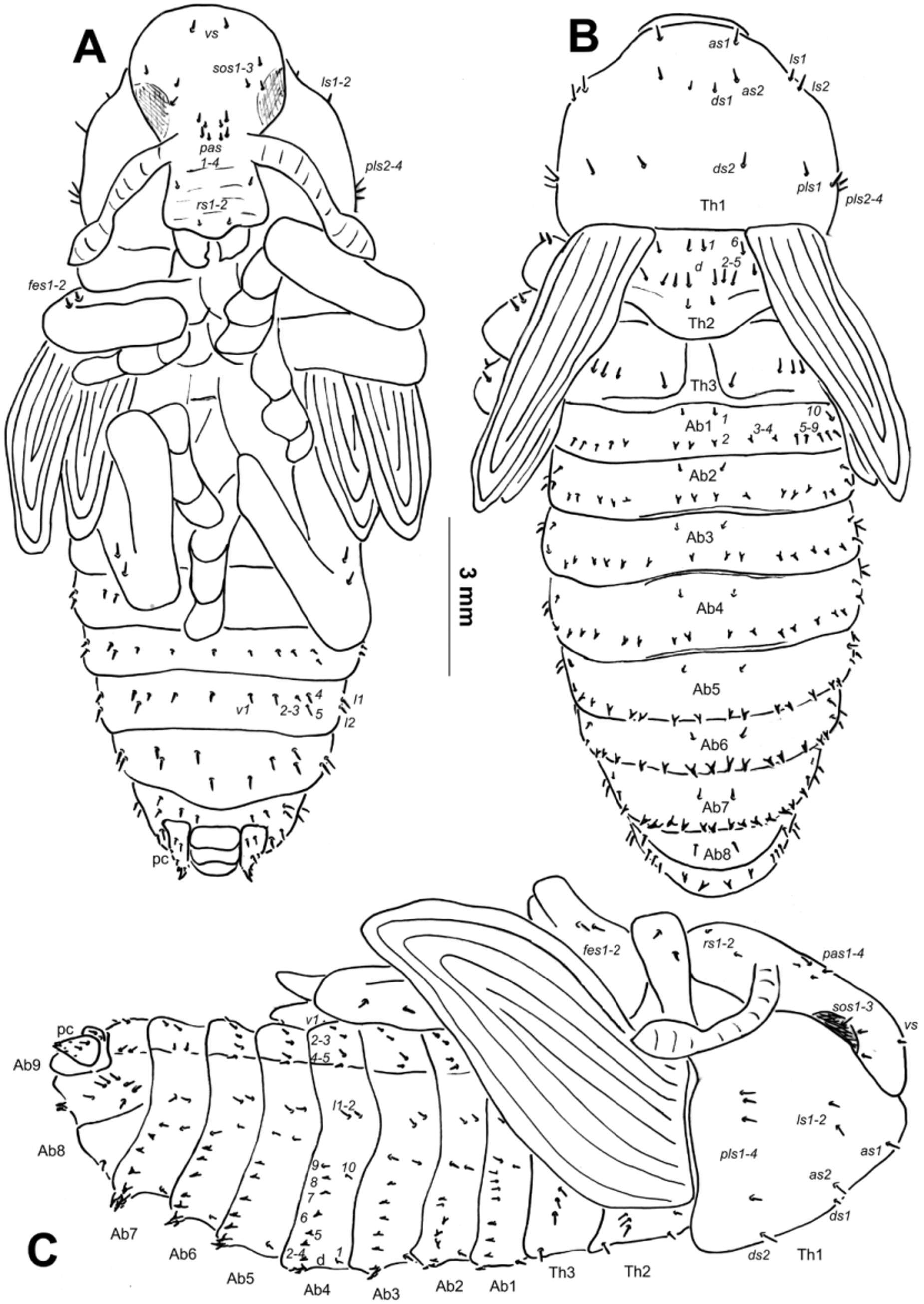
MORPHOLOGY ( Figs. 3 View FIGURE 3 A–C, 4F). Body length: 13.0– 17.5 mm (♂ 13.5–17.5 mm; ♀ 13.0 mm), at the widest region: 4.8–6.0 mm. The widest place in the body is commonly between the apex of the meso- or metafemora. Body stocky. Cuticle smooth. Rostrum short and wide, approximately 1.5 times as long as wide, extended to procoxae. Antennae relatively long and stout. Pronotum from 1.3 to 1.4 times as wide as long. Mesonotum and metanotum of almost equal length. Abdominal segments I–IV of almost equal length; abdominal segment V semicircular, next abdominal segments diminish gradually to the end of the body. Abdominal segments VI–IX distinctly smaller than other abdominal segments. Gonotheca (abdominal segment IX) in females ( 1 specimen) divided. Sexual dimorphism in weevils is visible mainly in the length of rostrum and in the structure of abdominal segment IX: gonotheca of ♂ undivided, of ♀ divided.
CHAETOTAXY ( Figs. 3 View FIGURE 3 A–C). Setae short to very short, unequal in length, light yellow or orange, some setae on abdominal segments I–VIII distinctly get stronger and located on protuberances. Setae well visible. Head capsule includes only 1 vs, 3 sos and 4 pas. Rostrum with 2 rs. Setae on head capsule straight, as short as the remaining setae on thoracic and abdominal segments. Pronotum with 2 as, 2 ds, 2 ls and 4 pls, ds1 distinctly shorter than the remaining setae. Dorsal parts of mesothorax with 1 seta located posteromedially, 1 seta posterolaterally and 4(5) setae located along its anterior margin. Dorsal parts of metathorax with 4 setae located along its anterior margin. Each apex of femora with groups of 2 fes. Dorsal parts of abdominal segments I–VIII each with 2 setae located posteriorly ( d1, d10) and 8 pairs ( d2–9) located along theirs anterior margins. Setae d2–4 (on abdominal segment I), d2–6 and d8 (on abdominal segment II), d2–8 (on abdominal segment III–VII) short, thorn-like, located on protuberances. Remaining setae short, hair-like. Abdominal segments I–VII with groups of 2 lateral setae and 5 ventral setae (4 setae on abdominal segment VII). Dorsal part of abdominal segment VIII with 2 setae located posteriorlly ( d1, d10) and 8 ( d2–9) located along its anterior margin; d2, and d3 thorn-like, located on protuberances; remaining setae short. Abdominal segment VIII with groups of 2 lateral setae and 4 short ventral setae. Abdominal segment IX with 2 ventral microsetae and 1 short, thin seta. Pseudocerci short, triangular, with 2 spines, and 3 ventral very short and 2 micro, thin setae.
Comparison with larvae of other Cleonini. The larvae of ten cleonine taxa have already been described ( Hoffmann 1950, Scherf 1964, Zotov 2011, Stejskal et al. 2014). The detailed description of the pupa is similar for nine cleonine taxa ( Scherf 1964, Zotov 2011, Stejskal et al. 2014). The comparison of the larva and pupa of Adosomus roridus with those described by Scherf (1964) was somewhat difficult due to the use of different terminology for morphology and chaetotaxy and/or the absence of good quality drawings (more in Stejskal et al. 2014). The comparison of the larva with Leucomigus tesselatus (Fairmaire, 1849) described/ drawn by Hoffmann (1950) is somewhat challenging due to the existence of only a few drawings, and some positions of setae are suspicious (e.g., number of fs, cls and mbs; see Skuhrovec et al. 2014, 2015). The descriptions of five cleonine larvae and pupae by Zotov (2011) are of higher quality and very useful; however, the described characters are only useful for differential diagnosis. The detailed descriptions of these five cleonine taxa are missing. Despite these challenges, we were able to compare the morphology of all ten taxa (Table 1).
Table 1. Differential diagnosis of mature larvae and pupae of 11 species from the tribe Cleonini.
Adosomus Asproparthenis Bothynoderes B. declivis Coniocleonus Cleonis Cyphocleonus C. dealbatus Leucomigus ‘ Pachycerus Rhabdorrhynchus
roridus carinicollis affinis nigrosuturatus pigra achates tesselatus scabrosus ’ * karelinii Larva
Endocarina present present present present absent present present absent absent absent present Number of des 5 5 4 4 5 4 5 5 5 5 5 Number of fs 5 5 5 5 5 4 5 5 2 5 5
2 2 2 2 2 not 2 3 2 3 3
Number of les presented
3 not presented not presented not 2 not not presented not presented not presented not presented not presented
Number of ves presented presented
Number of cls 2 3 2 2 2 2 2 2 3 3 2
in a triangle in a triangle in a line in a line in a triangle in a in a line in a triangle in a line in a line in a line
Position of lrms1-3 triangle
Number of als 4 3 3 3 4 4 3 6 3 3 3 Number of mds 2 1 1 1 1 2 1 2 2 2 2 Number of ligs 3 2 2 2 3 3 2 2 3 3 3 Number of mbs 1 1 1 0 1 0 1 1 2 1 0 Number of pds on 6 4 6 6 6 7 6 not presented 5 5 6. seg I- VII
Pupa
Number of setae on 6(7) 3 3 6 4 3 Unknown Unknown 4 6 mesonotum
Number of fes 2 2 2 3 2 2 Unknown Unknown not presented 2 Number of setae on 10 8 6 9 7 9 Unknown Unknown 5-7 10 dorsum of Abd. seg.
This larva was probably misidentified by Scherf (1964) and more likely belongs to the genus Rhabdorrhnychus (see the text for details).
May (1993) considered the increased number of pds on meso- and metathorax and abdominal segments I–VII and the increased number of epipharyngeal lining setae ( als) (i.e., higher than the most frequent number of setae in weevils) (for details, see Stejskal et al. 2014) as diagnostic for the mature larva of the subfamily Lixinae . Descriptions of mature larvae from the Lixini ( Larinus species: Zotov 2009a, 2010; Gosik & Skuhrovec 2011; Lixus species: Scherf 1964; Lee & Morimoto 1988; Nikulina 2001, 2007; Zotov 2009a, b; Nikulina & Gültekin 2011; Gosik & Wanat 2014; Skuhrovec & Volovnik in press; Rhinocyllus conicus: May 1994 ) fit this diagnosis, as do all known species from the Cleonini tribe (Stejskal et al. 2014). Gosik & Wanat (2014) compared the larvae and pupae of genera Lixus and Larinus and determined their differential characters. The precise key, detailed generic study of the Cleonini tribe and the comparison of both tribes was not possible because of our limited knowledge of the immature stages.

No known copyright restrictions apply. See Agosti, D., Egloff, W., 2009. Taxonomic information exchange and copyright: the Plazi approach. BMC Research Notes 2009, 2:53 for further explanation.
|
Kingdom |
|
|
Phylum |
|
|
Class |
|
|
Order |
|
|
Family |