
Mexico xiei Liu and Jia, 2021
|
publication ID |
https://doi.org/ 10.1080/00222933.2021.1931516 |
|
persistent identifier |
https://treatment.plazi.org/id/03F487FC-F704-762B-909F-FB6EF9C227BC |
|
treatment provided by |
Plazi |
|
scientific name |
Mexico xiei Liu and Jia |
| status |
sp. nov. |
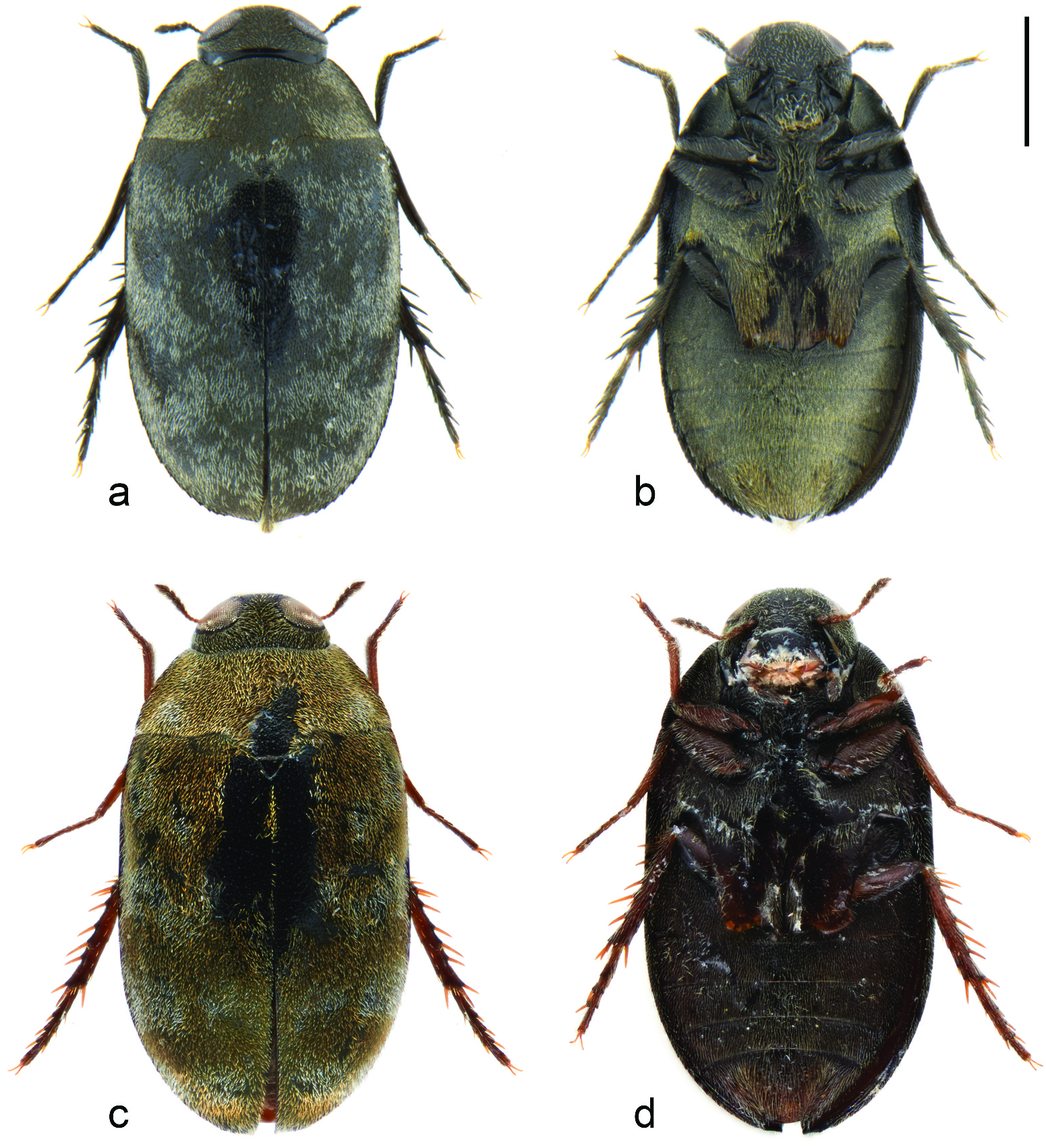
Mexico xiei Liu and Jia , sp. nov.
( Figures 1 View Figure 1 (a–b), 2)
Etymology
The new species is named after the family name of Professor Qiang Xie, the collector, who donated the specimens to the authors.
Type locality
China, Sansha Prefecture, Xisha, Yongxing Island.
Type material
Holotype. ♂, CHINA, Sansha Prefecture , Xisha, Yongxing Island (中国, 三沙市, 西沙, 永兴 岛), seaside coral reefs. 18 April 2019, 16.833N, 112.333E. Qiang Xie leg ( SYSBM) GoogleMaps . Paratypes: 10 specimens, same data as holotype ( SYSBM).
Diagnosis
This species mostly resembles M. taiwanus , M. masamii , M. ogasawaraensis, M. morrisoni and M. palauensis in the shape of the median lobe, with subapical projections distinctly divided from the apex of the median lobe.It can be distinguished from the first four species by the left subapical projections curved outwardly, and from the last species by the robust apex of the median lobe.
Description
Colour and form. Body small-sized and ovate, length 1.6–2.0 mm, width 0.8–1.1 mm. Dorsum and venter both black and slightly convex, covered with short, dense brown and silver setae.
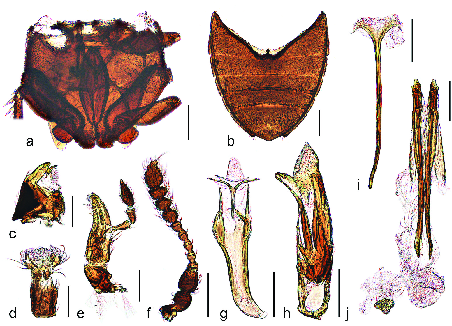
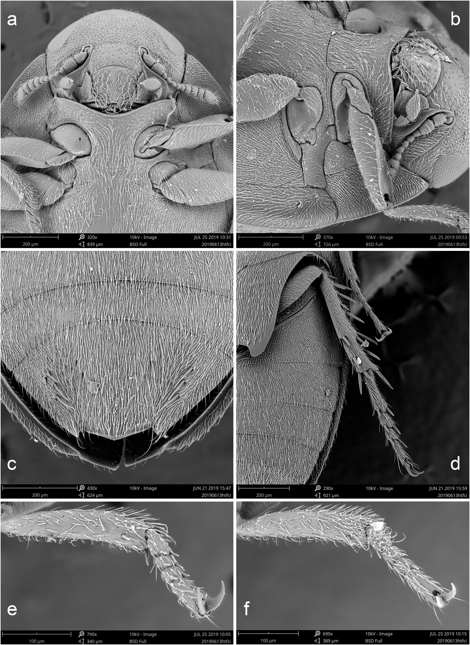
Head. Strongly hypognathous with mouthparts mostly concealed by labrum and prosternum ( Figure 3 View Figure 3 (a)), surface with dense microsculptures, occipital incisions absent; genal areas with distinct antennal grooves ( Figure 3 View Figure 3 (b)), glabrous anteriorly and punctured posteriorly; frontoclypeal suture distinct and straight; clypeus rectangular, with truncate apical margin. Eyes anterolaterally located, large and finely faceted, well separated ( Figures 1 View Figure 1 (a,b), 3(a)). Antennal insertions exposed, laterally attaching the frontoclypeal suture ( Figure 3 View Figure 3 (a)). Antennae ( Figure 2 View Figure 2 (f)) 11-segmented, with 4-segmented and slightly asymmetrical antennal club, covered with relatively long setae on basal and apical segments; scape and pedicel enlarged, antennomere 3 stout, antennomeres 4–7 narrow, antennomeres 8–10 dilated and gradually shortened, last segment fusiform with narrowed apex. Labrum ( Figure 3 View Figure 3 (a)) large and sub-trapezoid, longer than wide. Mandibles ( Figure 2 View Figure 2 (c)) stout and sub-triangular, with large basal turbercle on outer edge, apex bidentate; inner edge membranous, with long, dense setae; mola present and large. Maxilla ( Figure 2 View Figure 2 (e)) slender, maxillary palps 4-sgemented, last segment enlarged and apically acuminate; galea and lacinia both elongate and sub-triangular. Labium ( Figure 2 View Figure 2 (d)) with 3-segmented labial palps, second segment enlarged and terminal segment narrowed; ligula broad. Mentum rectangular, longer than wide. Ventral side with gular area highly reduced. Cervical sclerites present and large.
Thorax. Pronotum ( Figure 1 View Figure 1 (a)) transverse, about 0.4 times as long as wide, widest posteriorly; lateral margins slightly curved, posterior margin slightly sinuate with median lobe; anterior angles broadly acute and forwardly projecting; disc slightly convex with dense and fine punctations, covered with dense brownish setae. Pronotal epipleuron broad and slightly depressed. Prosternum with area before procoxae about 0.8 times as long as the length of coxal cavities; prosternal process wide and parallel-sided, apical margin nearly straight. Notosternal suture complete. Procoxae transverse, trochantins partly exposed ( Figure 3 View Figure 3 (a)); procoxal cavities widely separated, with a small anterolateral incision, externally broadly opened and internally narrowly opened. Scutellum small and triangular. Elytra slightly elongated, 1.4–1.5 times as long as wide, with about 12 tiny teeth along anterolateral edges and one small apical projection; disc slightly shiny, with dense and fine punctations, covered with dense brownish and silver setae that form four sinuate transverse stripes as in Figure 1 View Figure 1 (a); epipleuron ( Figures 1 View Figure 1 (b), 3(c)) broad at base, extending to the apical projection. Hind wings present. Mesoventrite short and transverse, with large anterior depression to accept prosternal process; mesanepisternum and mesepimeron fused ( Figure 3 View Figure 3 (b)). Metaventrite strongly transverse at base, apex with deep incision; metendosternite with stalk highly sclerotised, large and fusiform, fused with metaventrite by transverse carinae; lateral arms relatively long, with complicated ventral projections connected with apex of stalk ( Figure 2 View Figure 2 (a)); metanepisternum highly reduced and only partly exposed from ventral side, triangular ( Figure 3 View Figure 3 (b)). Mesocoxae slightly transverse, trochantins exposed; mesocoxal cavities laterally open to mesepimeron ( Figure 3 View Figure 3 (b)), separated by distance greater than width of cavities. Metacoxae strongly transverse and oblique, sub-contiguous; metacoxal plates ( Figures 1 View Figure 1 (b), 3(d)) large, broad and sharply narrowed to lateral edges. Fore and mid legs with femora slightly enlarged, tibiae slender; mid tarsi with basal three segments relatively short and bearing pair of tiny spines on apex ( Figure 3 View Figure 3 (d,f)), last tarsomere elongated with small falciform claws. Hind legs ( Figure 3 View Figure 3 (d)) with large trochanters, femora slightly expanded; tibiae slender with a few short to long spines along the outer and inner edges, apex with a single short spur; tarsi with claws nearly the same length as tibiae, first tarsomere longest, each of basal three segments with pair of apical spines, last tarsomere narrow and slightly elongate with pair of simple claws.
Abdomen ( Figure 2 View Figure 2 (b)) 5-segmented with basal three ventrites connate, lateral edges expanded with narrow flanges, covered with dense depressed yellowish setae. Ventrite 1 long, nearly separated by the oblique metacoxae at middle, intercoxal process reduced; ventrite 2–4 sub-equal in length; last ventrite elongate with pair of small posterolateral projections, surface with three pairs of long and thick bristles laterally and two pairs of thinner bristles on the inner side ( Figure 3 View Figure 3 (c)).
Male genitalia with tergite IX bifurcate, tergite VIII lamellate and asymmetrical as in Figure 2 View Figure 2 (g). Aedeagus ( Figure 2 View Figure 2 (h)) with basal piece sub-annular; lateral lobes short and sub-triangular, freely articulated, bearing two long apical setae on each side; median lobe trifurcate with lateral projections asymmetrical, apex of median lobe broad and subtriangular. Female genitalia with spiculum ventrale ( Figure 2 View Figure 2 (i)) elongated and bifurcate; ovipositor ( Figure 2 View Figure 2 (j)) with reduced and scleritized gonocoxite, baculus extremely long, paraproct membranous and stylus absent; spermatheca sclerotised.
Sexual dimorphism not distinct, tibiae and basal tarsomeres of fore leg in male with dense and relatively long setae, which are much sparser in females ( Figure 3 View Figure 3 (e,f)).
Habitats
Living on wet seaside reefs.
No known copyright restrictions apply. See Agosti, D., Egloff, W., 2009. Taxonomic information exchange and copyright: the Plazi approach. BMC Research Notes 2009, 2:53 for further explanation.
|
Kingdom |
|
|
Phylum |
|
|
Class |
|
|
Order |
|
|
Family |
|
|
Genus |