
Heteromysis ( Heteromysis ) gulfarii, Wittmann & Abed-Navandi & Dubois & Chevaldonné, 2021
|
publication ID |
https://doi.org/10.11646/zootaxa.4980.3.3 |
|
publication LSID |
lsid:zoobank.org:pub:83C3E976-6AE1-4EA2-B7E8-301196255D80 |
|
DOI |
https://doi.org/10.5281/zenodo.5041486 |
|
persistent identifier |
https://treatment.plazi.org/id/7414BC4A-BCCF-4E52-BEF9-DC4E40D474B8 |
|
taxon LSID |
lsid:zoobank.org:act:7414BC4A-BCCF-4E52-BEF9-DC4E40D474B8 |
|
treatment provided by |
Plazi |
|
scientific name |
Heteromysis ( Heteromysis ) gulfarii |
| status |
sp. nov. |
Heteromysis ( Heteromysis) gulfarii sp. nov.
http://zoobank.org/ urn:lsid:zoobank.org:act:
Figs 1–4 View FIGURE 1 View FIGURE 2 View FIGURE 3 View FIGURE 4
Holotype. Adult male with 4.1 mm body length ( NHMW –27016), aquaria of the ‘ Gulfarium Marine Adventure Park’, Fort Walton Beach, Florida, USA, Feb. 2020, leg. Emlyn MacKenzie.
Paratypes. 2 F ad. 3.7 mm, 2 M ad. 3.7–3.9 mm ( USNM –1642204) in vial, plus 1 M ad. 3.8 mm ( NHMW – 27017), 1 F ad. 3.7 mm ( NHMW –27015) on slides, same sample as for holotype; 1 M ad. 3.0 mm, 3 F ad. 3.5–4.5 mm ( MNINGA MYS 440 ) in vial, collection data as for holotype .
Material sequenced. 18S and COI barcodes here provided for 1 F ad. from same sample as for holotype, deposited in GenBank under accession numbers MW591699 View Materials and MW596477 View Materials .
Type locality. Not defined because the locality of origin ought to be indicated according to Art. 76.1.1. of the nomenclatorial code ( ICZN, 1999). The species was first discovered from the public aquarium center ‘Gulfarium Marine Adventure Park’ (Fort Walton Beach, Florida, USA).
Derivatio nominis. The species name is a noun with neutral ending in genitive singular, using the same gender and declination as for the Latin noun aquarium, referring to the ‘Gulfarium Marine Adventure Park’, from where the mysids were obtained.
Diagnosis. Carapace normal, rostrum triangular with narrowly rounded to acute tip, 0.2–0.5 times length of terminal segment of antennular trunk, covering <1/3 of eyestalks. Antero-lateral edges of carapace well rounded.
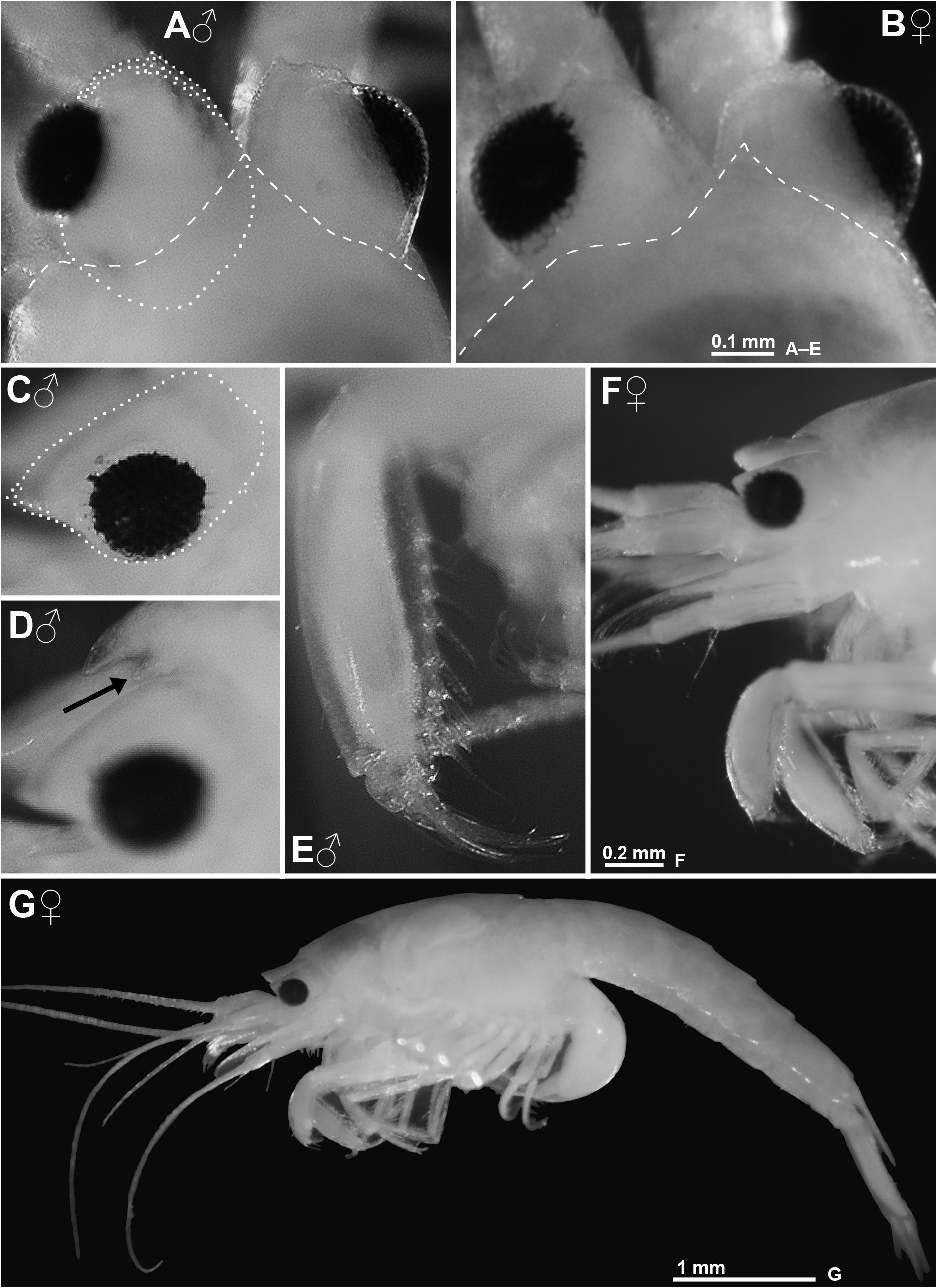
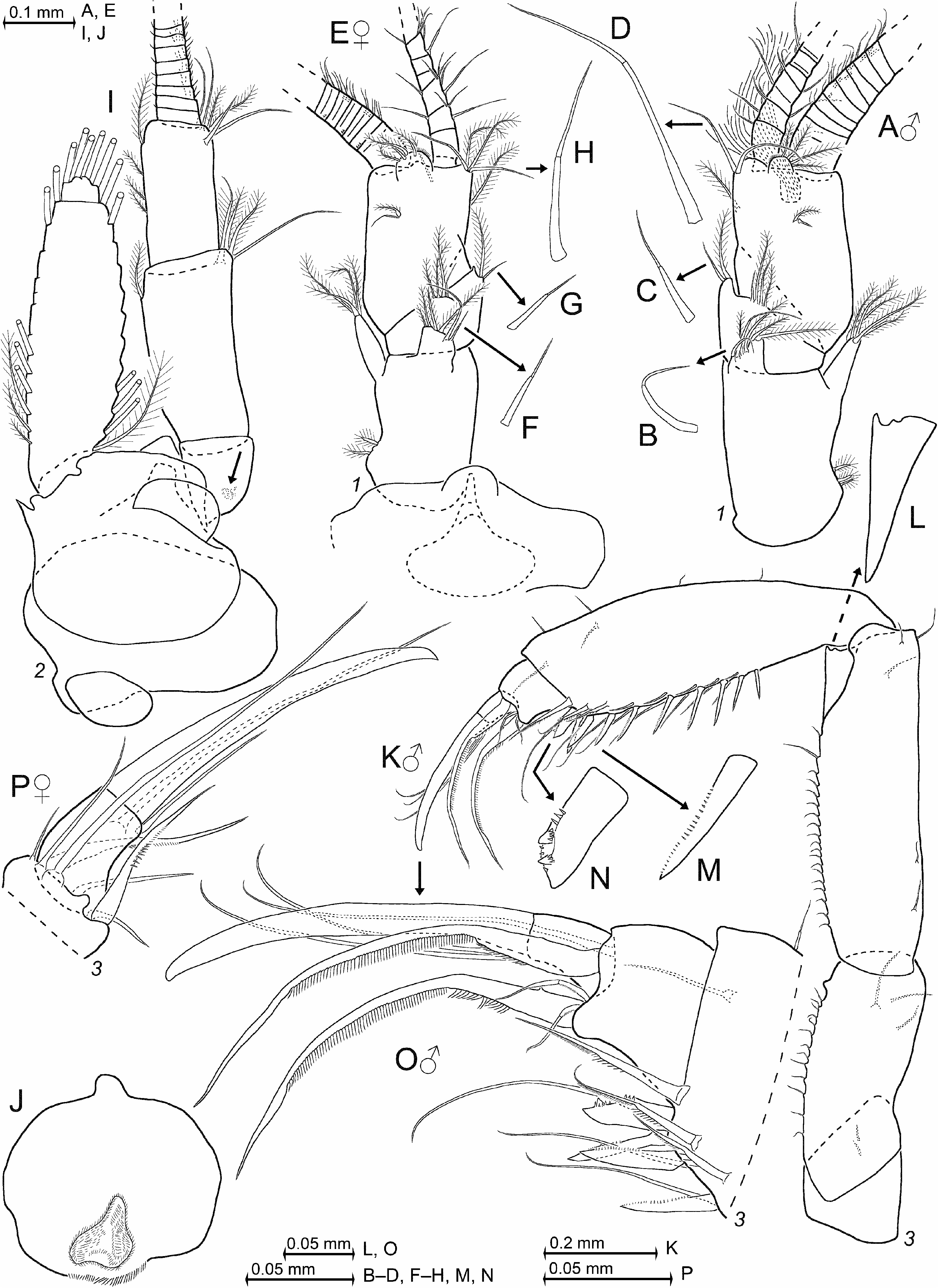
Eyes ( Fig. 1A–C View FIGURE 1 ) subquadrate in dorsal view. Eyestalk with flat upper face, anterior margin wrinkled, distolateral edge without spiniform extension. Stalk wedge-shaped in lateral view (oblique in Fig. 1C View FIGURE 1 ), posteriorly broad, from there anteriorly narrowing to form a triangular projection with acute or narrowly rounded tip; overall dorsoventrally compressed by a factor of 1.6–1.8 (length by maximum height). The margins of this projection converge at 50–60 angular degrees in males ( Fig. 1C View FIGURE 1 ), 80–90 in females ( Fig. 1F View FIGURE 1 ). Small cornea located mid-laterally, where it extends over <20% eye surface; its ommatidia compound, structure normal, functional. Cornea calotte-shaped in dorsal view ( Fig. 1A View FIGURE 1 ), diameter 0.6–0.9 times length of terminal segment of antennular trunk. Cornea ovate to circular in lateral view ( Fig. 1C View FIGURE 1 ), dorsoventrally compressed by a factor of 1.0–1.4.
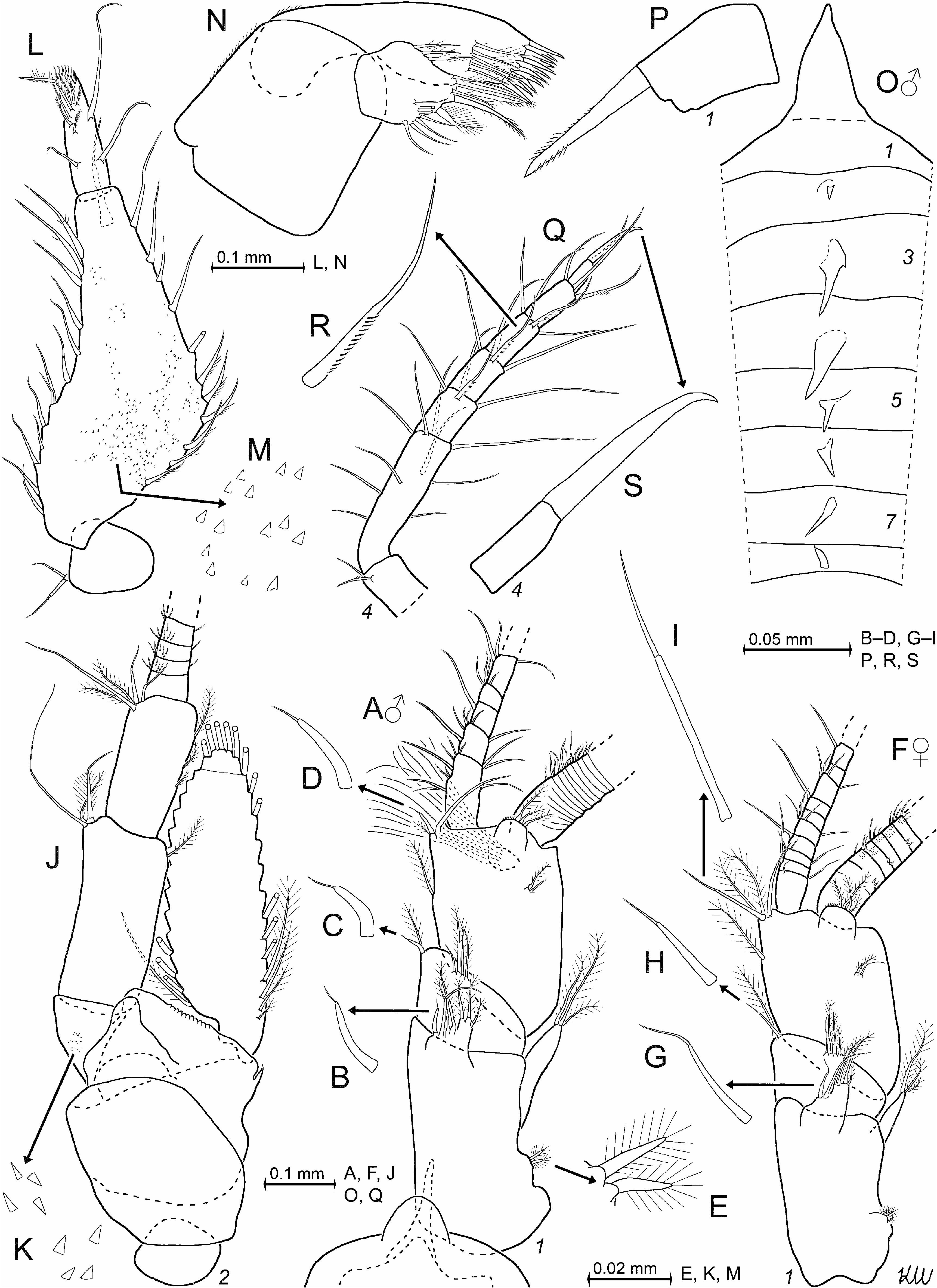
Antennulae ( Fig. 2A–I View FIGURE 2 ). Antennular trunk ( Fig. 2A, F View FIGURE 2 ) with setose mid-dorsal apophysis close to terminal margin of basal segment and with another apophysis on terminal segment; median segment without apophysis. Trunk with diverse types of setae, in part ( Fig. 2B–D, G, H View FIGURE 2 ) spine-like. Besides other setae, each segment with dimorphic whip seta, stouter in males ( Fig. 2B–D View FIGURE 2 ) compared to females ( Fig. 2G–I View FIGURE 2 ); one such whip seta on dorsal apophysis of basal segment, one on disto-mesial edge of the median segment, and a third one on disto-mesial edge of terminal segment. Appendix masculina small, with dense tuft of long setae.
Antennae and mouthparts. Antennal sympod ( Fig. 2J View FIGURE 2 ) with small spiniform extension on lateral face. Antennal scale reaching only to 40–80% length of terminal segment of antennular trunk in both sexes. Scale length 3.1–3.2 times maximum width. Scale with small apical segment. Mouthparts ( Fig. 2L–N View FIGURE 2 ) normal. Labrum caudally bulbous, rostrally broadly subtriangular with small rounded, anterior protrusion (as in Fig. 6J View FIGURE 6 ).
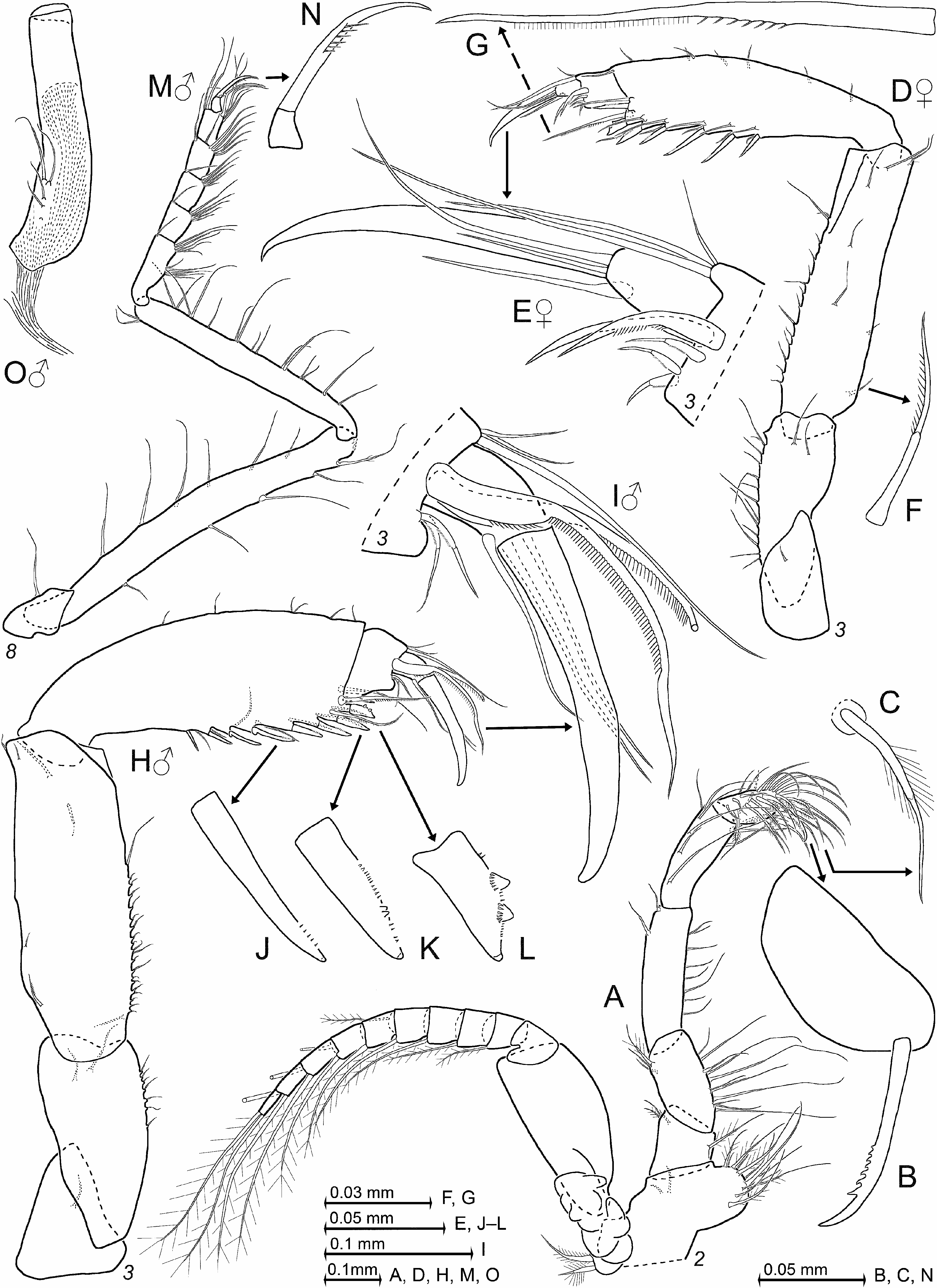
Thorax. Only the males with median processes from thoracic sternites 2–8 ( Fig. 2O View FIGURE 2 ; the anterior lobe of sternite 1 not counted as a process of that kind). Flagellum ( Fig. 3A View FIGURE 3 ) of thoracic exopods 1–8 with 8, 9, 9, 9, 9, 9, 9, and 8–9 segments, respectively. Carpopropodus ( Figs 2Q View FIGURE 2 ; 3A, D, H, M View FIGURE 3 ) of thoracic endopods 1–8 with 2, 2, 2, 4–5, 5, 5–6, 5–6, and 5–6 segments, respectively. Claw of endopod 3 ( Fig. 3E, I View FIGURE 3 ) weakly curved, measured along its median line 3–4 times dactylus length and 23–35% carpopropodus. Claws 2 ( Fig. 3B View FIGURE 3 ) and 4 ( Fig. 2S View FIGURE 2 ) with 50–70% length of claw 3, strong, apically more strongly curved; claw 1 ( Fig. 2P View FIGURE 2 ) with 40% length of claw 3, straight, strong; claws 5–8 ( Fig. 3N View FIGURE 3 ) with 30–50% length of claw 3, strongly curved, slender. Claw of endopods 1, 2, 5–8 subapically serrated, claws 3, 4 smooth. Penes 0.7–0.8 times length of ischium 8, and 1.0–1.1 times merus 8. Penes tube-like, terminally truncate, with 3–5 setae at about 1/3 length from basis ( Fig. 3O View FIGURE 3 ).
Gnathopods ( Fig. 3D, H View FIGURE 3 ). Thoracic endopod 3 with most of mesial margin of ischium and merus rugose by small (minute) projections, each bearing a small whip seta at its tip. Merus disto-mesially with triangular ridge (as in Fig. 6L View FIGURE 6 ), flanking the basis of the carpus on caudal face in both sexes when the subchela is closed. Carpus, propodus, and dactylus separated by distinct sutures. Carpopropodus length is 12–15% body length in females, 16–23% in males. Carpopropodus swollen, its length 3.3–4.5 times maximum width ( Fig. 3D, H View FIGURE 3 ); average stoutness about the same in both sexes (compare panels E, F in Fig. 1 View FIGURE 1 ). Carpus with 6–8 spines arranged along distal half of mesial margin. Propodus without spines; its paradactylary setae modified by comb-like series of barbs (cilia), dimorphic, about as long as claw in males ( Fig. 3I View FIGURE 3 ; due to more basal insertion not reaching to tip of claw), less strongly modified and only half the length of claw in females ( Fig. 3E View FIGURE 3 ).
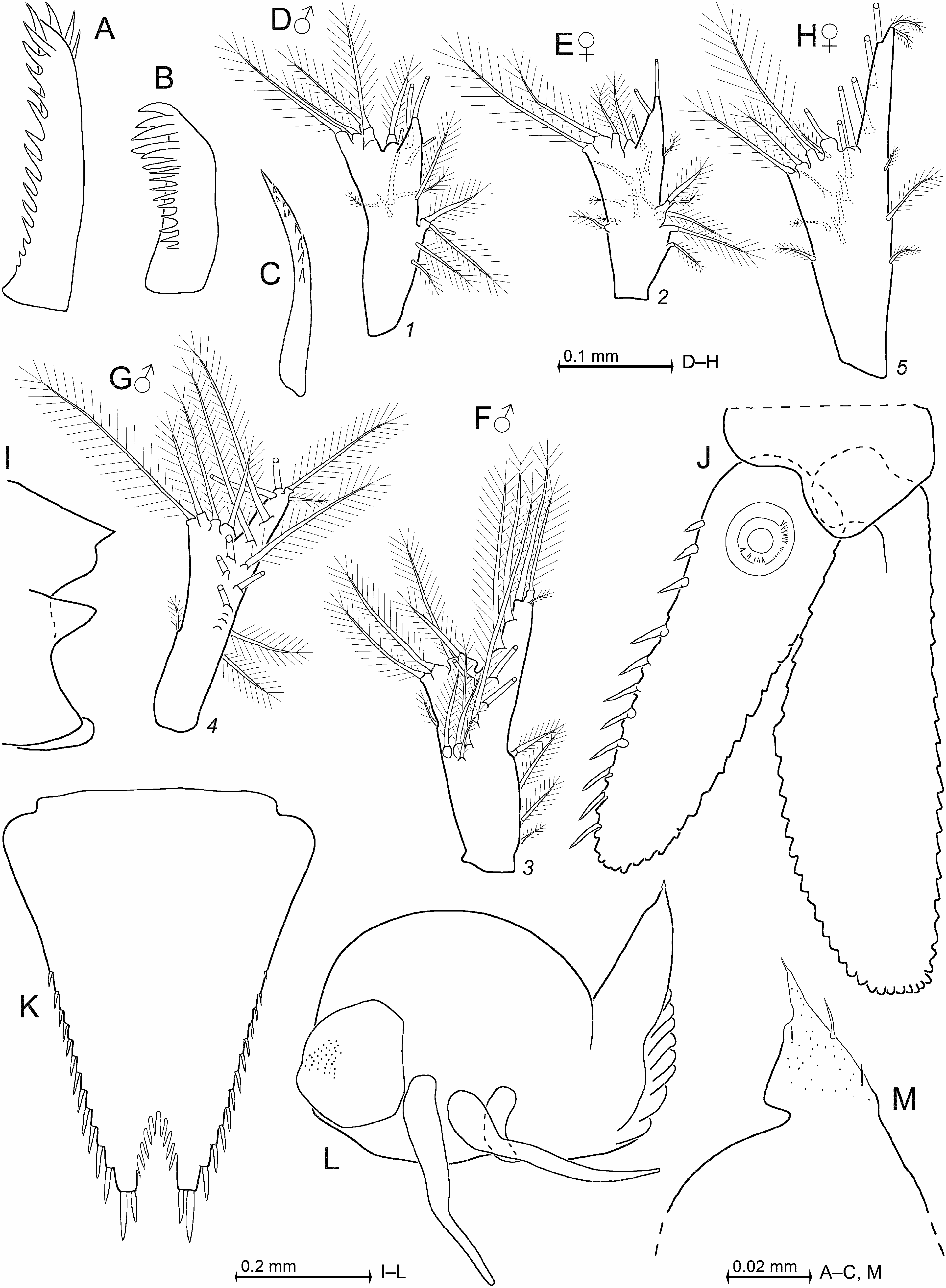
Pleon. Pleopods ( Fig. 4D–H View FIGURE 4 ) non-dimorphic, reduced to small, setose, obscurely bilobate plates without spines. Uropods ( Fig. 4J View FIGURE 4 ) normal, entire; exopod reaches with 13–26% its length beyond endopod. Endopod armed with linear series of 10–11 spines along mesial margin from statocyst to 4–14% endopod length below apex. Telson ( Fig. 4K View FIGURE 4 ) subtriangular, length 1.3–1.4 times maximum width, and 0.7–0.9 times exopod of uropod. Each lateral margin of telson with 11–13 spines on distal 50–60%, proximal portion smooth. Disto-lateral lobes each with two spines on narrowly truncate apex; latero-apical spines are 10–14% telson length; medio-apical spines are 0.5–0.7 times length of latero-apical spines. Proximally narrowly rounded, U-shaped to V-shaped apical cleft penetrates 17–19% telson length. Cleft armed with 8–9 acute laminae along proximal 60–80% of its margins, distal portions smooth.
Description. All features of the diagnosis plus the above listed features shared by the three here described species. General appearance moderately robust ( Fig. 1G View FIGURE 1 ). Body size of adults is 3.5–4.5 mm (n = 6) in females and 3.0– 4.1 mm (n = 5) in males. Cephalothorax comprises 27–42% body length, pleon without telson 40–54%, telson 12–17%, carapace without rostrum 24–34%, and rostrum 2–5%. Frons with triangular subrostral process ( Fig. 1D View FIGURE 1 ). Posterior margin of carapace leaves 0.5–1.0 ultimate thoracic somite mid-dorsally exposed.Abdominal somites 1–5 measure 0.4–0.6, 0.5–0.9, 0.6–0.9, 0.5–0.7, and 0.6–0.7 times the length of somite 6, respectively.
Eyes ( Fig. 1A–D View FIGURE 1 ). Anterior margin of eyestalk more strongly wrinkled in males than in females. Stalk with field of minute scales on ventral face near cornea in both sexes. Cornea diameter 0.5–0.7 times the length of eyestalk.
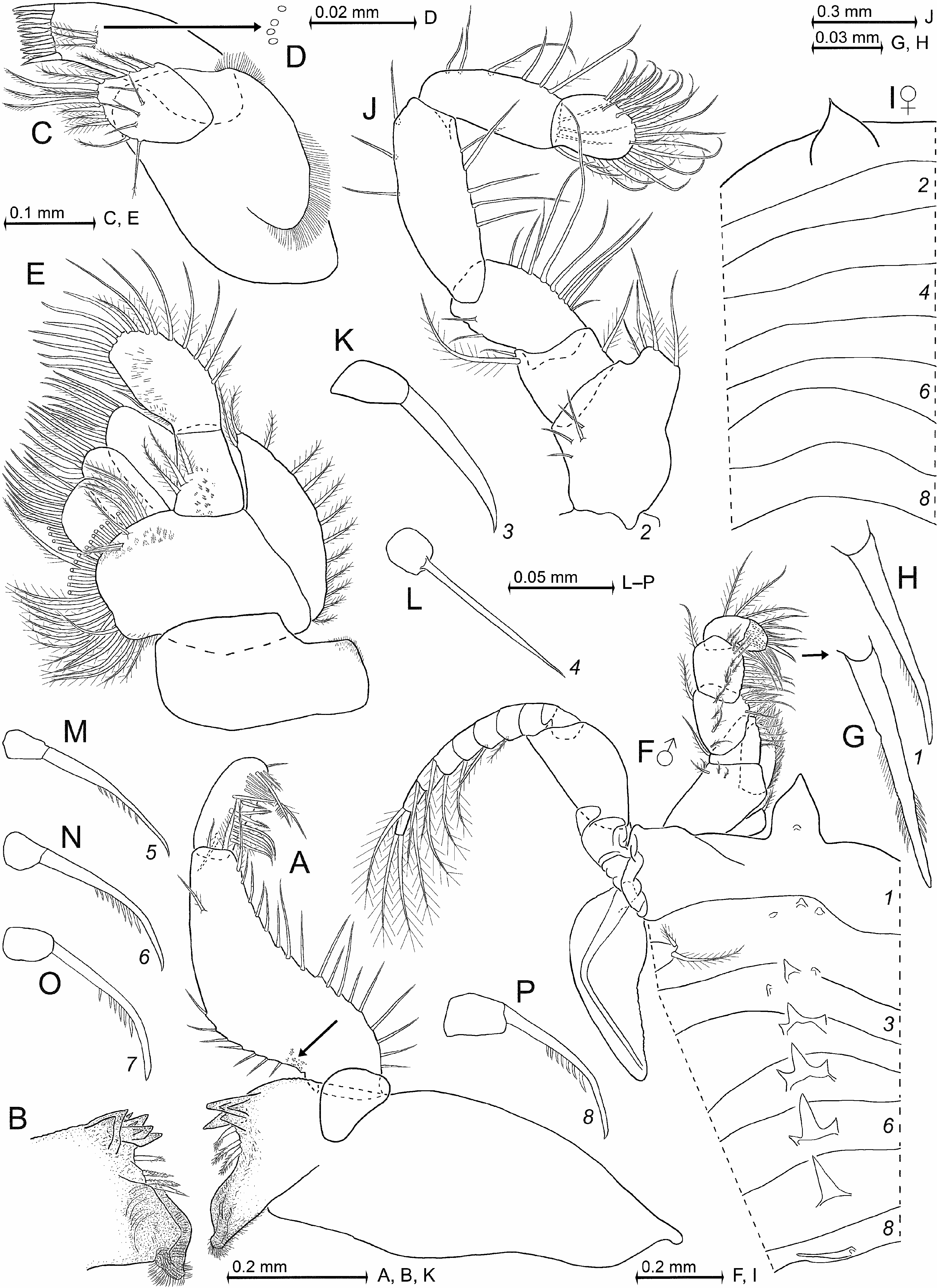
Antennulae ( Fig. 2A–H View FIGURE 2 ). Trunk extends 40–50% its length beyond eyes. Measured along dorsal midline, the basal segment is 43–49% trunk length, median 16–25%, and terminal 31–41%. Basal segment on basal half of its lateral face with 2–3 small setae bearing long barbs ( Fig. 2E View FIGURE 2 ). Its dorsal apophysis with four barbed setae and two whip setae; the mesial whip seta dimorphic ( Fig. 2B View FIGURE 2 versus Fig. 2G View FIGURE 2 ) with stout handle, cord shorter than handle; the laterally adjoining whip seta non-dimorphic, longer, slender, with cord about as long as handle. Lateral lobe of basal segment with three barbed setae only. Median segment with seta group formed by 3–4 barbed setae plus one short whip seta about mid-dorsally, closely behind distal margin. Additional set formed by a plumose seta and a shorter whip seta on disto-mesial edge of median segment; this whip seta with cord shorter than handle in both sexes, whereby its handle stouter in males ( Fig. 2C View FIGURE 2 ) than in females ( Fig. 2H View FIGURE 2 ). Terminal segment 1.2–1.4 times longer than wide in both sexes. Its mid-dorsal apophysis with four small barbed setae, with small cilia lining the distomesial margin; no spiniform anterior projection. Disto-mesial edge of terminal segment with two large whip setae facing opposite directions: the mesial, inward directed whip seta even more dimorphic compared to its counterpart on median segment, longer and more slender in females ( Fig. 2I View FIGURE 2 ), but short with stout handle (resembling flagellate spine typical of the subgenus Olivemysis ) in males ( Fig. 2D View FIGURE 2 ). In both sexes, lateral antennular flagellum is thicker than mesial one by a factor of 1.4–1.5 when measured near basis of flagella. Male lobe setose, inserts ventrally close to terminal margin of antennular trunk, broadly rounded, length (antero-posterior extension) is 16–22% width of terminal segment of trunk, and lobe width is 19–26%. Epi-antennular process forms an unevenly to evenly rounded shield; hypo-antennular process a long, slender, triangular projection with acute ( Fig. 2A View FIGURE 2 ) or blunt tip.
Antennae ( Fig. 2J, K View FIGURE 2 ). Sympod dorsally with terminally rounded, tongue-like process, and caudally with bulbous lobe containing end sac of antennal gland. Ventrally with crescent-shaped shield (dashed line below drawing plane in Fig. 2J View FIGURE 2 ) proximally behind antennal peduncle; shield 2.0–2.6 times longer than its maximum width. Basal segment contributing 17–20% to peduncle length, second 43–49%, and third 33–35%. Basal segment dorsally with field of triangular scales ( Fig. 2K View FIGURE 2 ). Antennal scale extends to 60–80% length of third segment of antennal peduncle. Scale setose all around, with slightly convex lateral margin and more strongly convex mesial margin. Short, broad apical segment separated by a tiny transverse suture. Apical segment with 5–8% total scale length; distally bearing five plumose setae.
Mandibles ( Fig. 2L–M View FIGURE 2 ). Mandibular palp with segments 1–3 contributing 13–15%, 64–67%, and 19–23% to palp length, respectively. Proximal segment with two whip setae on lateral margin, no additional setae. Median segment 2.3–2.5 times longer than its maximum width; 13–16 whip setae distally increasing in length almost all along lateral margin, the distal 2–4 setae with sparsely barbed handle; 11–13 basally thick setae along subbasal to subterminal portions of mesial margin; most setae of proximal third are unilaterally barbed in both sexes; one large whip seta close to disto-mesial edge (below drawing plane in Fig. 2L View FIGURE 2 ). Proximal 2/3 of median segment with large field of minute triangular scales ( Fig. 2M View FIGURE 2 ) on rostral face in both sexes. Terminal segment strongly setose. Pars molaris with well-developed grinding surface in both mandibles. Pars incisiva with four teeth, digitus mobilis with 3–4 teeth, and pars centralis with four spiny teeth.
Maxillula and maxilla ( Fig. 2N View FIGURE 2 ). Distal segment of maxillula terminally with 11–14 strong, smooth spines, the innermost spine being thickest and longest. This segment subterminally with five setae barbed on their distal half; two pores (size as in Fig. 10D View FIGURE 10 ) a short distance laterally of the outer (= most ventral) seta. Endite of maxillula terminally with three large, distally spiny setae, mesially accompanied by one proximally thick, barbed seta; both margins of the endite with numerous less thick setae. Maxilla with 14–16 plumose setae all along lateral margin of exopod, the two apical setae larger than the remaining ones. Basal segment of endopod with four basally thick, barbed setae. Terminal segment 1.3–1.9 times longer than wide.
Thoracic sternites ( Fig. 2O View FIGURE 2 ). Male sternites 2–8 with (sub)-triangular processes. Processes 3, 4 larger than processes 2, 5–8. Sternite 1 with large anterior lobe whose distal half is triangular with narrowly rounded apex in both sexes.
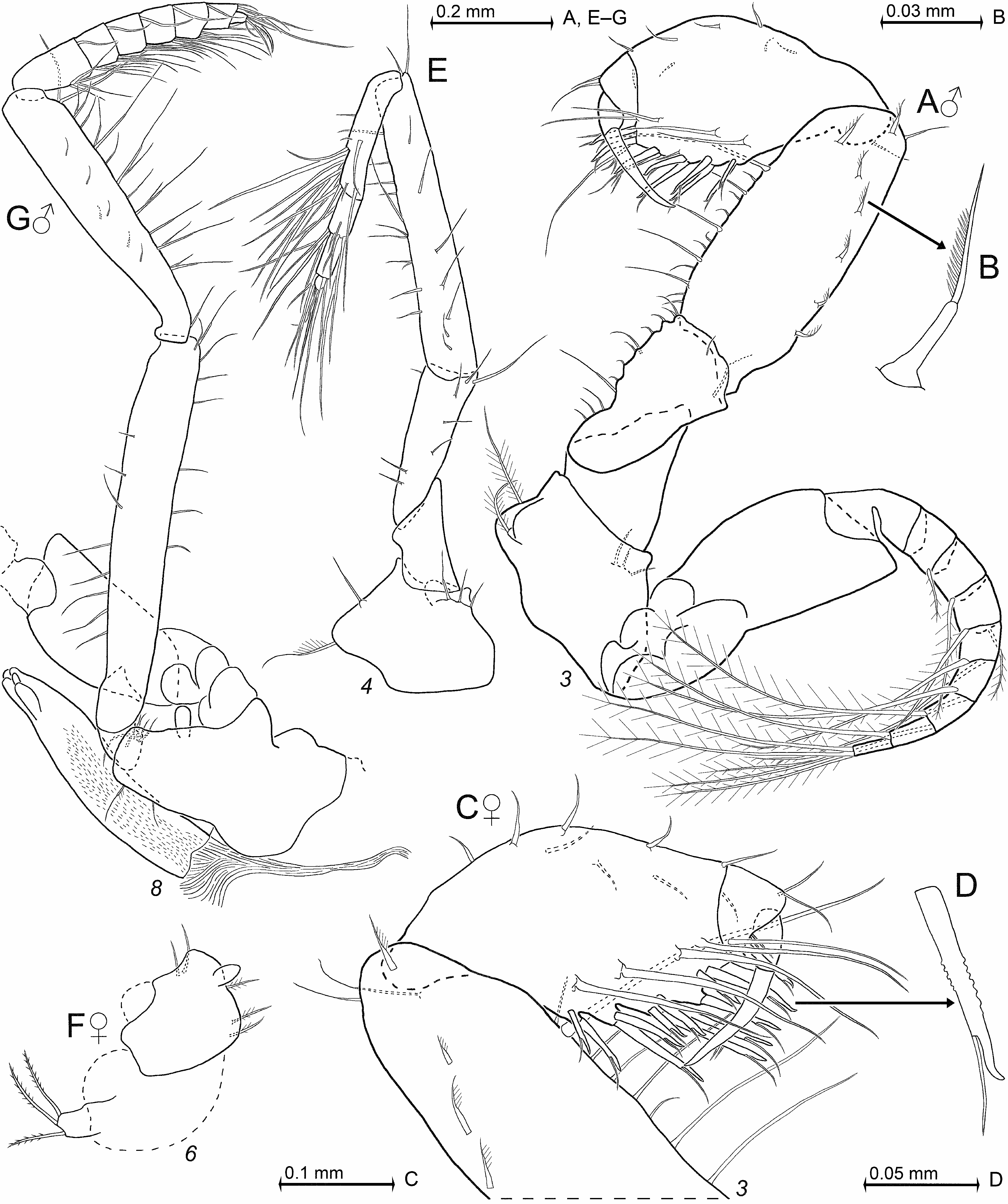
Thoracopods general ( Figs 2Q–S View FIGURE 2 , 3 View FIGURE 3 ). Length of flagella as well as of basal plates ( Fig. 3A View FIGURE 3 ) increase from exopod 1 to 6, and remain subequal among exopods 6 to 8. Basal plates weakly expanded, length 1.8–2.3 times maximum width in both sexes. Lateral margin of plates ends in a broadly rounded edge. Thoracopod 1 with large, leaf-like, in most cases smooth epipod. One out of three specimens with a seta on proximal third of epipod, distal 2/3 of this seta sparsely furnished with short, stiff bristles. Length of endopods increases in series of thoracopods 1, 2, 4–8, 3. Basis of endopods 4–8 with a small, lappet-like apophysis (as in Fig. 11E, G View FIGURE 11 ) on rostral face below endopod; no such apophysis in endopods 1–3. Ischium becomes increasingly slender from endopods 1 to 5 (compare panels A, D, M in Fig. 3 View FIGURE 3 ), and length of ischium increases in series of endopods 1, 2, 4, 3, 5; both these measurements remain (sub)-equal among endopods 5–8. Ischium shorter than merus in endopods 1–4, but longer than merus in endopods 5–8. Thoracic endopods 1–3 ( Figs 2P View FIGURE 2 ; 3B, E View FIGURE 3 ) each with dactylus larger than that of endopods 4–8 ( Figs 2S View FIGURE 2 , 3N View FIGURE 3 ). Combined praeischium plus ischium of endopod 2 ( Fig. 3A View FIGURE 3 ) are 0.9–1.0 times merus length, carpopropodus plus dactylus 1.0–1.1 times merus. Dactylus very large, with dense brush formed by great numbers of normal setae and 10–12 modified setae, the latter apically bent, bearing two symmetrical series of stiff barbs on either side in subbasal to median portions ( Fig. 3C View FIGURE 3 ). Distal 1–3 segments of carpopropodus of endopod 4 ( Fig. 2Q View FIGURE 2 ) each with modified seta bearing minute teeth (modified barbs) along its thickened handle ( Fig. 2R View FIGURE 2 ), no such setae in endopods 5–8. When stretched anteriorly, endopod 8 reaches to mandibles or up to eyes; when stretched posteriorly to pleonite 6 or up to telson.
Gnathopods ( Figs 1E–F View FIGURE 1 , 3D–L View FIGURE 3 ). Thoracic endopods 3 form a powerful subchela. Basis with indistinct endite. Ischium 1.9−2.7 times as long as wide; merus 2.7−4.3 times as long as wide and 1.2−1.7 times length of ischium. Mesial margin of ischium with 3–13 whip setae implanted on small (minute) projections; merus with 5–18 such setae on tips of such projections. Lateral face of merus close to its basis with whip seta bearing a ‘comb’ of minute cilia ( Fig. 3F View FIGURE 3 ) along its cord in both sexes; one shorter, smooth whip seta in submedian position and a longer, smooth whip seta at the disto-lateral edge. Carpopropodus 0.9–1.2 times merus length, 1.6–2.5 times ischium. The proximal 4–6 spines of carpus in linear series ( Fig. 3D, H View FIGURE 3 ). The two distal spines closely set obliquely side by side: the caudal one ( Fig. 3K View FIGURE 3 ) ‘normal’, the rostral one shorter, wider, and flattened ( Fig. 3L View FIGURE 3 ). Only the most proximal spines are smooth; most of the more distal spines with fine serration on their distal margin ( Fig. 3J–K View FIGURE 3 ). Modified seta closely behind disto-mesial edge of carpus in both sexes. This seta with analogous but weaker modification ( Fig. 3G View FIGURE 3 ) compared with the paradactylary setae of males ( Fig. 3I View FIGURE 3 ). A short distance proximally from this seta there is a shorter seta with similar but even weaker modification in both sexes (seta visible but its modification too small for visualization in Fig. 3D, H View FIGURE 3 ).
Marsupium. Oostegite 1 near basis with 3–8 setae which are microserrated along their distal half; oostegite 2 with 2–3 such setae. Ventral and rostral portions of outer face of oostegite 2 with 15–24 small whip setae with longer barbed setae in between; no such setae on oostegite 1.
Pleopods ( Fig. 4D–H View FIGURE 4 ). The pleopods show no consistent size difference between sexes. Length without setae increases generally from first to fifth pleopods; intermediate pleopods not always in continuous series due to individual variation in size; pleopod 5 always the longest. All setae are plumose or barbed in both sexes.
Tail fan ( Fig. 4I–K View FIGURE 4 ). Scutellum paracaudale (sub)-triangular with acute or narrowly rounded apex ( Fig. 4I View FIGURE 4 ). Exopod of uropods ( Fig. 4J View FIGURE 4 ) with clearly convex mesial margin and weakly convex, almost straight lateral margin. Exopod extends by 32–43% its length beyond telson; endopod 11–44% (partly due to telson inserting more rostrally). Distal 8–9 spines of endopod are subequal, proximal 2–3 spines shorter. Endopod basally with large statocyst, statolith diameter 46–105 µm (n = 8 statoliths from four specimens). Statoliths discoidal with shallow fundus and distinct tegmen. Statolith formula 2 + (2–3) + (4–7) + (4–7) = 14–18 (n = 8). Telson ( Fig. 4K View FIGURE 4 ) length 0.9–1.1 times endopod of uropod. Spines on lateral margins continuously increasing in size towards the tip. Apical cleft deeper than wide, its laminae shorter than the spines on distal fourth of lateral margins of telson.
Larvae ( Fig. 4L–M View FIGURE 4 ). Eight mounted nauplioid larvae at substage N3 with smooth cuticle all around except for tip of abdomen, which bears a few setae ( Fig. 4M View FIGURE 4 ); no caudal furca. Remaining features in Fig. 4L View FIGURE 4 are typical of the state of development.
Foregut ( Fig. 4A–C View FIGURE 4 ). Lateralia anteriorly with apically pronged and apically coronate spines with smooth shaft, and a mesial group of long spines armed with loose series of small denticles along shaft. Lateralia more caudally with separate group of three spines unilaterally serrated by dense series of comparatively long teeth ( Fig. 4B View FIGURE 4 ). Dorsolateral infoldings with two larger, apically pronged, unilaterally serrated spines ( Fig. 4A View FIGURE 4 ). Each lateral margin of the superomedianum, in addition to setae and normal spines, with 6–8 comparatively large spines serrated on their distal half ( Fig. 4C View FIGURE 4 ).
Gut contents of two specimens were mostly masticated, unidentifiable material and a few diatoms; no crustacean remains identified.
No known copyright restrictions apply. See Agosti, D., Egloff, W., 2009. Taxonomic information exchange and copyright: the Plazi approach. BMC Research Notes 2009, 2:53 for further explanation.
|
Kingdom |
|
|
Phylum |
|
|
Class |
|
|
Order |
|
|
Family |
|
|
SubFamily |
Heteromysinae |
|
Tribe |
Heteromysini |
|
Genus |
|
|
SubGenus |
Heteromysis |