
Hydrocassis jengi Satô, 1998
|
publication ID |
https://doi.org/ 10.1080/00222933.2011.602805 |
|
DOI |
https://doi.org/10.5281/zenodo.10537029 |
|
persistent identifier |
https://treatment.plazi.org/id/03F587F0-FF85-FFFB-FE59-851FFBC546C4 |
|
treatment provided by |
Felipe |
|
scientific name |
Hydrocassis jengi Satô, 1998 |
| status |
|
Hydrocassis jengi Satô, 1998 View in CoL
Figures 1B View Figure 1 , 4 View Figure 4 , 5 View Figure 5 , 12A,B View Figure 12
Material examined
Japan: 1 L2 ( SEHU), Kinsakubaru, Amami-Ôshima I., Kagoshima pref., 30 April 1999 , no collector data; 1 L2 ( EUMJ), Akatsuchi-yama, Uken-son, Amami-Ôshima I., Kagoshima pref., 2 May 1999 , no collector data.; 1 L2 ( SEHU), upstream of Naon-kawa R., Yamato-son, Amami-Ôshima I., Kagoshima pref., 30 July 2008, J. Fujiwara leg.
Description of general morphology of second instar
Slide preparations of two specimens were examined.
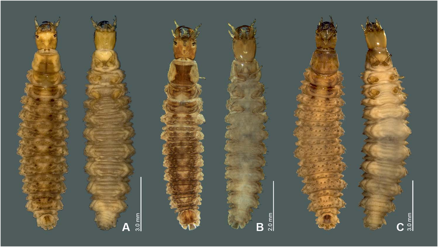
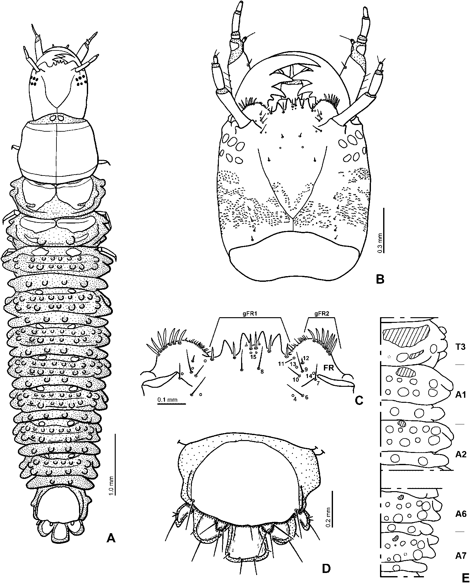
Colour ( Figure 1B View Figure 1 ). Dorsal surface of head light brown, lighter in median and anterior parts of frontale and around stemmata, with two pairs of dark brown spots medioposteriorly; light brown ventrally. Pronotum light yellowish brown, median portion darkened; meso- and metathoracic terga and abdominal segments light yellowish brown to dark brown, lighter medially and laterally, so looking as two dark longitudinal lines along the body; sclerotized parts darker than median part of abdomen; dorsal sclerite of abdominal segment 8 dark brown in medioposterior part. Ventral surface of thorax and abdomen greyish white to yellowish white, proscutum and legs darkened.
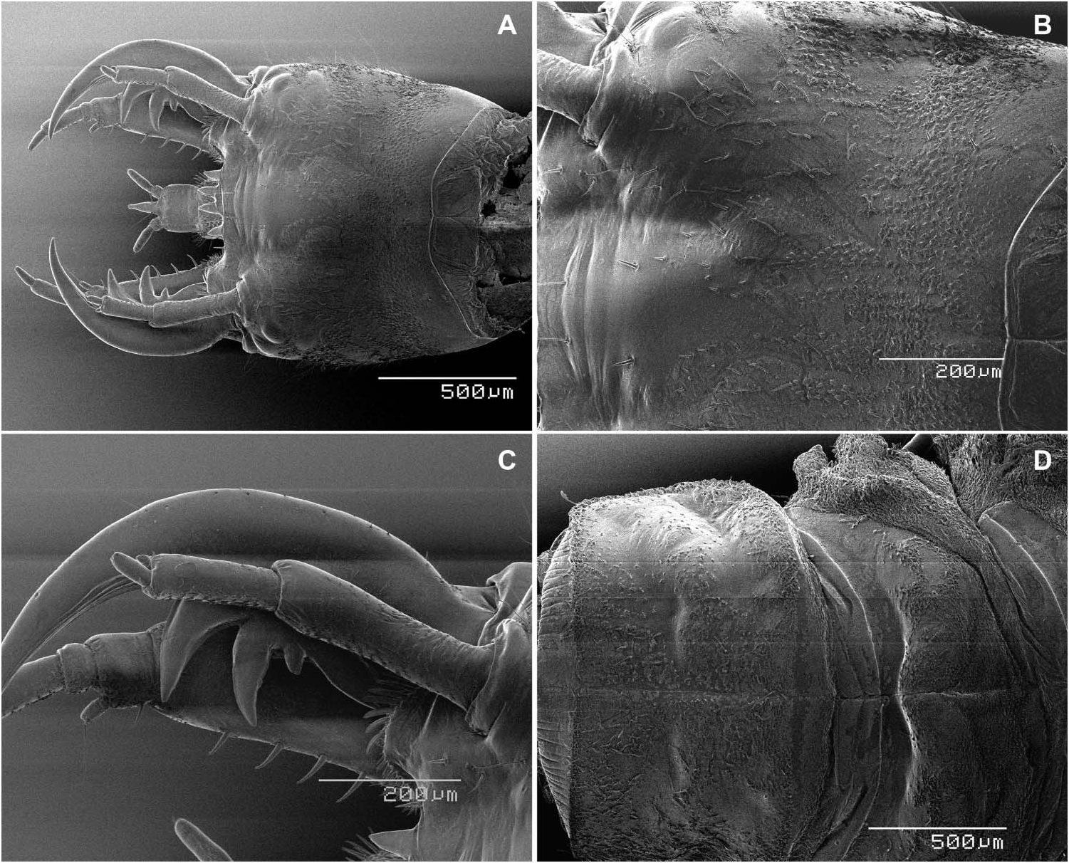
Head. Posterior half to two-thirds of head capsule bearing dense, small but strong tooth-like cuticular projections on dorsal and lateral surfaces behind stemmata ( Figure 4B View Figure 4 ). Nasale with four teeth. Epistomal lobes not projecting further than nasale, each lobe with dense, rather short to short spine-like cuticular projections on inner margin ( Figure 4C View Figure 4 ).
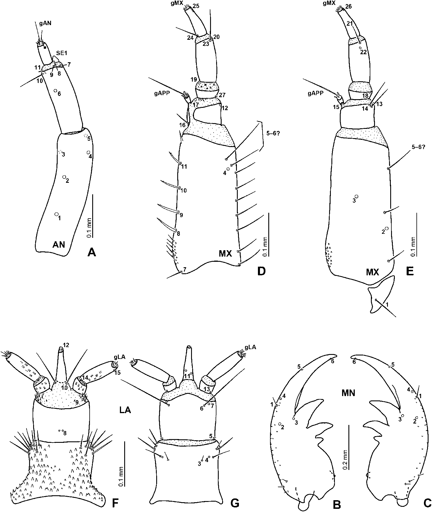
Antenna ( Figure 5A View Figure 5 ). Ventral and lateral surfaces of antennomeres 1 and 2 covered with fine hair-like cuticular projections, the projections on antennomere 2 denser than those on antennomere 1. Antennomere 1 the longest, longer than antennomeres 2 and 3 combined. Antennomere 2 about 0.6 times as long as antennomere 1.
Abdomen. Segment 1 with three transverse rows of tubercles behind the sclerites ( Figure 4E View Figure 4 ); the first row with four rather large tubercles on each side, outer one larger than inner three; second row with four tubercles on each side, inner three more closely situated than outer one, the size of the tubercles from median to lateral one,: rather large (L), L, rather L, L; third row with three large tubercles on each side, outer one projecting laterally. Arrangement of tubercles on segments 2 to 5 similar to that on segment 1 but first row with five tubercles, tubercles somewhat larger than those on segment 1: L, rather L, L, L, L. Segment 6 similar to segments 2 to 5 but tubercles of first row smaller than those on segments 2 to 5; inner four rather large, outer one large; outer two closely located. Segment 7 with three transverse rows of tubercles, the first row with five tubercles on each side, outer two closely located: rather L, very small (S) (hardly recognizable, absent on left side in specimen examined this study), rather L, rather L, L; second row with four tubercles, inner three closely located: S, L, very S (hardly recognizable, absent on left side in specimen examined for this study), L; third row with two tubercles each side, outer one projecting laterally.
Description of chaetotaxy of head of second instar
Frontale. Lateral part bearing numerous rather short secondary setae behind sensilla FR4–6 (e.g. Figure 9B View Figure 9 ). Each epistomal lobe with group of six to seven moderately long setae (gFR2), inner two slightly shorter than outer setae.
Parietale. Bearing many secondary sensilla, on dorsal and lateral parts.
Mandible ( Figure 5B, C View Figure 5 ). With several small secondary setae on lateral part; basal part of mandible with seven rather short secondary setae, three on dorsal, four on ventral face.
Maxilla ( Figure 5D, E View Figure 5 ). Stipes with 10–11 long secondary setae on outer face; one of them very long, close to MX 5–6.
Labium ( Figure 5F, G View Figure 5 ). Mentum with nine stout secondary setae in each anterolateral corner; a trichoid secondary seta situated ventrally, close to and outside LA4.
Habitat
Running water. A larva was found in the root of a tree in the bank of a stream (Naonkawa River) (J. Fujiwara, personal communication).
Identification
Hydrocassis jengi is the only species of the genus distributed in the Amami Islands, southeastern Japan. We identified the larval instar by presence or absence of secondary sensilla and comparing the size of their head capsule with those of H. lacustris and H. uncinata , of which the larvae and adults of each species are almost the same size.

| EUMJ |
Ehime University |
No known copyright restrictions apply. See Agosti, D., Egloff, W., 2009. Taxonomic information exchange and copyright: the Plazi approach. BMC Research Notes 2009, 2:53 for further explanation.