
Hydrocassis Fairmaire, 1878
|
publication ID |
https://doi.org/ 10.1080/00222933.2011.602805 |
|
DOI |
https://doi.org/10.5281/zenodo.10537027 |
|
persistent identifier |
https://treatment.plazi.org/id/03F587F0-FF93-FFFD-FE22-8398FE6F402F |
|
treatment provided by |
Felipe |
|
scientific name |
Hydrocassis Fairmaire, 1878 |
| status |
|
Genus Hydrocassis Fairmaire, 1878 View in CoL
( Figures 1–13 View Figure 1 View Figure 2 View Figure 3 View Figure 4 View Figure 5 View Figure 6 View Figure 7 View Figure 8 View Figure 9 View Figure 10 View Figure 11 View Figure 12 View Figure 13 )
Species examined
Hydrocassis jengi Satô, 1998 (L2), Hydrocassis lacustris (Sharp, 1884) (L1–3), Hydrocassis uncinata Ji and Schödl, 1998 (L2–3).
Diagnosis
Larvae of Hydrocassis can be distinguished from those of other known genera of the tribe Sperchopsini by the following combination of character states: (1) frontal lines fused at base ( Figures 2A View Figure 2 , 6B View Figure 6 ); (2) nasale with four to five teeth ( Figures 2C View Figure 2 , 4C View Figure 4 , 6C View Figure 6 , 10C View Figure 10 ); (3) clypeolabrum almost symmetrical ( Figures 2C View Figure 2 , 6C View Figure 6 ); (4) epistomal lobes with rather slender setae ( Figure 4C View Figure 4 ); (5) mandibles with three inner teeth ( Figure 5B, C View Figure 5 ); (6) inner surface of stipes with five setae in all instars ( Figures 3C View Figure 3 , 5D View Figure 5 , 7D View Figure 7 ); (7) dorsal sclerite of abdominal segment 8 not subdivided medially ( Figure 4D View Figure 4 ).
Description of general morphology
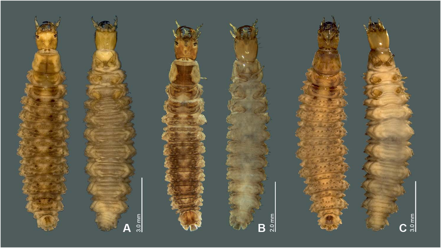
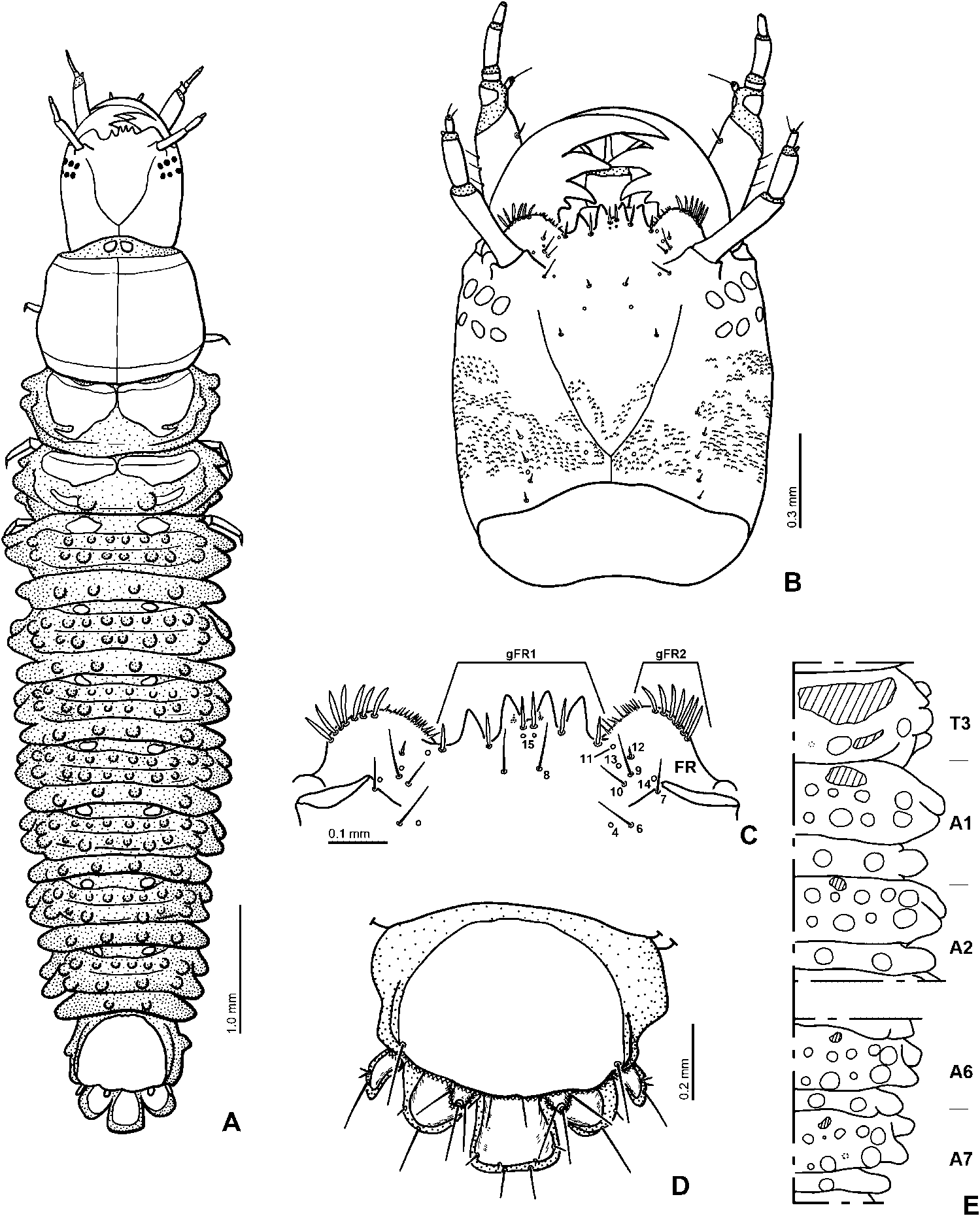
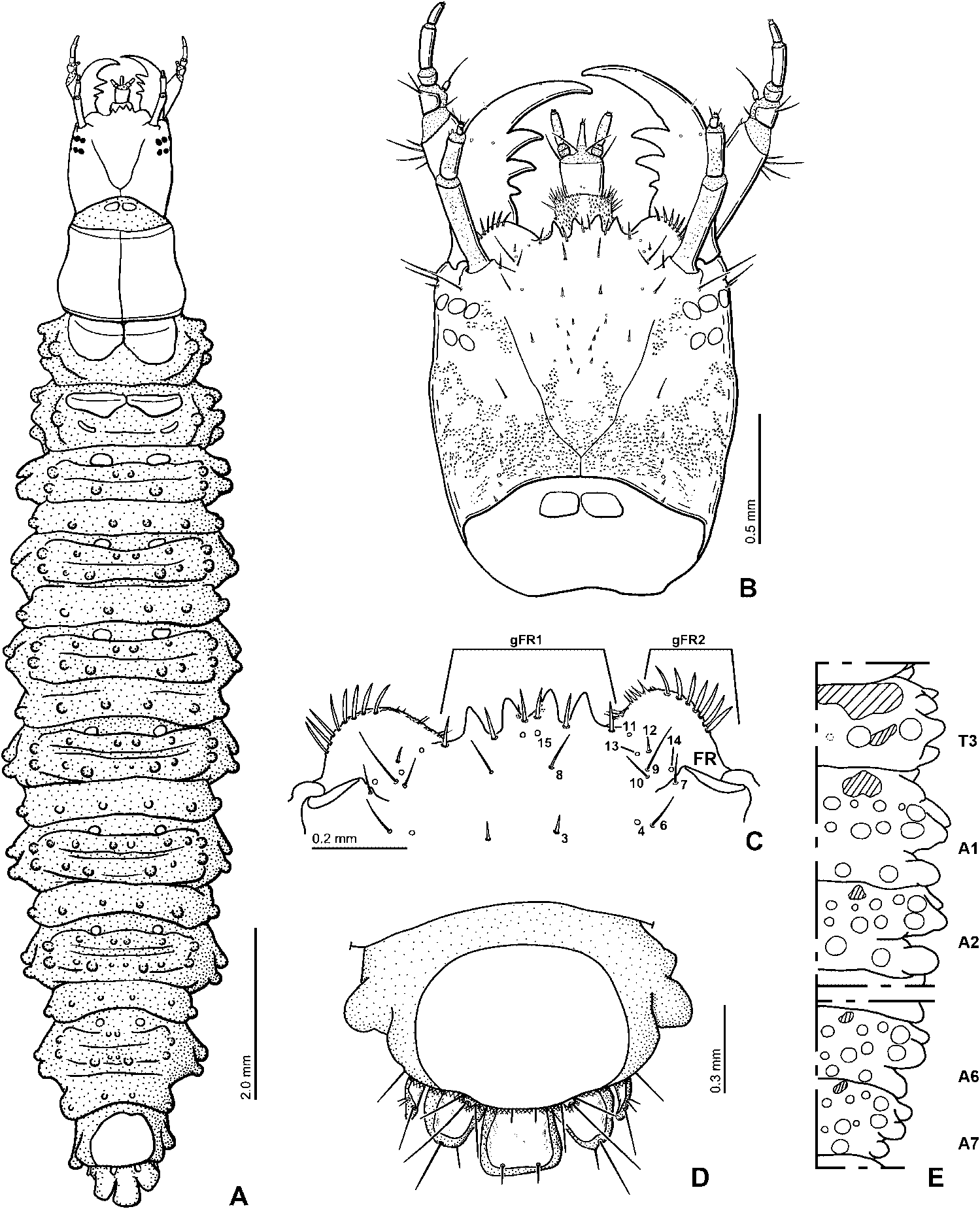
Body rather slender, widest between abdominal segments 3 and 4 ( Figure 1 View Figure 1 ). Lateral sides of meso- and metathorax and abdominal segments 1 to 8 with lateral projections on each segment ( Figures 4A View Figure 4 , 6A View Figure 6 , 10A View Figure 10 ). Nine pairs of spiracles, one on anterolateral portion of mesothorax and eight on abdomen; mesothoracic and first seven abdominal spiracles non-functional, biforous; last pair annular, large and functional, enclosed within spiracular atrium.
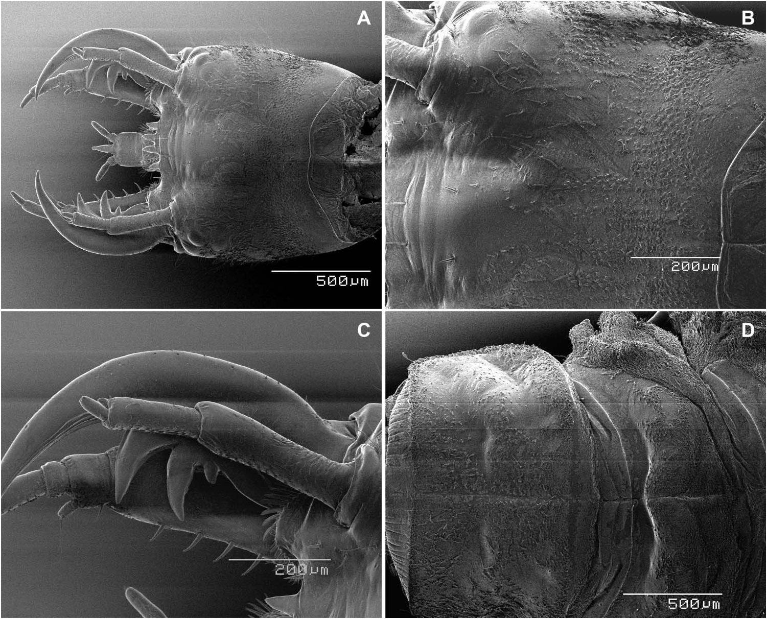
Head. Head capsule subquadrate ( Figures 4B View Figure 4 , 6B View Figure 6 , 10B View Figure 10 ). Frontal lines almost V-shaped, fused at base of head capsule; coronal line short. Dorsal and lateral surfaces of head capsule bearing dense, small, strong to rather strong, tooth-like cuticular projections (e.g. Figures 8C View Figure 8 , 9B View Figure 9 ). Six stemmata on each anterolateral corner of head capsule. Clypeolabrum slightly asymmetrical. Nasale with four to five teeth, median tooth small or absent ( Figures 6C View Figure 6 , 10C View Figure 10 ). Epistomal lobes rounded, almost symmetrical, projecting about as far as nasale or slightly lower than nasale; inner part of each lobe with spine-like cuticular projections.
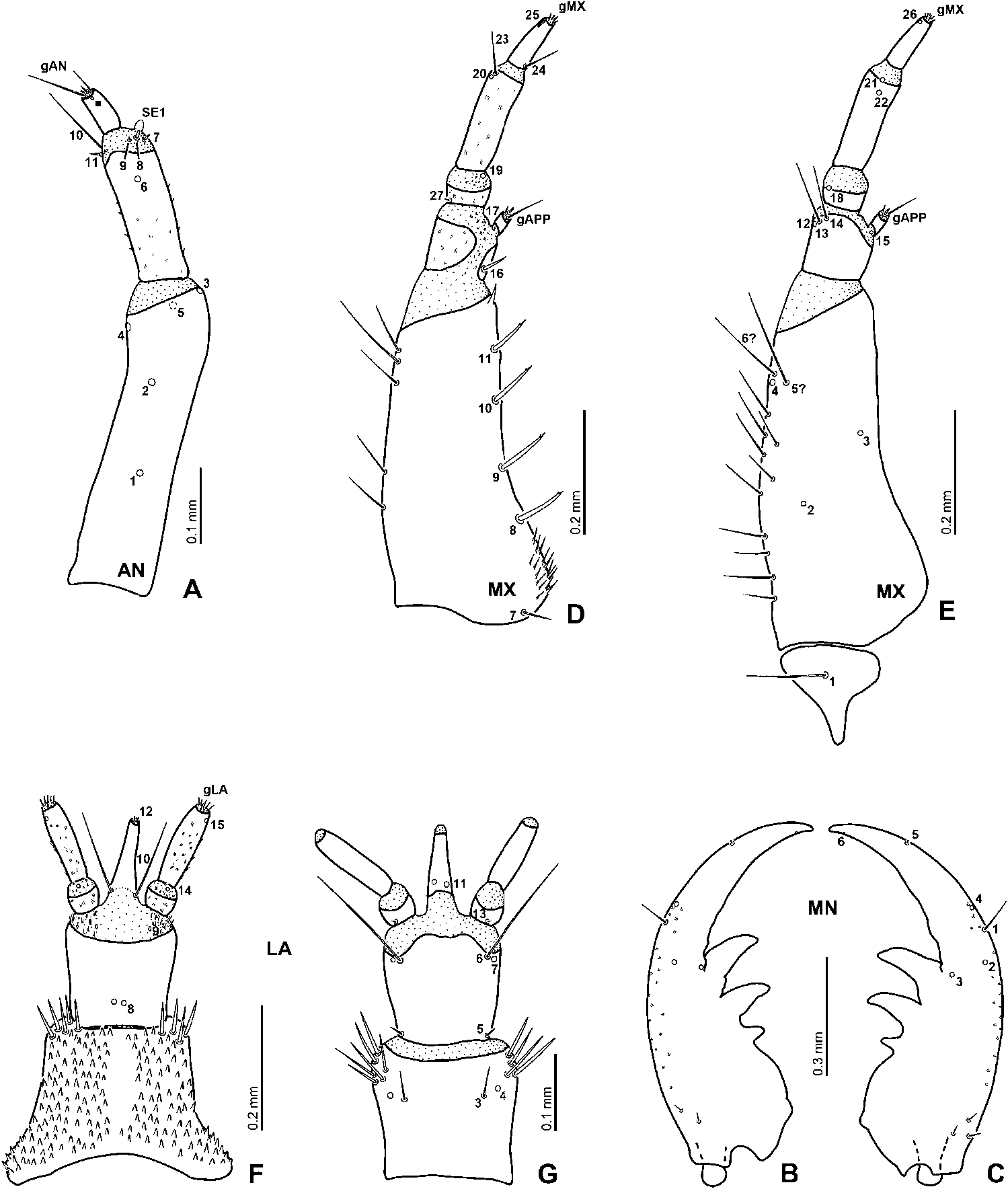
Antenna. Three-segmented, rather short and stout (first instar, Figure 3A View Figure 3 ) to moderately long and slender (third instar, e.g. Figure 7A View Figure 7 ). Antennomere 1 the longest, longer than antennomeres 2 and 3 combined (third instar) to almost about as long as antennomere 2 (first instar). Antennomere 3 the shortest, small. Surface of antenna with small, spin-like cuticular projections (e.g. Figure 9C View Figure 9 ).
Mandibles. Symmetrical, with three inner teeth, two distal ones large, basal one small (e.g. Figure 5B–C View Figure 5 ).
Maxilla. Counting cardo, six-segmented, distinctly longer than antenna (e.g. Figures 3C,D View Figure 3 , 5D,E View Figure 5 ). Cardo large, irregularly shaped, subtriangular to subquadrate. Stipes the longest, longer than palpomeres 1–4 combined, with one spine-like cuticular projection on apex of inner surface, and narrow and dense cuticular projections in basal part of inner surface, between sensilla MN7 and MN8. Maxillary palpus four-segmented; palpomere 3 the longest, palpomere 2 the shortest; palpomere 1 the widest, palpomere 4 the narrowest; palpomere 1 incompletely sclerotized on dorsal surface, bearing small hair-like cuticular projections on anterior part of dorsal surface of intersegmental membrane between palpomeres 1 and 2 (e.g. Figure 7D View Figure 7 ); inner process sclerotized; dorsolateral surface of intersegmental membrane of palpomeres 2 and 3 bearing fine hair-like cuticular projections; palpomere 3 slightly longer than palpomeres 1 and 2 combined, narrower than palpomere 2.
Labium. Well developed ( Figures 2B View Figure 2 , 3E–F View Figure 3 , 7F–G View Figure 7 ). Submentum fused to head capsule, large, subpentagonal, wider than mentum ( Figure 2B View Figure 2 ). Mentum trapezoidal in dorsal view, with short, stout tooth-like cuticular projections on dorsal surface except on median part. Prementum subquadrate, bearing fine hair-like cuticular projections on dorsolateral part of intersegmental membrane between prementum and labial palpi. Ligula shorter than palpi, mostly sclerotized. Labial palpi moderately short, slender; palpomere 1 small; palpomeres and intersegmental membrane between palpomere 1 and 2 densely covered with fine hair-like cuticular projections ( Figure 5F View Figure 5 ).
Thorax. Prothorax trapezoidal, wider than head capsule. Proscutum formed by one large plate divided by fine sagittal line, covered with short setae (Figure, 9D). Presternum subpentagonal ( Figure 12A View Figure 12 ), with long, almost complete sagittal line on sclerite. Mesothorax with three dorsal sclerites on each side; medioanterior sclerite small, narrow; anterolateral sclerite small; posterior sclerite large, subdivided by fine sagittal line, and with transverse ridge on anterior part; one small tubercle outside dorsal sclerite; four strong lateral projections on each side visible from dorsal view. Metathorax with two dorsal sclerites on each side; anterior sclerite large, subdivided by sagittal line; posterior sclerite narrow, smaller than anterior one, behind tubercle; lateral sides of posterior sclerites with two large projections; four strong to rather weak lateral projections on each side visible from dorsal view. Legs ( Figure 12B–D View Figure 12 ): rather short, barely visible in dorsal view, five-segmented, bearing numerous stout setae.
Abdomen. Ten-segmented, tapering towards posteriad, segments 8 to 10 forming spiracular atrium. Segments 1 to 7 similar in size and shape, each subdivided by transverse folds and bearing several tubercles ( Figure 4E View Figure 4 ). Segment 1 with three lateral projections on each side dorsally, and with one pair of small dorsal sclerites medioanteriorly, the sclerites larger than those on segments 2 to 7, and three transverse rows of tubercles behind the sclerites; sclerites larger than those on segments 2 to 7. Segments 2 to 6 almost similar to segment 1. Segment 7 with one pair of small dorsal sclerites medioanteriorly, with three transverse rows of tubercles, third row of the tubercles with two tubercles on each side, outer one projected laterad.
Spiracular atrium ( Figure 4D View Figure 4 ). Segment 8 with a large, oval dorsal plate, rather densely covered with short setae, posterior edge of the plate weakly rounded. Segment 9 trilobed, largely sclerotized on dorsum, partially covered by segment 8, with a pair of short, one-segmented urogomphi; urogomphi with three very long setae; procercus incompletely sclerotized, with two setae. Segment 10 reduced.
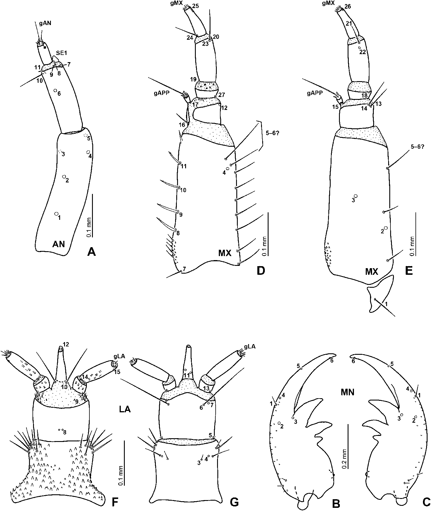
Description of primary chaetotaxy of head
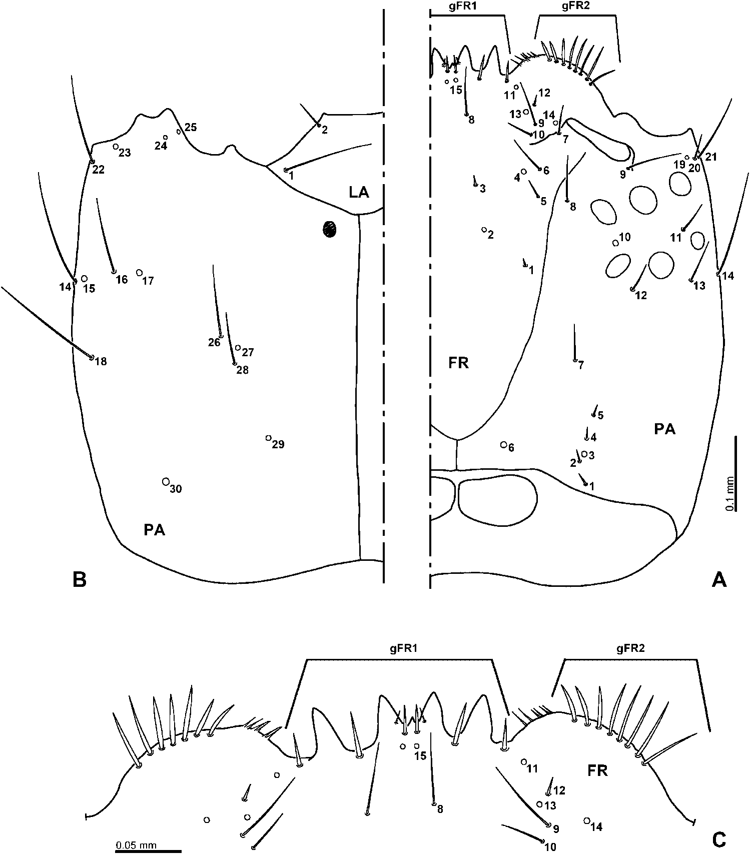
(Based on first instar larvae of H. lacustris ; slide preparations of eight specimens were examined.) Frontale with 52–54 sensilla ( Figure 2A, C View Figure 2 ). Central part with three pairs of sensilla (FR1–3) divergent posteriad; FR1 trichoid, short seta, situated close to frontal line; FR2 pore-like, situated mesally of line connecting FR1 and FR3; FR3 trichoid, moderately stout seta, situated more anteriorly and slightly more mesally than FR2. FR4–6 situated posteromesally to antennal socket, forming a triangular group; FR4 pore-like; FR5 rather short seta; FR6 rather long seta. FR7 rather short seta situated on inner margin of antennal socket. FR8 long seta situated posteriorly to two median teeth of nasale. Nasale with a group of eight setae (gFR1) ( Figure 2C View Figure 2 ); six setae rather short, stout, intercalated between teeth; one short seta situated ventrally on inner margin of each large median tooth. Each epistomal lobe with a group of seven to eight moderately long, stout setae (gFR2), inner three slightly shorter than outer ones. FR15 pore-like, placed posteriorly to median setae of gFR1. FR9–10 and FR14 situated mesally to antennal socket; FR9 pore-like, located between FR10 and FR12; FR10 rather long, trichoid seta, situated posteriorly to FR9; FR14 porelike, located laterally of FR9–10 and FR12, close to FR7. FR11 placed in inner part of epistomal lobe, anteriorly to FR9–10; FR11 pore-like, situated posterolaterally to outer seta of gFR1; FR13 pore-like, placed between FR11 and FR9; FR12 short seta, located laterally of line connecting FR11 and FR13.
Parietale. With 30 sensilla each ( Figure 2A, B View Figure 2 ). Dorsal surface with a group of sensilla (PA1–5) located posteriorly at mid-width, forming a slightly irregular longitudinal row; PA3 pore-like, situated slightly anteriorly to PA2; PA1–2 and PA4–5 short setae. PA6 pore-like, situated posteromesally, close to joint of frontal and coronal lines. PA7 moderately long seta close to frontal line, situated laterally of line connecting PA6 and PA8. PA8 moderately long seta situated between frontal line and inner stemma of anterior row. PA9 long seta, situated on outer edge of antennal socket. PA10 pore-like, located between two innermost stemmata. PA11 rather long seta, situated anteromesally to outer stemma of posterior row. PA12 and PA13 rather long, trichoid setae situated approximately at mid-length of parietale, behind PA10–11 and stemmata. Anterolateral corner of epicranium with three sensilla (PA19–21); PA19 pore-like, situated more dorsally than others; PA20 rather short, trichoid seta, situated between PA19 and PA21; PA21 long, trichoid seta. Anterior one-third of lateroventral surface of parietale with four sensilla (PA14–17); the sensilla forming a slightly irregular transverse row; PA14 long seta; PA15 pore-like, situated close to PA14; PA16 long seta, situated between PA15 and PA17; PA17 pore-like; PA15–17 situated more ventrally than PA14. PA18 long seta, situated at mid-length of lateral surface of parietale, posteriorly to PA15–16, more ventrally than PA14. PA22–25 located ventrally along anterolateral portion of parietale; long seta PA22 and pore-like sensillum PA23 situated close to anterolateral corner; PA24–25 pore-like, close to inner margin of mandibular acetabulum; PA24 pore-like, located between PA23 and PA25, close to PA25. Median part of parietale with three sensilla (PA26–28), PA26 long seta, situated more anteriorly than remaining sensilla, PA27 pore-like, situated medially of line connecting PA26 and PA28; PA28 rather long seta, located more posteriorly than remaining sensilla. One pore-like sensillum (PA29) situated in basal one-third of parietale on ventral side behind PA28. PA30 pore-like, situated ventrally in about basal one-quarter of parietale.
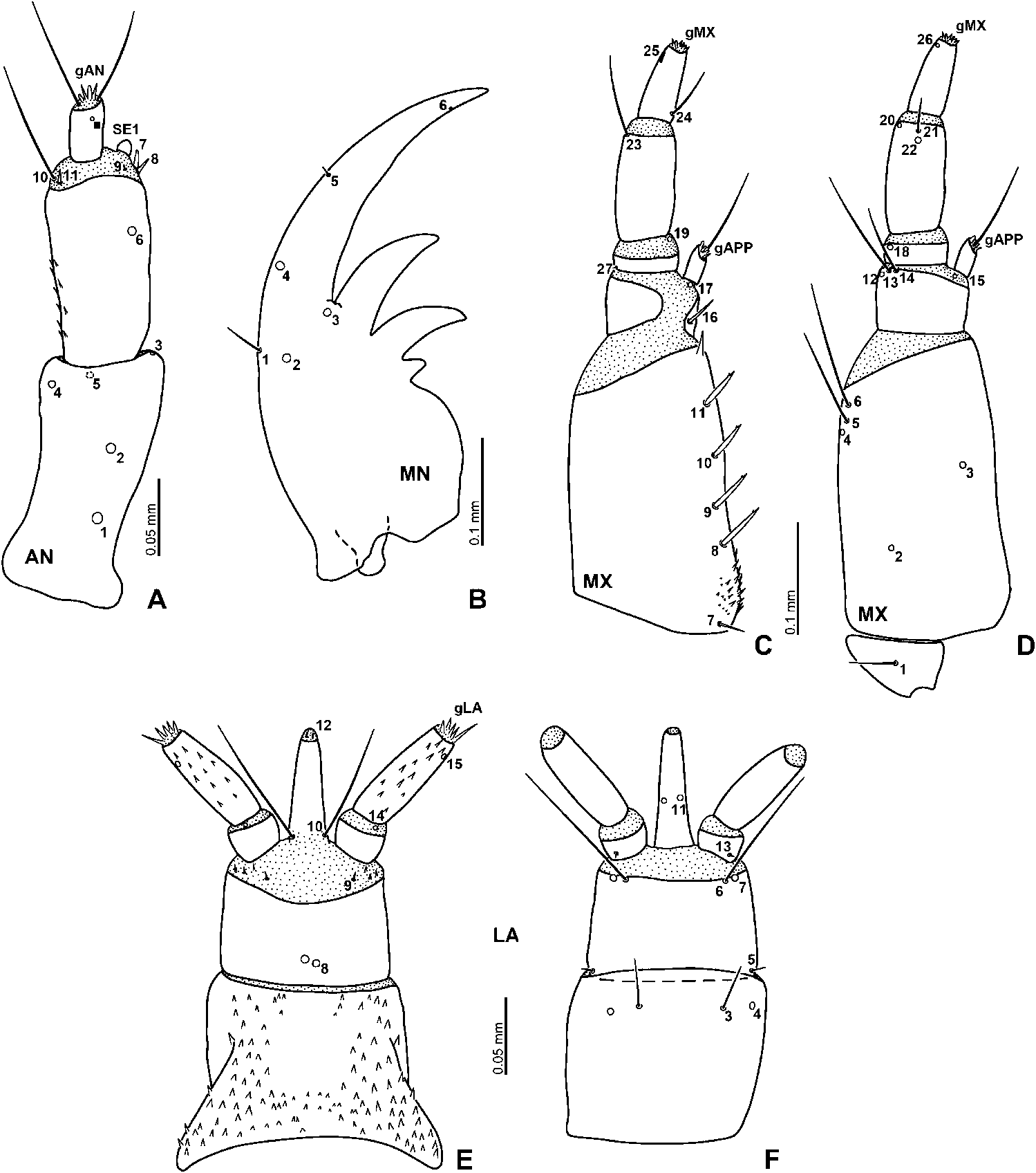
Antenna ( Figure 3A View Figure 3 ). Antennomere 1 with five pore-like sensilla (AN1–5); AN1 situated in basal one-third; AN2 situated in distal one-third; AN3 located apically on inner face, indistinct; AN4 located subapically on outer face; AN5 located subapically on ventral surface. Antennomere 2 with one pore-like sensillum (AN6) situated dorsally in distal two-fifths. Five setae (AN7–11) situated on intersegmental membrane between antennomeres 2 and 3; AN7–8 small, stout, situated on outer face of antenna next to sensorium (SE1); AN9 small, situated mesally of AN7–8. AN10–11 on inner face of antenna; AN10 long and trichoid, AN11 short, both setae close to each other. SE1 small, much shorter than antennomere 3, about as long as AN7 and AN8. Antennomere 3 with one pore-like additional sensillum placed dorsally close to distal margin of sclerite, and six sensilla on apical membranous area (gAN): two long setae, two minute conical setae, and two minute setae.
Mandible ( Figure 3B View Figure 3 ). Bearing two setae (MN1 and MN5) and four pore-like sensilla (MN2–4 and MN6) situated on dorsal to lateral surface. MN1 rather long on outer face. MN2–4 forming a triangular group at mid-length on dorsal surface; MN2 more closely attached to MN1 than to remaining sensilla; MN3 situated mesally of line connecting MN2 and MN4, MN4 on outer face. MN5 short seta, placed on outer margin in distal one-quarter of mandible. MN6 pore-like, very small, situated subapically on inner face.
Maxilla ( Figure 3C, D View Figure 3 ). Cardo with one moderately long, trichoid seta ventrally ( MX 1). Stipes with a row of five setae ( MX 7–11) situated dorsally along inner surface; MX 7 moderately short, shorter than MX 8–11, situated basally; MX 8–11 rather stout, spiniform, with subapical tooth, equidistant from each other. Ventral surface of stipes with two pore-like sensilla ( MX 2 and MX 3) situated in basal 0.3 and distal 0.4, respectively; one pore-like sensilla ( MX 4) and two long trichoid setae ( MX 5 and MX 6) situated subapically on outer surface of sclerite; MX 4 situated posteriorly to MX 5 and MX 6, MX 5 between MX 4 and MX 6. Palpomere 1 with one rather stout, spiniform seta ( MX 16) situated basally on inner face, and with one pore-like sensillum ( MX 12) and two setae ( MX 13–14) situated lateroventrally along distal margin of sclerite; MX 12 situated more dorsally than MX 13 and MX 14; MX 13 long seta, between MX 12 and MX 14; MX 14 moderately long seta. Two pore-like sensilla ( MX 15 and MX 17) on membrane below inner appendage, MX 17 dorsal, MX 15 ventral. Appendage with one long and four minute setae apically (gAPP). Palpomere 2 with two pore-like sensilla ( MX 18 and MX 19) and one minute seta ( MX 27); MX 18 situated lateroventrally on anterior margin of sclerite; MX 19 situated apically on inner face of intersegmental membrane between palpomeres 2 and 3; MX 27 situated basally on outer face of sclerite. Palpomere 3 with four sensilla ( MX 20–23); MX 21–22 rather short seta and pore-like sensillum respectively, both situated subapically at mid-width of ventral surface of sclerite; MX 22 located posteriorly to MX 21; pore-like sensillum ( MX 20) and moderately long seta ( MX 23) situated apically on outer face of sclerite; MX 23 dorsal, MX 20 ventral. Palpomere 4 with proximal rather long seta ( MX 24) on inner face of sclerite, and with digitiform sensillum ( MX 25) and pore-like sensillum ( MX 26) situated subapically on outer face, MX 25 dorsal, MX 26 ventral. Apical membranous area of palpomere 4 with six minute setae (gMX).
Labium ( Figures 2B View Figure 2 , 3E, F View Figure 3 ). Submentum with two pairs of setae (LA1–2); LA1 long, on lateral corners; LA2 moderately short, on anterior corners ( Figure 2B View Figure 2 ). Mentum with two pairs of sensilla (LA3–4) situated ventrally in distal one-fifth of sclerite; LA3 rather long seta, located more mesally than pore-like sensillum LA4. Dorsal surface of prementum with one pair of pore-like sensillum (LA8) and small seta (LA9); LA8 situated subbasally at midwidth of sclerite; LA9 on lateral part of membrane between prementum and palpi. Ventral surface of prementum with three pairs of sensilla (LA5–7); LA5 rather short seta, situated proximo-laterally; long seta LA6 and porelike sensillum LA7 situated laterally on distal margin of sclerite; LA6 situated more mesally than LA7. Long seta (LA10) situated dorsally at base of ligula. Ligula with two pairs of pore-like sensilla (LA11–12); LA11 situated ventrally close to mid-length of ligula; LA12 situated dorsally in apical membranous area. Labial palpomere 1 with a minute seta (LA13) on base of ventral surface of palpomere. Pore-like sensillum LA14 situated dorsally on intersegmental membrane between palpomeres 1 and 2. Palpomere 2 with one pore-like sensillum (LA15), situated subapically on outer face; apical membranous area with several small setae (gLA).
Variation
Hydrocassis shows a weak intra-specific variation (e.g. Figures 6C View Figure 6 , 10C View Figure 10 ) in the following characters: (1) Number of teeth of nasale: in some specimens the small median tooth is reduced, therefore in this case, the number of teeth on nasale is four; (2) number of setae of epistomal lobe (gFR2): it ranges from six to nine based on the specimens examined in this study.


No known copyright restrictions apply. See Agosti, D., Egloff, W., 2009. Taxonomic information exchange and copyright: the Plazi approach. BMC Research Notes 2009, 2:53 for further explanation.
|
Kingdom |
|
|
Phylum |
|
|
Class |
|
|
Order |
|
|
Family |