
Mortoniella (Mortoniella) monopodis, Blahnik & Holzenthal, 2017
|
publication ID |
https://doi.org/ 10.5281/zenodo.5170203 |
|
publication LSID |
lsid:zoobank.org:pub:AB1A57F0-7CB4-4830-920B-DF219740A596 |
|
DOI |
https://doi.org/10.5281/zenodo.5186280 |
|
persistent identifier |
https://treatment.plazi.org/id/03F687A7-FFED-F811-FF01-BD6644A8FBEF |
|
treatment provided by |
Felipe |
|
scientific name |
Mortoniella (Mortoniella) monopodis |
| status |
sp. nov. |
Mortoniella (Mortoniella) monopodis , new species
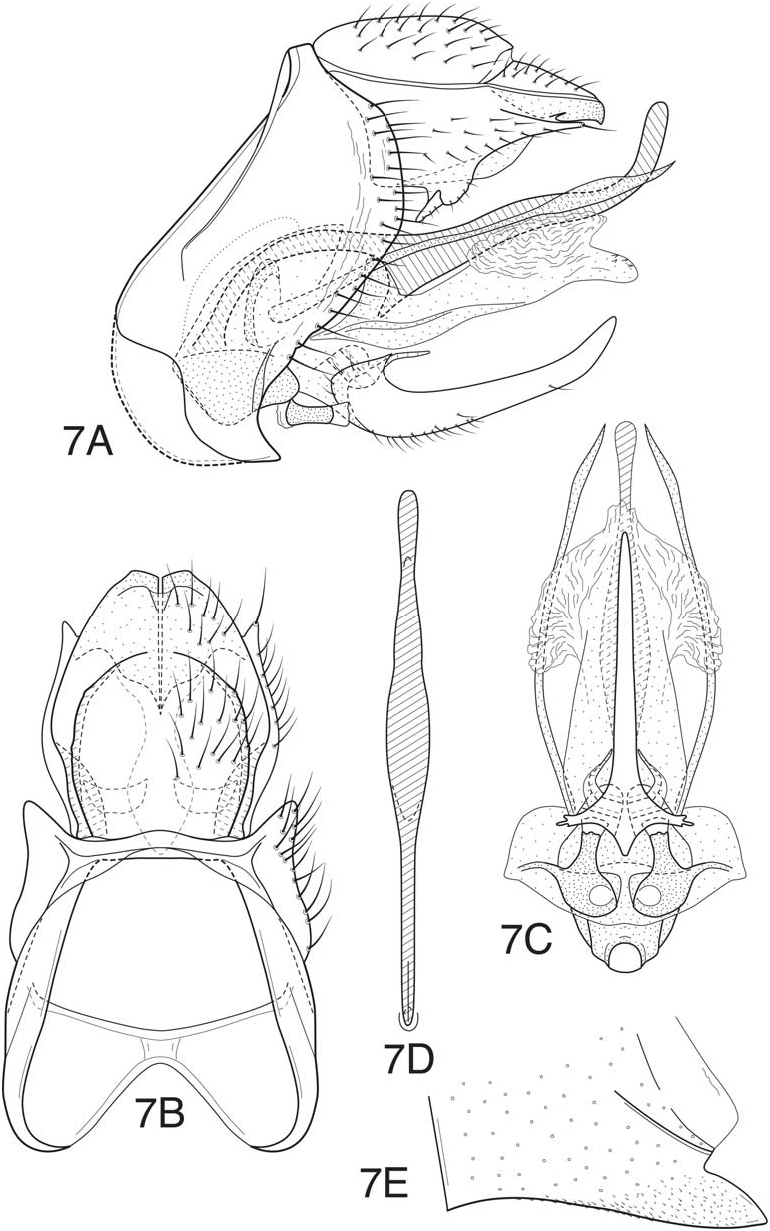
Fig. 7 View Figure 7
This species is easily diagnosed by the very elongate, narrow, mesal projection of the inferior appendages. It is unlikely to be confused with any other described species. The left side of segment IX is somewhat deformed in the holotype specimen and the illustration shows the contour of the opposite side. The posterolateral margins of segment IX in this species are rounded and less angular than most other species in the bilineata subgroup, and the ventral lobe or lobes of the phallicata somewhat less developed, but otherwise the species conforms well to the generalized features of the group.
Adult —Length of forewing: male 4.8-5.0 mm. Forewing with forks I, II, and III present, hind wing with forks II, III, and V. Spur formula 0:4:4. Overall color dark brown. Legs same color, tibial spurs slightly darker, not greatly contrasting with legs. Palps and basal segments of antenna blackishbrown, base of antenna contrasting with subsequent light brown segments, apex of antenna dark brown (like wings). Forewing with 2 distinct white wing bars, 1 at anastomosis and 1 on proximal part of wing, approximately midway between base and anastomosis.
Male genitalia —Ventral process of segment VI prominent, posteriorly projecting, relatively wide basally, length about 1 to 1½ times width at base. Tergum VIII relatively narrow, subtending ventral margin of segment IX, membranous connection to tergum IX elongate. Segment IX with anterolateral margin rounded and produced in ventral half, posterolateral margin with broadly rounded projection in dorsal half; segment deeply mesally excised dorsally and ventrally, forming lateral lobes, separated dorsomesally by more than ½ width of segment. Tergum X moderately elongate, lateral margins rounded, laterally with acute, finger-like, lateral lobes, each with prominent apical seta; apex of tergum distinctly sclerotized, rounded laterally, weakly truncate at extreme apex, with ventrolateral margins incurved and converging mesally to form linear “seam,” apicodorsally with lightly sclerotized connection near apex (mesal notch nearly absent); tergum ventromesally with paired, lightly sclerotized, rounded and compressed, ventromesal lobes in basal half, each with short setae. Inferior appendages with very short rounded dorsolateral lobes, and with single elongate, narrow ventromesal lobe; mesal lobe, as viewed laterally, upcurved apically, very uniformly tapering and acute apically, as viewed ventrally. Mesal pockets of inferior appendage with short, posteriorly-directed, spine-like, apicoventral projections. Paramere appendage elongate, narrow, slightly widened preapically, apex acute, appendage extending about same length as dorsal phallic spine; fused basal segments of appendages articulating near base of dorsal phallic spine. Phallobase with evident rounded, laterally compressed, dorsomesal apodeme. Dorsal phallic spine, as viewed laterally, curved and arched basally, linearly extended in middle, and sinuously upturned in about apical 1/5, apex of spine rounded; base of spine narrow, curved and stalk-like, abruptly widened on ventral margin in basal ½, forming very acute ventral projection; spine, as viewed dorsally, narrow throughout, only slightly widened in middle, rounded apically. Phallicata with sclerotized basodorsal projection, articulating with angular ventral projection of dorsal phallic spine; phallicata ventrally with sclerotized lobes, lateral margins compressed and slightly widened near base, narrowed apically, forming single apicomesal projection, mesal projection with several small spines (more extensively developed in paratype specimen). Endophallic membrane with well- developed, pleated and membranous, lateral lobes; phallotremal spines absent.
Holotype male (pinned)— COLOMBIA: Chocó: km 130, 86 km E Quibdo, 17.ii.1983, OS Flint, Jr ( UMSP000157314 View Materials ) ( NMNH).
Paratype — ECUADOR: Imbabura: Reserva Los Cedros, Río los Cedros, 00.30359° N, 78.78233° W, 1312 m, 18-19.x.2011, Holzenthal, Rios, Encalada, Acosta– 1 male (pinned) ( UMSP).
Etymology —The name monopodis is taken from the Greek words mono for one and podos for foot and referring to the single elongate mesal projection of the inferior appendages.
No known copyright restrictions apply. See Agosti, D., Egloff, W., 2009. Taxonomic information exchange and copyright: the Plazi approach. BMC Research Notes 2009, 2:53 for further explanation.