
Zimbabwecepheus maidii, Jordaan, 2017
|
publication ID |
https://doi.org/ 10.11646/zootaxa.4324.2.5 |
|
publication LSID |
lsid:zoobank.org:pub:A9F81De2-D3De-45Da-8111-D22B12055278 |
|
DOI |
https://doi.org/10.5281/zenodo.6016120 |
|
persistent identifier |
https://treatment.plazi.org/id/03F7879B-474B-B36D-FF29-FD65A42EFE07 |
|
treatment provided by |
Plazi |
|
scientific name |
Zimbabwecepheus maidii |
| status |
gen. nov. |
Zimbabwecepheus maidii gen. nov., sp. nov.
( Figs.15–50 View FIGURES 15 – 16 View FIGURES 17 – 27 View FIGURES 28 – 34 View FIGURES 35 – 44 View FIGURES 45 – 50 )
Etymology. The specific epithet is dedicated to Mrs Maidi Lili Schroetlin Beling, Paraguayan plastic artist, who managed to survive in a harsh world, and was respected for her qualities and remarkable work. Exceptional wife, inseparable companion, recently passed away.
Material examined. Holotype Female. “ Zimbabwe Umtali . II.1969. LEG R.MUSSARD; material deposited in the Collection of the MHNG; preserved in 70% ethanol”. ( Umtali is now Mutare, Zimbabwe) . Two Paratype adult females, same locality and date as Holotype; deposited in Collection of MHNG; preserved in 70 % ethanol. Material studied with SEM: three specimens, not deposited.
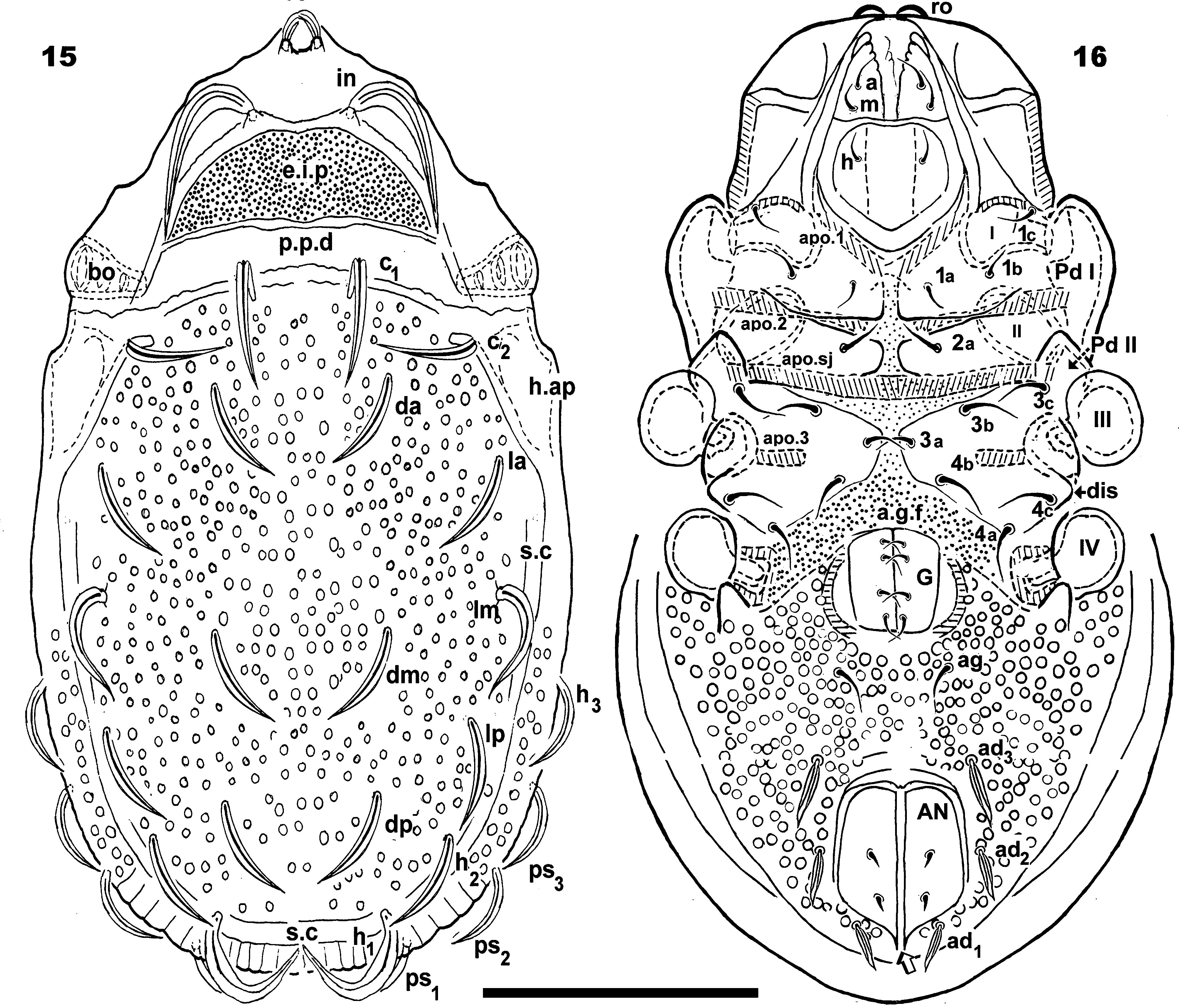
Diagnosis. Prodorsum. Trapezoidal; slightly elevated interlamellar process; large, anteriorly situated setae in initially directing forward, but then curving backward; in setae inserted antiaxially to medial plane, slightly internally to ro insertion level; ro setae curved, directing to medial zone; apical tips distanced from each other; sensillus uncinate. Smooth bothridial ring with bothridial tooth; lamellar tip not observed; lamellar furrow not discernible.
Notogaster. In dorsal view, anterior polyedral and posterior oval; convex in lateral view; d.sj narrow, slightly rectilinear; notogastral anterior depression absent.
Fourteen pairs of setae: c 1, c 2, da, dm, dp, la, lm, lp, h 1, h 2, h 3, p 1, p 2, p 3; c 2 setae directing laterally to medial zone; other setae directing backwards; all setae more or less similar length. Circumgastric depression clearly visible anterior to p 1, p 2, p 3, h 3 setae. Easily observed humeral apophysis; excavated V-shaped depression present.
Lateral region. Truncated lamellar tips; tutorium (tu) a prominent lamina with curving margin. Bothridium cup-shaped, opening directing downwards.
Ventral region. Slightly elevated epimera delimited by shallow furrow. Epimera 3–4 fused, epimeral chaetotaxy 3-1-3-3. Genital plate small relative to anal plate; four pairs of genital setae present in a simple line; all setae of more or less equal length. Aggenital setae observed posterior to genital opening. Three pairs of adanal seta; ad 3 distanced from ag setae. Clearly visible lyrifissures iad situated laterally and between ad 3 and ad 2.
Description. Measurements. SEM 528 µm (539–519) x 228 µm (223–231) (measurements on three specimens). Light microscopy: 536 µm (542–534) x 301 µm (297–305).
Shape. Elongate oval ( Figures 15–17 View FIGURES 15 – 16 View FIGURES 17 – 27 ).
Colour. Specimens without cerotegument, light brown to brown when observed in reflected light.
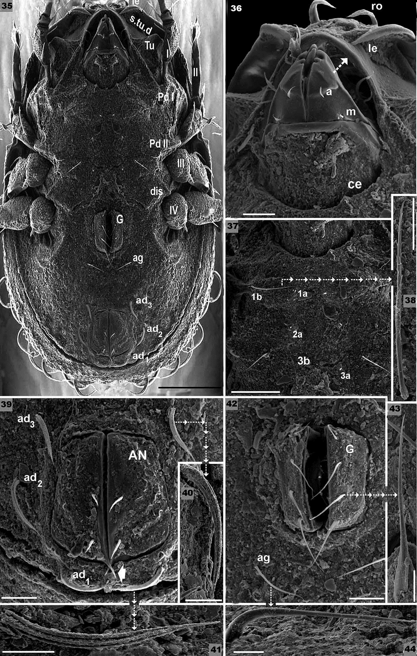
Cerotegument. Present: consistently thick layer with adhering soil particles, present all over body, femora I, II, and trochanter and femora III, IV. Rugous: prodorsum, anterior notogastral zone; c 1, c 2 setae insertion zones ( Figure 17 View FIGURES 17 – 27 ) and femora I, II and trochanter and femora III, IV; tuberculate rugous-porous: notogaster behind c 1, c 2 setal insertions ( Figure 17 View FIGURES 17 – 27 ), h.ap; amorphous: subcapitular zone surrounding h setae, epimeral zone, genital and anal plates (39–42). Absent: anterolateral border of lamellae (Lam) ( Figure 31 View FIGURES 28 – 34 ), bothridial ring (bo.ri) ( Figure 31 View FIGURES 28 – 34 ), anterior subcapitular zone ( Figure 36 View FIGURES 35 – 44 ); tibiae and tarsi ( Figures 45, 46 View FIGURES 45 – 50 ); posterior zone of femur I and posterior superior zone of femur II ( Figure 45, 46 View FIGURES 45 – 50 ).
Setation. Elongate lanceolate with two elevated medial veins: ro, in ( Figure 24 View FIGURES 17 – 27 ); elongate lanceolate with one elevated medial vein: notogastral setae ( Figure 27 View FIGURES 17 – 27 ); ad setae ( Figure 26 View FIGURES 17 – 27 ); both sides serrate, finely dentate: le setae ( Figures 19, 20, 22 View FIGURES 17 – 27 )(observations from different angles were necessary to understand the shape and characteristics of these setae); flabellate: ge ( Figures 42, 43 View FIGURES 35 – 44 ), ag (figure 44); barbate: epimeral ( Figure 38 View FIGURES 35 – 44 ); simple: subcapitular ( Figure 36 View FIGURES 35 – 44 ), anal ( Figure 39 View FIGURES 35 – 44 ).
Prodorsum. Trapezoidal in dorsal view ( Figures 15 View FIGURES 15 – 16 , 17 View FIGURES 17 – 27 ), slightly convex in lateral view ( Figure 28 View FIGURES 28 – 34 ), trapezoidal in frontal view ( Figure 30 View FIGURES 28 – 34 ). slightly elevated interlamellar process (e.i.p) ( Figure 28 View FIGURES 28 – 34 ); setae in situated anteriorly on e.i.p; large twisting in setae, initially directing forward, but later curving backward; in setae inserted antiaxially to medial plane and slightly internally to ro insertion level ( Figure 30 View FIGURES 28 – 34 ); clearly visible ro setae, curved, directing to medial zone; apical tips distanced from one another ( Figures 18 View FIGURES 17 – 27 , 30 View FIGURES 28 – 34 ); le setae laterally ( Figures 19, 20 View FIGURES 17 – 27 , 33 View FIGURES 28 – 34 ); ro setal insertion at same level as le setal insertion. Sensillus (si) uncinate ( Figure 31 View FIGURES 28 – 34 ). Smooth, well defined bothridial ring (bo.ri) with bothridial tooth ( Figure 31 View FIGURES 28 – 34 ); p.p.d narrow, clearly visible. Rostral margin rounded to hexagonal ( Figure 30 View FIGURES 28 – 34 ). Lamellae running laterally; lamellar tips absent.
Notogaster. Shape: in dorsal view anterior part polyedral and posterior part oval ( Figures 15 View FIGURES 15 – 16 , 17 View FIGURES 17 – 27 ); convex in lateral view ( Figure 28 View FIGURES 28 – 34 ); d.sj narrow, curving slightly, well delimited; notogastral anterior depression (n.a.d) not present.
Fourteen pairs of setae: c 1, c 2, da, dm, dp, la, lm, lp, h 1, h 2, h 3, p 1, p 2, p 3; c 2 setae directed laterally to medial zone ( Figures 15 View FIGURES 15 – 16 , 17 View FIGURES 17 – 27 , 30 View FIGURES 28 – 34 ); other setae directing backward ( Figures 15 View FIGURES 15 – 16 , 17 View FIGURES 17 – 27 ), all seate more or less the same length. Easily discernible circumgastric depression (s.c) observed in notogastral posterior part, in front of p 1, p 2, p 3, h 3 setae ( Figures 15 View FIGURES 15 – 16 , 17 View FIGURES 17 – 27 ). Humeral apophysis (h.ap) easily visible ( Figures 28, 31 View FIGURES 28 – 34 ); excavated V-shaped depression present ( Figure 28 View FIGURES 28 – 34 ).
Lateral region ( Figure 28 View FIGURES 28 – 34 ). Lamellae (lam) with truncated tips clearly discernible. Tutorium (tu): a prominent lamina, margin curved ( Figure 28 View FIGURES 28 – 34 ). Deep supratutorial depression (s.tu.d) running parallel to and between lamellae and tutorium; large pocket-shaped depression (a.tu.d) in anterior position. Pedotectum I, prominent extended lamina covering acetabulum I, rounded apex. Pd II small, ovoid lamina; small, triangular discidium (dis) visible, rounded apex.
Cup-shaped bothridia; bothridial opening directing downwards ( Figures 28, 31 View FIGURES 28 – 34 ); smooth bothridial ring (bo.ri), incomplete with bothridial tooth (bo.to) well discernible. Barbed, cylindrical sensillus, arching to the top ( Figure 31 View FIGURES 28 – 34 ). Long, extended humeral apophysis (h.ap), rounded apex, basally curved; anterior tip overlapping posterior bothridial part. Clearly observed large depression (dep) behind leg IV.
Ventral region ( Figures 16 View FIGURES 15 – 16 , 35 View FIGURES 35 – 44 ). Slightly elevated epimera delimited by shallow furrow (bo.1, bo.2, bo.sj). Epimera 3–4 fused, small epimeral furrow (bo.3); apo.1, apo.2, apo.sj and apo.3 clearly discernible. Epimeral chaetotaxy 3-1-3-3. Discidum well discernible; a.g.f clearly visible situated anterior to genital plate.
Genital plate small relative to anal plate; four pairs of genital setae in a simple line; all setae of more or less equal length; aggenital (ag) setae situated posterior to genital opening. Three pairs of adanal seta; ad 3 distant from ag setae. Sharply tipped polyhedral anal plate, two pairs of anal setae. Lyrifissures iad well discernible, situated laterally and between ad 3 and ad 2. Depressions (dep) clearly visible, situated laterally to genital and anal openings.
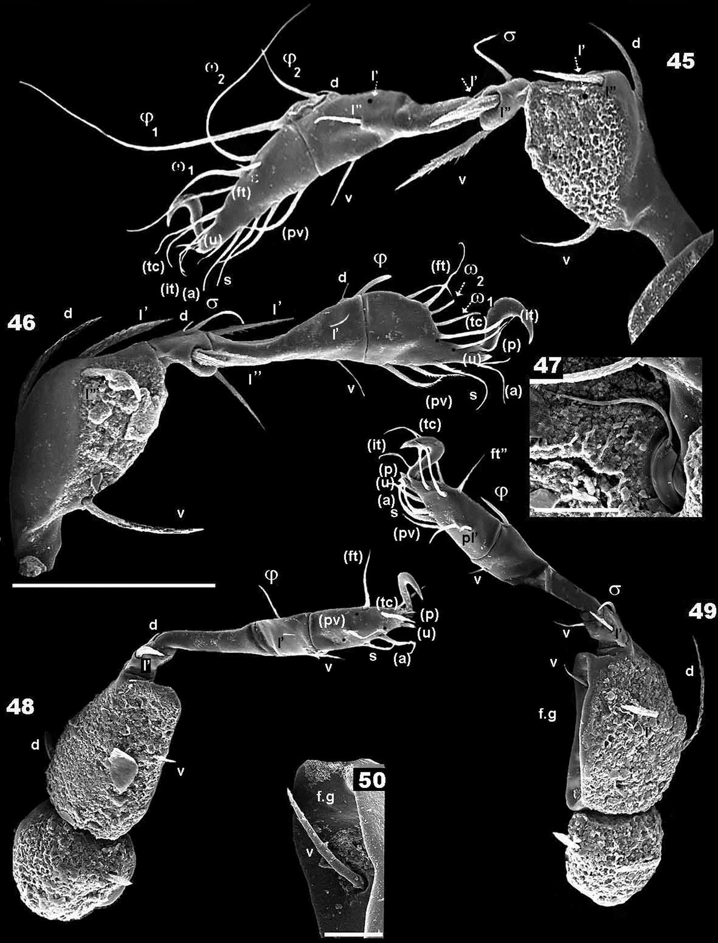
Legs ( Figures 45, 46, 48, 50 View FIGURES 45 – 50 , Table II). All legs monodactyle. Setal formulae I(1-3-2-3-16 -1) (1-2-2); II(1-4-3- 3-15 -1) (1-1-2); III(2-3-1-2-14 -1) (1-1-0); IV(1-2-2-3-13 -1) (0-1-0). Trochanter of Leg II with one clearly discernible seta ( Figure 47 View FIGURES 45 – 50 ). Femoral groove (Femur III) large ( Figure 50 View FIGURES 45 – 50 ).
Remarks. The cerotegumental layer impeded clear observation of the c.s.s and the f.l.p. Observation of notogastral setae was complicated, due to their length and the fact that they are twisted; sticky residue on the setal surface also impeded clear observation.


| MHNG |
Museum d'Histoire Naturelle |
No known copyright restrictions apply. See Agosti, D., Egloff, W., 2009. Taxonomic information exchange and copyright: the Plazi approach. BMC Research Notes 2009, 2:53 for further explanation.
|
Kingdom |
|
|
Phylum |
|
|
Class |
|
|
Order |
|
|
SubOrder |
Oribatida |
|
Family |
|
|
Genus |