
Prometheoarchaeum syntrophicum
|
publication ID |
https://doi.org/10.1038/s41586-019-1916-6 |
|
DOI |
https://doi.org/10.5281/zenodo.5920940 |
|
persistent identifier |
https://treatment.plazi.org/id/03F887AB-FFFD-FFE0-761F-FDF68FF0FA12 |
|
treatment provided by |
Plazi |
|
scientific name |
Prometheoarchaeum syntrophicum |
| status |
candidatus |
‘ Candidatus Prometheoarchaeum syntrophicum ’ strain MK-D1 for the isolated archaeon (see Supplementary Note 3 for reasons why the provisional Candidatus status is necessary despite isolation).
Cell biology, physiology and metabolism
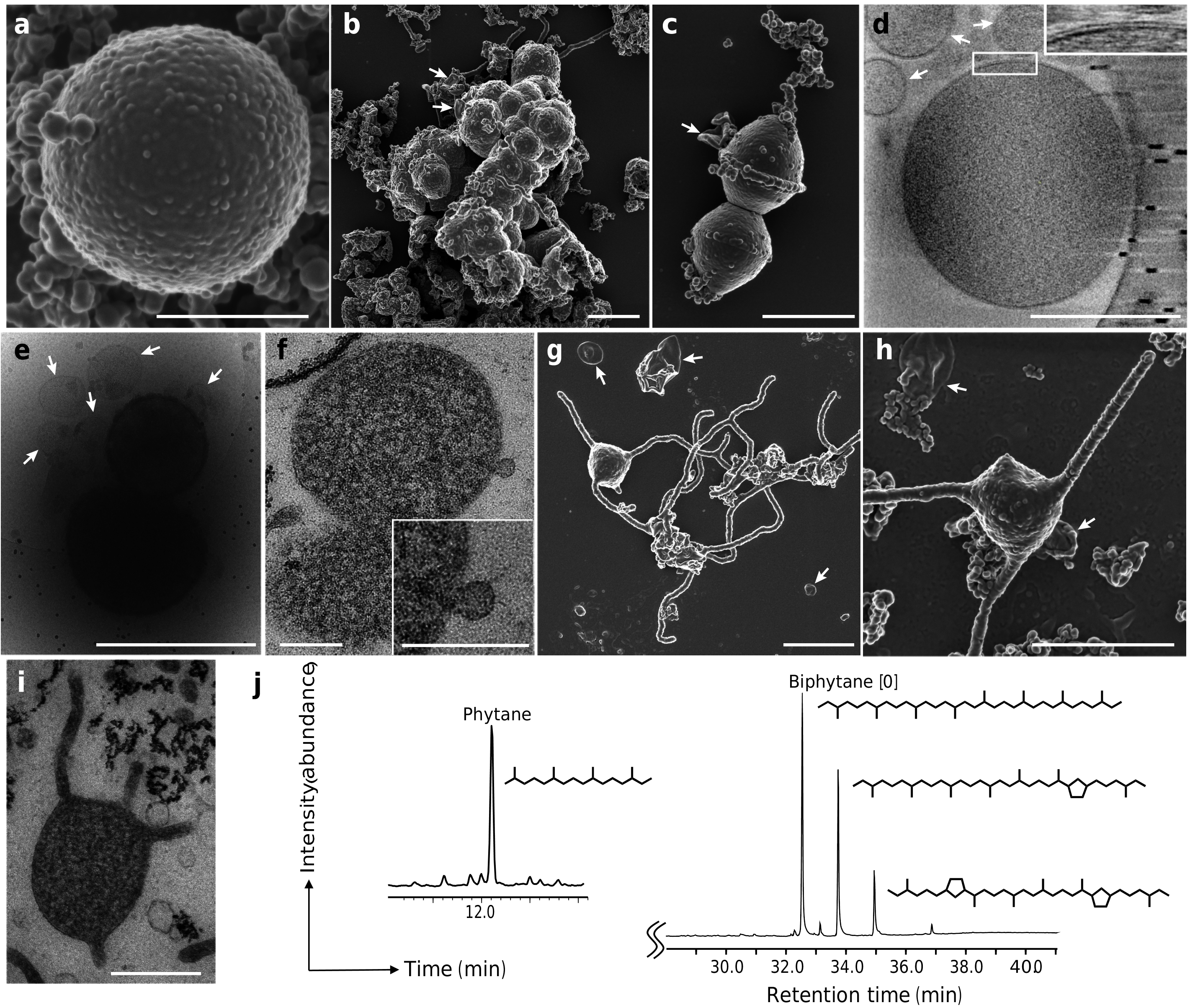
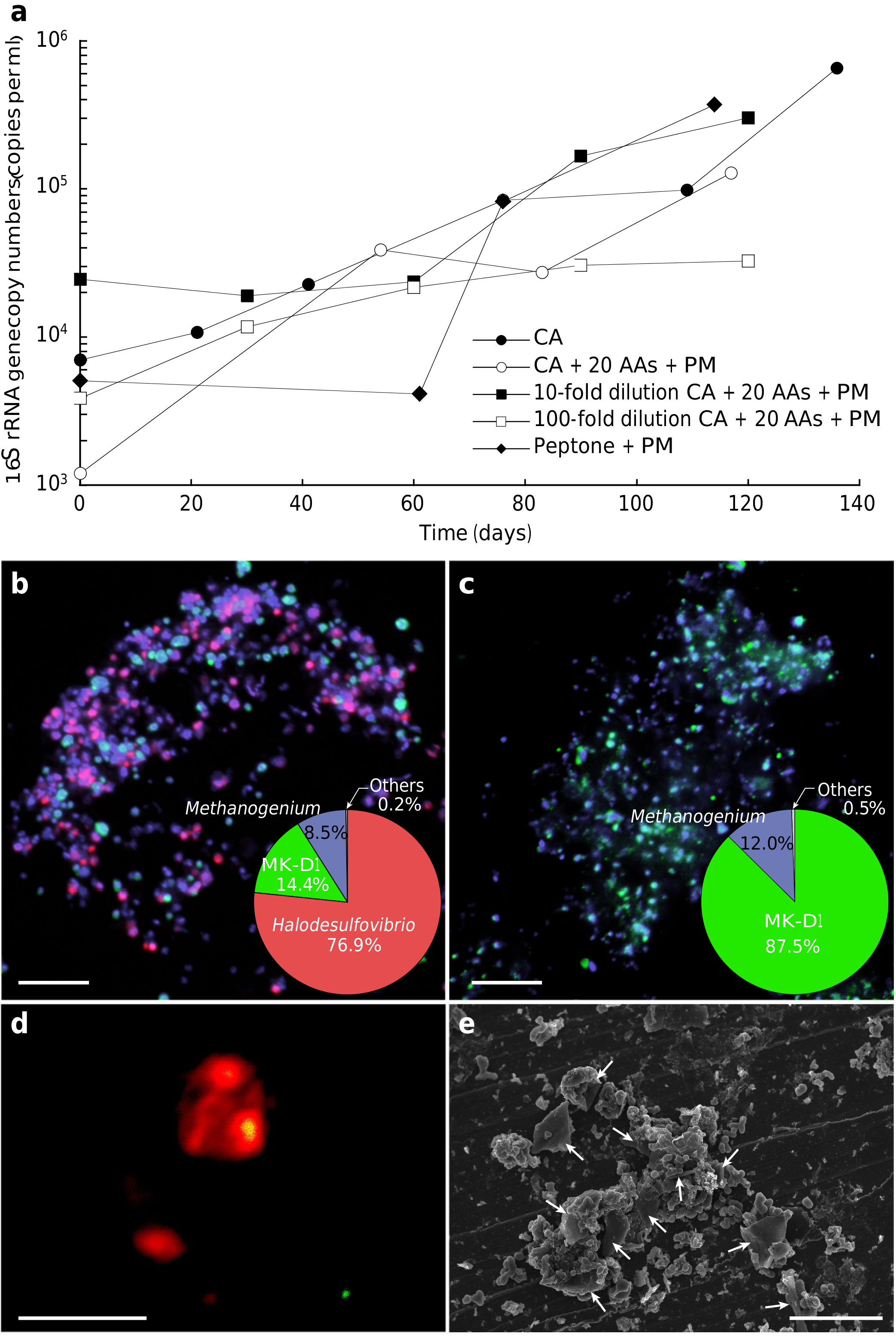
We further characterized MK-D1 using the pure co-cultures and highly purified cultures.Microscopy analyses showed that the cells were small cocci (approximately300–750nm in diameter (average,550 nm)),and generally formed aggregates surrounded by extracellular polymer substances (EPS) ( Fig.3a,b View Fig and Extended Data Fig.3 View Fig ),consistent with previous observations using FISH 15, 17. MK-D1 cells were easily identifiable given the morphological difference from their co-culture partner Methanogenium (highly irregular coccoid cells of ≥2 µm; Fig. 1d, e View Fig ). Dividing cells had less EPS and a ring-like structure around the cells ( Fig. 3c View Fig ). Cryo-electron microscopy (cryo-EM) and transmission electron microscopy (TEM) analyses revealed that the cells contain no visible organelle-like inclusions ( Fig. 3 View Fig d–f and Supplementary Videos 1–6), in contrast to previous suggestions 6. For cryo-EM, cells were differentiated from vesicles on the basis of the presence of cytosolic material (although DNA and ribosomes could not be differentiated),EPS on the cell surface and cell sizes that were consistent with observations by SEM and TEM analyses (Supplementary Videos 4–6). The cells produce membrane vesicles (50–280 nm in diameter) ( Fig. 3 View Fig b–f) and chains of blebs ( Fig. 3c View Fig ). MK-D1 cells also form membrane-based cytosol-connected protrusions of various lengths that have diameters of 80–100 nm,and display branching with a homogeneous appearance unlike those of other archaea ( Fig.3 View Fig g–i; confirmed using both SEM and TEM).These protrusions neither form elaborate networks (as in Pyrodictium 18) nor intercellular connections ( Pyrodictium , Thermococcus and Haloferax 18 – 20), suggesting differences in physiological functions.The MK-D1 cell envelope may be composed of a membrane and a surrounding S-layer, given the presence of four genes that encode putative S-layer proteins (Supplementary Fig. 1 View Fig ), stalk-like structures on the surface of the vesicles ( Fig. 3e View Fig and Extended Data Fig.3f, g View Fig ) and the even distance between the inner and outer layers of the cell envelope ( Fig.3d View Fig ). Lipid composition analysis of the MK-D1 and Methanogenium co-culture revealed typical archaeal isoprenoid signatures—C 20 -phytane and C 40 -biphytanes with 0–2cyclopentane rings were obtained after ether-cleavage treatment( Fig.3j View Fig ). Considering the lipid data obtained from a reference Methanogenium isolate (99.3% 16S rRNA gene identity; Supplementary Fig. 2 View γ-amino-butyryl-CoA ), MK-D1 probably contains C 20 -phytane and C 40 -biphytanes with 0–2 rings. The MK-D1 genome encoded most of the genes necessary to synthesize ether-type lipids—although geranylgeranylglyceryl phosphate synthase was missing—and lacked genes for ester-type lipid synthesis (Supplementary Tables 3, 4).
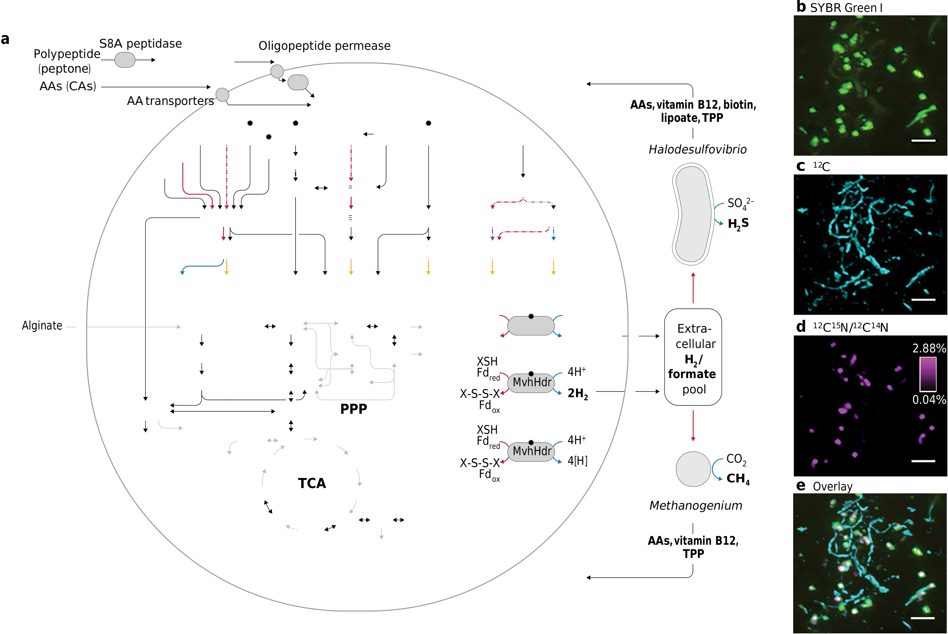
MK-D1 can degrade amino acids anaerobically, as confirmed by monitoring the depletion of amino acids during the growth of pure co-cultures (Extended Data Fig. 1b, c View Fig ). We further verify the utilization of amino acids by quantifying the uptake of a mixture of 13 C- and 15 N-labelled amino acids through nanometre-scale secondary ion mass spectrometry (NanoSIMS) ( Fig. 2 View γ-amino-butyryl-CoA b–e). Cell aggregates of MK-D1 incorporated amino-acid-derived nitrogen, demonstrating the capacity of MK-D1 to utilize amino acids for growth.Notably,the 13 C-labelling of methane and CO 2 varied depending on the methanogenic partner, indicating that MK-D1 produces both hydrogen and formate from amino acids for interspecies electron transfer(Extended Data Table 2). Indeed, addition of high concentrations of hydrogen or formate completely suppressed growth of MK-D1 (Extended Data Table 3). The syntrophic partner was replaceable—MK-D1 could also grow syntrophically with Methanobacterium sp. strain MO-MB1 21 instead of Methanogenium ( Fig.2 View γ-amino-butyryl-CoA b–e). Although 14 different culture conditions were applied,none enhanced the cell yield,which indicates
specialization of the degradation of amino acids and/or peptides (Extended Data Table 3).
To further characterize the physiology of the archaeon,we analysed the complete MK-D1 genome (Extended Data Fig.2 View γ-amino-butyryl-CoA and Supplementary Tables 2–6). The genome only encodes one hydrogenase (NiFe hydrogenase MvhADG–HdrABC) and formate dehydrogenase (molybdopterin-dependent FdhA),suggesting that these enzymes mediate reductive H 2 and formate generation,respectively. MK-D1 represents, to our knowledge, the first cultured archaeon that can produce and syntrophically transfer H 2 and formate using the above enzymes.We also found genes encoding proteins for the degradation of ten amino acids.Most of the identified amino-acid-catabolizing pathways only recover energy through the degradation of a 2-oxoacid intermediate (that is,pyruvate or 2-oxobutyrate; Fig.2a View γ-amino-butyryl-CoA and Supplementary Table 4). MK-D1 can degrade 2-oxoacids hydrolytically (through 2-oxoacid-formate lyases) or oxidatively (through 2-oxoacid:ferredoxin oxidoreductases) to yield acyl-CoA intermediates that can be further degraded for ATP generation. In the hydrolytic path, the carboxylate group of the amino acid is released as formate that can be directly handed off to partnering methanogenic archaea or SRB. In the oxidative path, 2-oxoacid oxidation is coupled with release of amino acid carboxylate as CO 2 and reduction of ferredoxin,which can be re-oxidized through H + and/or CO 2 reduction to H 2 and formate,respectively (through the electron-confurcating NiFe hydrogenaseMvhADG–HdrABC or formate dehydrogenase FdhA). On the basis of 13 C-amino-acid-based experiments (Supplementary Note 4), MK-D1 can probably switch between syntrophic interaction through 2-oxoacid hydrolysis and oxidation depending on the partner(s).
Etymology. Prometheoarchaeum, Prometheus (Greek) :a Greek god who shaped humans out of mud and gave them the ability to create fire; archaeum from archaea (Greek):an ancient life.The genus name is an analogy between the evolutionary relationship this organism and the origin of eukaryotes,and the involvement of Prometheus in the origin of humans from sediments and the acquisition of an unprecedented oxygen-driven energy-harnessing ability.The species name, syntrophicum, syn (Greek):together with; trephein (Greek) nourish; icus (Latin) pertaining to. The species name refers to the syntrophic substrate utilization property of this strain.
Locality. Isolated from deep-sea methane-seep sediment of the Nankai Trough at 2,533 m water depth,off the Kumano area,Japan.
Diagnosis. Anaerobic,amino-acid-oxidizing archaeon,small coccus, around 550 nm in diameter,syntrophically grows with hydrogen- and formate-using microorganisms.It produces membrane vesicles,chains of blebs and membrane-based protrusions.

No known copyright restrictions apply. See Agosti, D., Egloff, W., 2009. Taxonomic information exchange and copyright: the Plazi approach. BMC Research Notes 2009, 2:53 for further explanation.
|
Kingdom |
|
|
Phylum |
|
|
Class |
|
|
Order |
|
|
Family |
|
|
Genus |