
Philophylla millei Han and Norrbom
|
publication ID |
https://doi.org/10.5281/zenodo.181893 |
|
DOI |
https://doi.org/10.5281/zenodo.6232726 |
|
persistent identifier |
https://treatment.plazi.org/id/03FE5164-FFA6-8F2B-FF31-7165FF03748A |
|
treatment provided by |
Plazi |
|
scientific name |
Philophylla millei Han and Norrbom |
| status |
sp. nov. |
Philophylla millei Han and Norrbom View in CoL , n. sp.
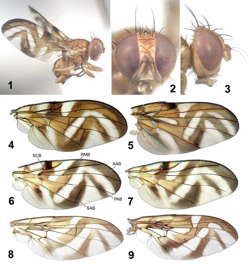
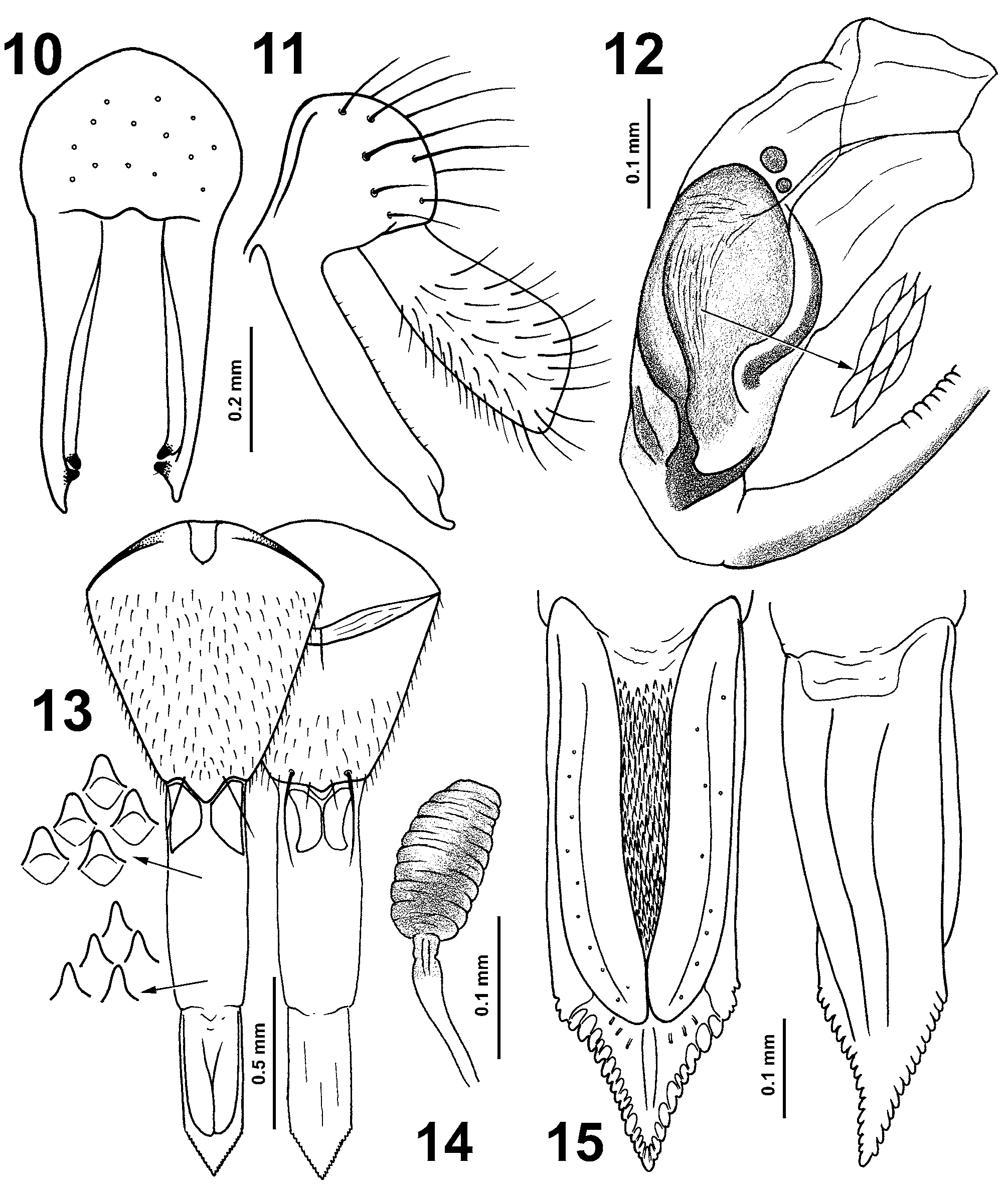
( Figs 1–15 View FIGURES 1 – 9. 1 – 3 View FIGURE 10 – 15 )
Anastrephoides View in CoL sp. Norrbom and Hancock, 2004: 67 (in New Caledonian list).
Type material. HOLOTYPE male: NEW CALEDONIA, Sarraméa Col d’Amieu, Malaise trap, 8.III.2006, C. Mille (the genitalia dissected and kept in a genitalia vial; left mid and hind legs removed for DNA extraction). PARATYPE female, 9.7 km NW Sarraméa, Malaise trap along Melaleuca Path, 15.I.1996, M.E. Irwin, D.W. Webb & E.I. Schlinger (the postabdomen dissected and kept in a genitalia vial). Both types are deposited in Museum National d’Histoire Naturelle, Paris.
Description. Body ( Fig. 1 View FIGURES 1 – 9. 1 – 3 ) almost entirely shiny to subshiny yellow brown with dark brown setae and setulae; wing length 3.9–4.2 mm; thorax length 1.9 mm. Head ( Figs 2, 3 View FIGURES 1 – 9. 1 – 3 ) yellow brown with ocellar tubercle dark brown, frons-head ratio (narrowest width of frons in dorsal view / width of head) 0.28, eye ratio (shortest eye diameter / longest eye diameter) 0.72–0.74, gena-eye ratio (genal height between ventral eye margin and ventral genal margin anterior to genal seta / longest eye diameter; gena measured with head tilted slightly dorsally so that gena is at its broadest) 0.08–0.10, arista-antenna ratio (length of arista / length of antenna excluding arista) 1.9–2.1; frons with 2 orbital and 3 evenly spaced frontal setae; medial vertical seta 0.7–0.8x as long as longest diameter of eye; lateral vertical seta 0.6x as long as medial vertical seta; postocellar seta 0.4x as long as medial vertical seta; paravertical seta hair-like, about 0.3x as long as medial vertical seta; ocellar seta 2.5x as long as ocellar tubercle; scape and pedicel yellow brown with short dark brown setulae; flagellomere 1 yellow brown; arista short pubescent, dark brown except for yellow brown basal thickened part; face more or less flat; parafacial narrow, 0.2x as wide as flagellomere 1; facial ridge with fine dark brown setulae; genal seta strong ( Figs 2, 3 View FIGURES 1 – 9. 1 – 3 ); postgena moderately swollen with relatively long dark brown setulae; postgenal seta strong; occiput flat, yellow brown; postocular setae extended 0.6x distance from upper eye margin to lower eye margin; supracervical setae dark brown; mouthparts short with labella yellow brown setulose and palpi dark brown setulose. Thorax entirely yellow brown with dark brown setae and setulae; scutum subshiny yellow brown, slightly microtrichose; 1 postpronotal, 2 scapular, 1 acrostichal, 1 dorsocentral, 1 intra-alar, 1 presutural supra-alar, 1 postsutural supra-alar, 1 postalar and 2 notopleural setae; dorsocentral seta slightly posterior to level of postsutural supra-alar seta; scutellum flat with sparse fine dark brown marginal setulae; basal scutellar seta 2.5x as long as scutellum; apical seta 1.7x as long as scutellum, crossing subapically; proepisternum densely covered with long dark brown setulae; ventral anepisternal seta slightly shorter than dorsal one; katepisternal seta well developed; mediotergite shiny yellow brown. Legs entirely yellow brown with dark brown setae and setulae; fore femur with 7 posteroventral setae; fore tarsomere 1 as long as remaining tarsomeres together; tibial spur 1.8x as long as wide; hind femur with 2 subapical dorsal setae; hind tibia with row of short anterodorsal setae. Wing ( Figs 1, 4 View FIGURES 1 – 9. 1 – 3 ) hyaline with orange brown to brown pattern, including: subbasal band from crossvein h to apex of cell bcu, cell bc and extreme base of cell br yellow, cell c brown on basal 1/3 and apical 1/5; brown subcostal band from pterostigma to BM-Cu, covering fork of Rs, becoming orange brown in cells br and dm where connected to radial-medial band so that apical half of br lacks hyaline area, also connected to subbasal band in cells br and bm; orange brown radial-medial band extending obliquely from costal margin in middle of cell r1, covering R-M, and turning posteriorly to reach wing margin in cell cu1 slightly distal to vein A1+Cu2, connected to orange brown to brown anterior apical band in cell r1 and to subcostal band in cells br and dm to form S-shaped mark; orange brown to brown posterior apical and subapical bands broadly connected to form inverted V-shaped mark, subapical band also broadly connected posteriorly to radial-medial band but separated anteriorly from anterior apical band. Wingthorax ratio (wing length / thorax length) 2.1–2.3, vein R4+5 ratio (distance along vein R4+5 between crossvein R-M and vein R4+5 apex / distance between crossvein R-M and basal node of vein R4+5) 2.9–3.0, vein M ratio (distance along vein M between crossveins R-M and DM-Cu / distance between crossveins R-M and BM-Cu) 1.0–1.1, subcosta-costa ratio (length of pterostigma / length of costal cell, both measured along vein C) 0.29– 0.32; R4+5 dorsally with 13–14 tiny setulae between node and R-M, 9–15 setulae apical to R-M; cell bm, cell bcu, anal lobe, and alula, except along fold, entirely microtrichose; posteroapical extension of cell bcu about half as narrow as width of wing veins, about 1.5x as long as crossvein BM-Cu.
Male abdomen subshiny yellow brown with dark brown setae and setulae, slightly longer than wide; epandrium ( Figs 10, 11 View FIGURE 10 – 15 ) yellow brown with yellow brown to brown setae and setulae; lateral surstylus yellow brown with more or less pointed apex in profile ( Fig. 11 View FIGURE 10 – 15 ), anterior lobe modified into prensiseta-like structure; medial surstylus with single prensiseta; prensiseta together with modified anterior lobe of lateral surstylus superficially appear as two prensisetae ( Fig. 10 View FIGURE 10 – 15 ); proctiger pale yellow with yellow brown setulae; medial sclerite of glans ( Fig. 12 View FIGURE 10 – 15 ) without internal pattern of granulation; dorsal sclerite of glans ( Fig. 12 View FIGURE 10 – 15 ) with faint internal sculpture pattern of narrowly rhombic cells dorsally.
Female abdomen entirely subshiny yellow brown with dark brown setae and setulae; preabdomen slightly longer than wide; oviscape ( Fig. 13 View FIGURE 10 – 15 ) slightly longer than wide, entirely yellow brown, dorsally with a pair of strong lateral marginal setae and ventrally with a pair of moderate sublateral marginal setae; eversible membrane medially with strong triangular denticles and posteriorly with smaller denticles; dorsal and ventral taeniae about 0.3x as long as total length of membrane; aculeus ( Fig. 15 View FIGURE 10 – 15 ) broad, parallel-sided with apical onethird tapered; tip triangular, with moderately large lateral serrations; medial membrane under ventral plates (= sternite 8) almost entirely covered with numerous anteriorly directed denticles; 3 spermathecae ( Fig. 14 View FIGURE 10 – 15 ) similar in size, brown, about twice as long as wide with numerous transerverse furrows; short apical portion of spermathecal duct yellowish brown.
Etymology. This species is named for Mr. Christian Mille, who acted upon our request to search for more specimens of this rare species. He ran traps in the area where the female had been found and collected the only known male (the holotype) which he promptly shipped so that DNA extraction as well as genitalic examination was possible.
Remarks. Adding P. millei to Philophylla could cause taxonomic confusion because its wing pattern is aberrant for the genus. Therefore, we provide here the following modifications of the diagnosis previously published for Philophylla and the section of the key to the genera of Trypetini involving the genus.
Generic diagnosis (modified from Han, 1999: 284): Philophylla can be defined as a monophyletic group by the following unequivocal synapomorphies: (1) oviscape dorsally with a pair of strong lateral marginal setae ( Han, 1999, fig. 11.9A); and (2) lateral surstylus with posterior lobe elongated and anterior lobe broadly flattened (all species for which male genitalia have been examined except P. m i l l e i) ( Han, 1999, figs 11.6A, B) or modified into a prensiseta-like structure ( P. m i l l e i; this state here interpreted as secondarily derived). Although dissection of genitalia is recommended to confirm generic placement, most species can be conveniently recognized as Philophylla by having one of four typical wing patterns ( Fig. 4 View FIGURES 1 – 9. 1 – 3 ; Han 1999, figs 11.5D– F).
Philophylla millei superficially resembles Anastrephoides matsumurai Shiraki , Myoleja korneyevi Han and Kütük , and M. sinensis (Zia) of the tribe Trypetini , especially in wing pattern ( Figs 4–7 View FIGURES 1 – 9. 1 – 3 ), but it can be distinguished from these species by its entirely orange brown cell br, which lacks the hyaline area present in the other species distal to the fork in vein Rs. Because wing patterns very similar to that shown in Han (1999, fig.
11.5F) occur also in some species of Anomoia , Hoplandromyia , and a few other genera, extra precaution is needed.
No known copyright restrictions apply. See Agosti, D., Egloff, W., 2009. Taxonomic information exchange and copyright: the Plazi approach. BMC Research Notes 2009, 2:53 for further explanation.
|
Kingdom |
|
|
Phylum |
|
|
Class |
|
|
Order |
|
|
Family |
|
|
Tribe |
Trypetini |
|
Genus |
Philophylla millei Han and Norrbom
| Han, Ho-Yeon & Norrbom, Allen L. 2008 |
Anastrephoides
| Norrbom 2004: 67 |