
Ctenocolum
|
publication ID |
https://doi.org/10.11646/zootaxa.3838.1.1 |
|
publication LSID |
lsid:zoobank.org:pub:1534C775-D28D-470F-9AEC-8BABB3D8FA56 |
|
DOI |
https://doi.org/10.5281/zenodo.6124227 |
|
persistent identifier |
https://treatment.plazi.org/id/03FF87F5-FFE6-FFEE-38AD-FAA2FA2A7365 |
|
treatment provided by |
Plazi |
|
scientific name |
Ctenocolum |
| status |
|
Key to the species of Ctenocolum
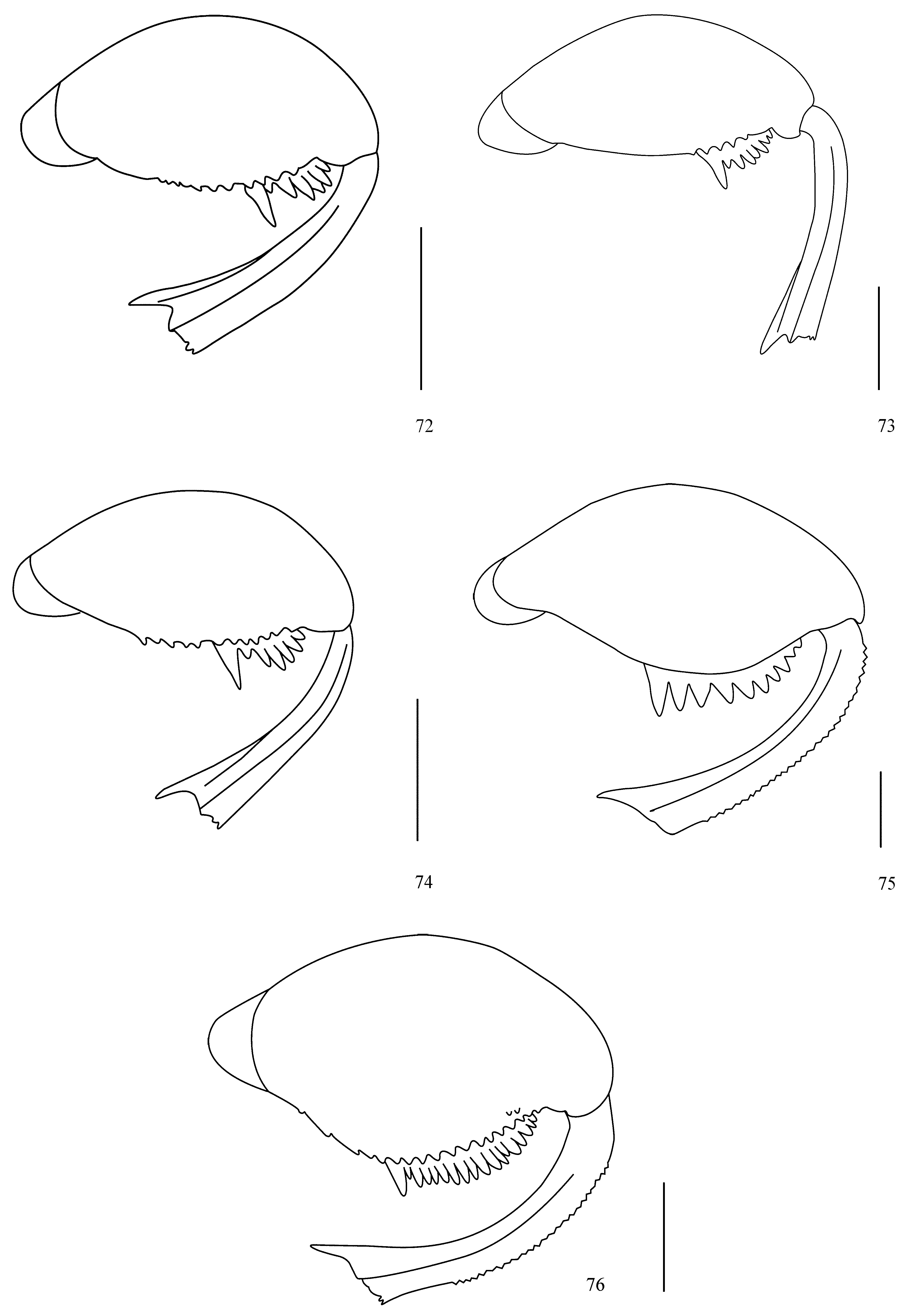
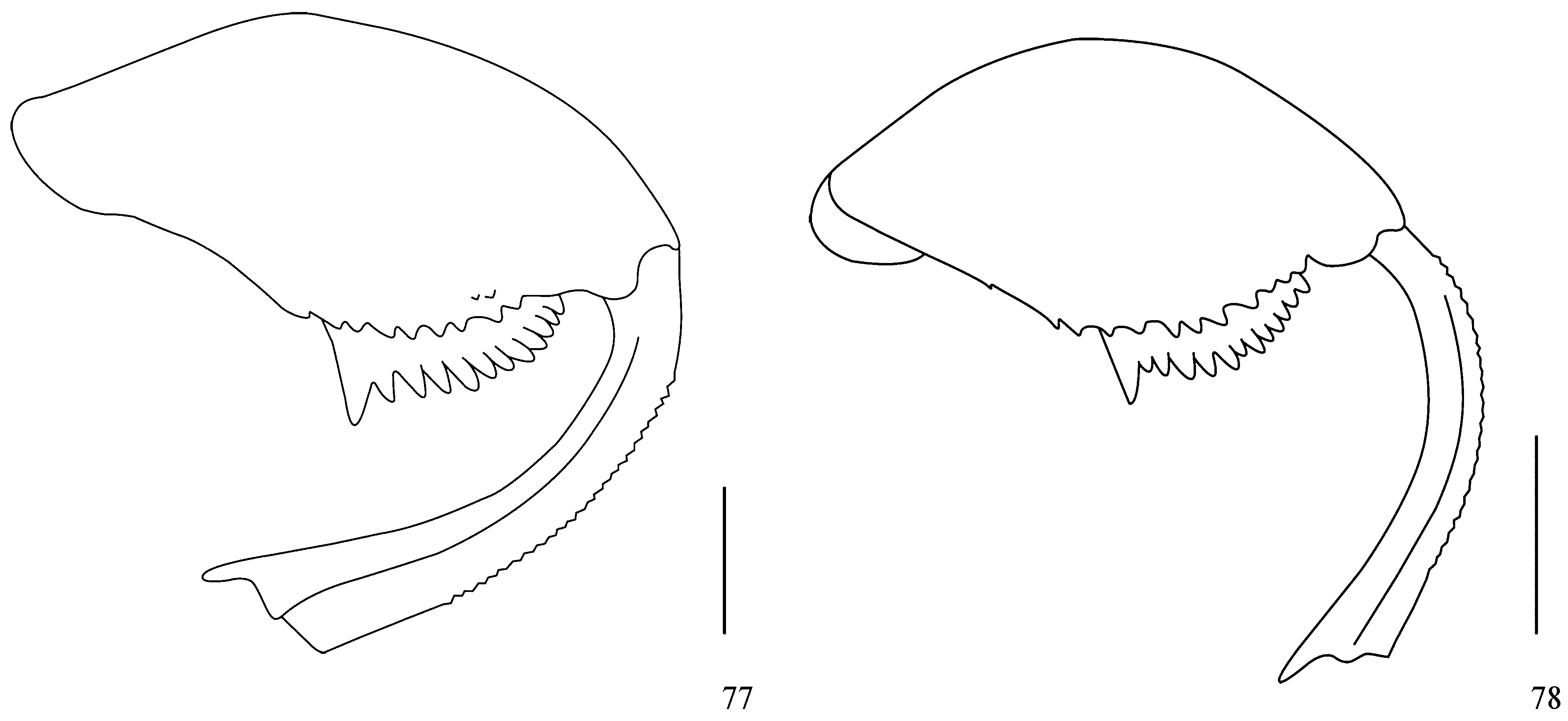
1. Hind femur with second tooth of pecten regular in profile until apex ( Figs. 75–78 View FIGURES 72 – 76 View FIGURES 77 – 78 ); hind tibia on outer surface with row of denticles ( Figs. 4 View FIGURES 1 – 7 , 75–78 View FIGURES 72 – 76 View FIGURES 77 – 78 ), apex lightly or moderately emarginated beside mucro ( Figs. 75–78 View FIGURES 72 – 76 View FIGURES 77 – 78 ) (Group tuberculatum )..... 2
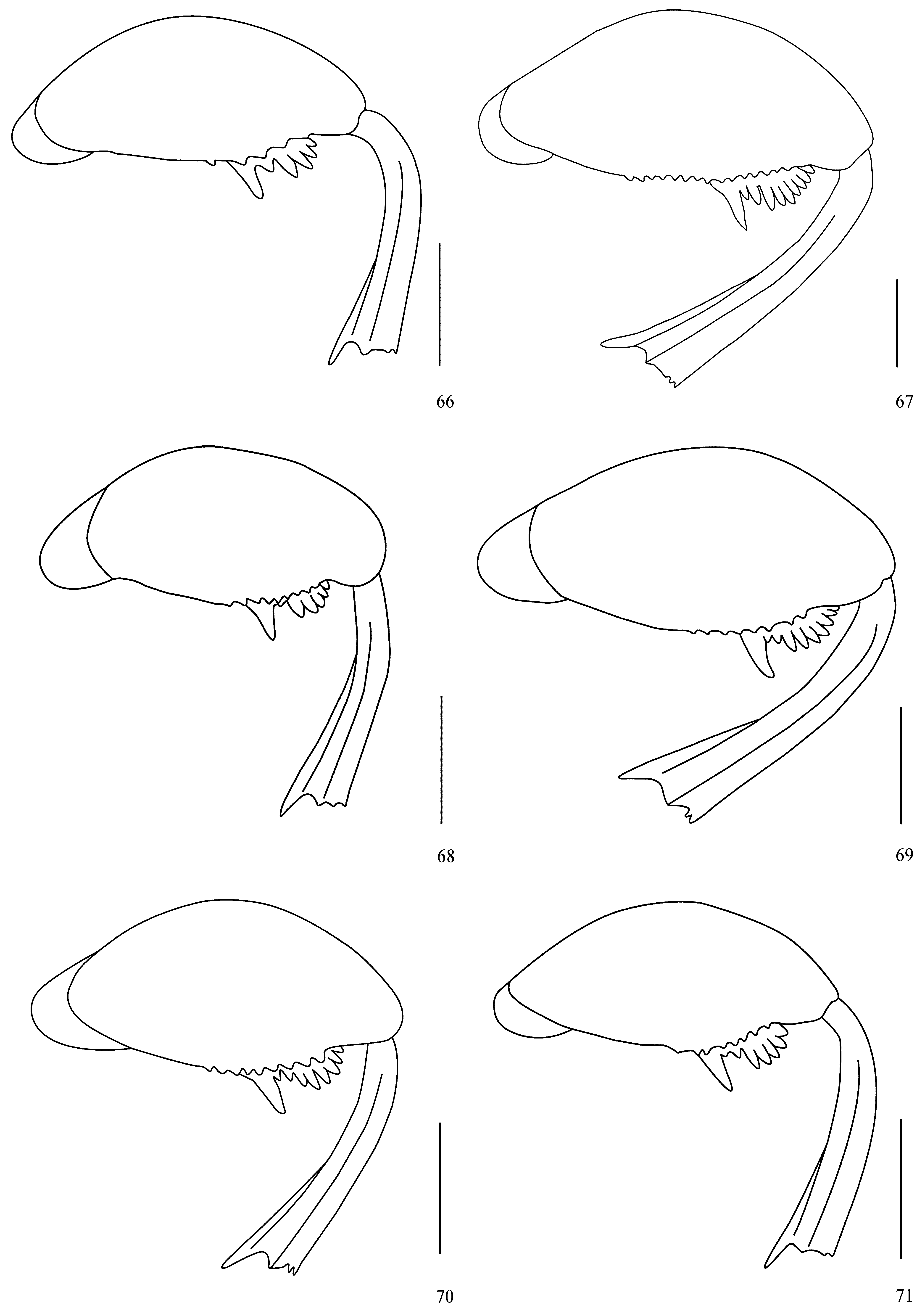
- Hind femur with second tooth of pecten gradually increasing in size until middle and decreasing towards apex ( Figs. 66–74 View FIGURES 66 – 71 View FIGURES 72 – 76 ); hind tibia on outer surface without row of denticles, apex strongly emarginated beside mucro ( Figs. 66–74 View FIGURES 66 – 71 View FIGURES 72 – 76 ) (Group podagricus )............................................................................................ 5
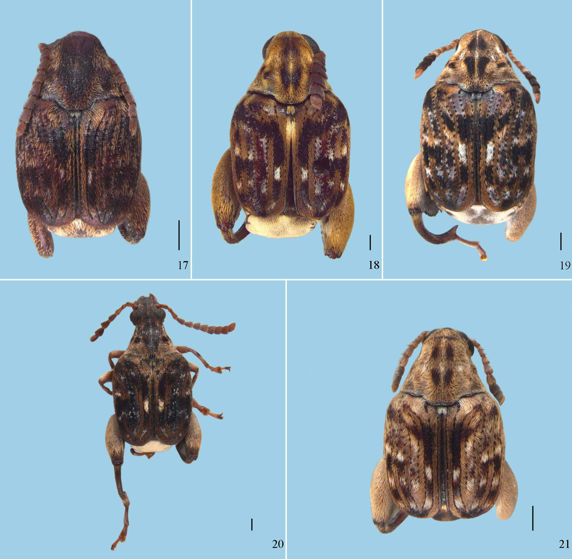
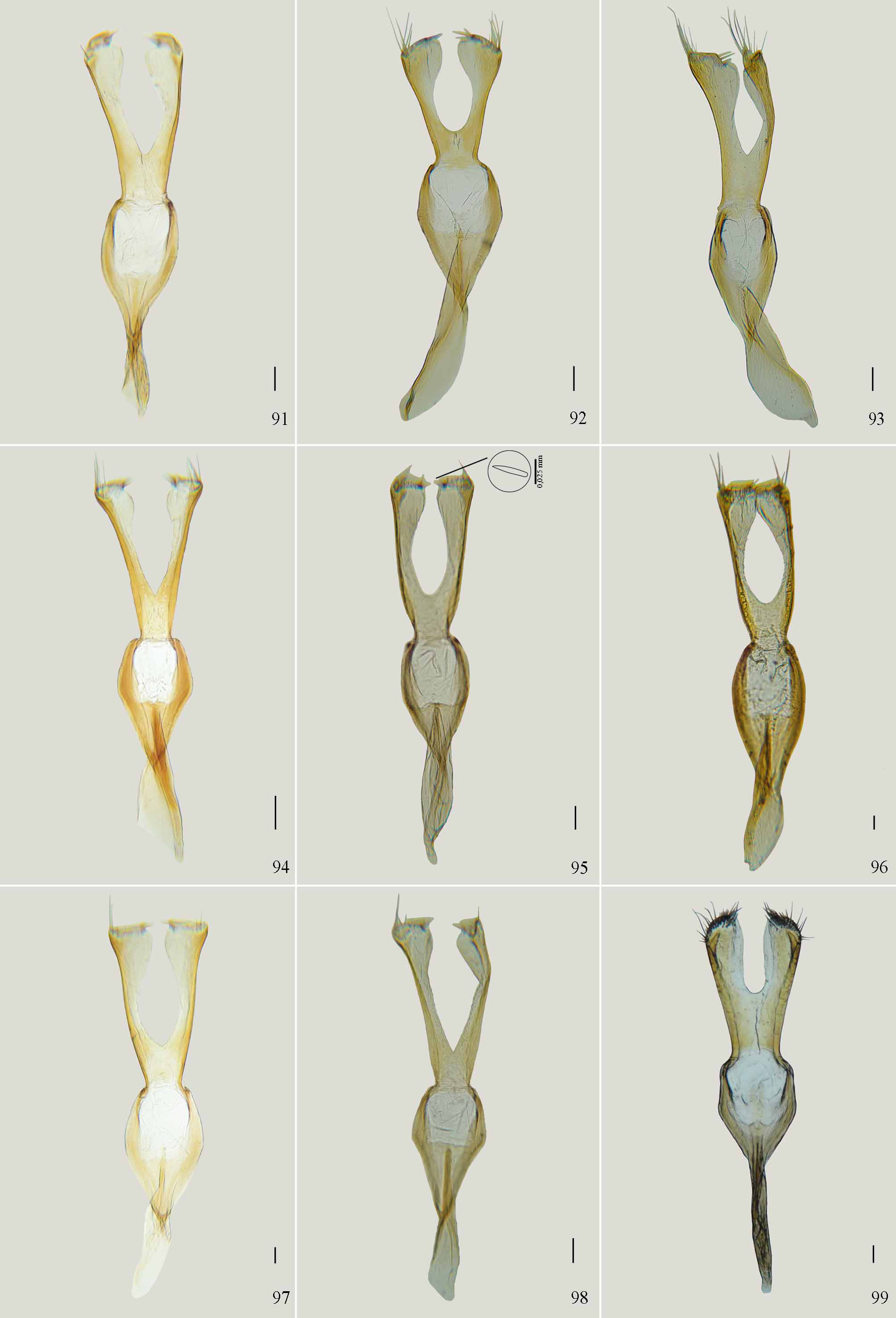
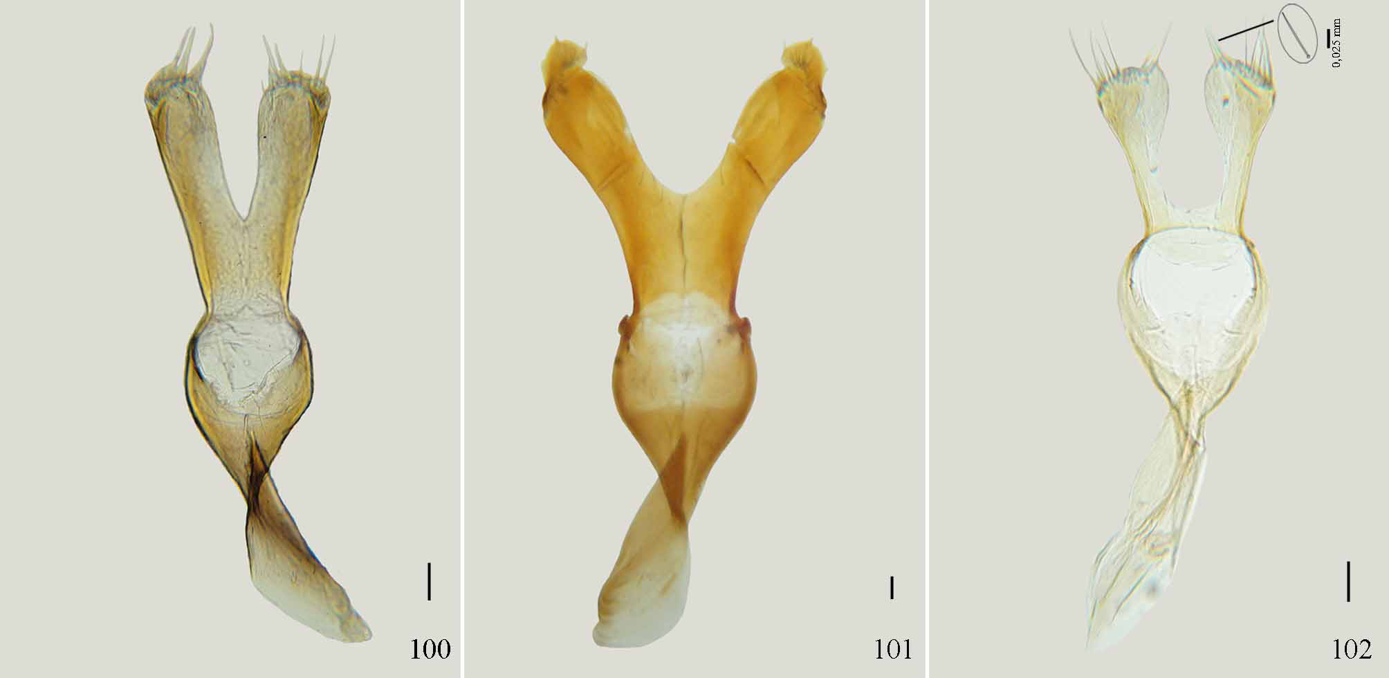
2 (1). Antennomeres 8–10 darker than the others ( Figs. 18, 19, 21 View FIGURES 17 – 21 , 63, 65 View FIGURES 61 – 65 ); internal sac at apex laterally with short tuft of setae ( Figs. 90 View FIGURES 85 – 90 ); tegmen, lateral lobe at apex without membranous projection ( Figs. 99–100, 102 View FIGURES 91 – 99 View FIGURES 100 – 102 ).............................3
- Antennomeres 8–10 the same color as the others ( Figs. 20 View FIGURES 17 – 21 , 64 View FIGURES 61 – 65 ); internal sac at apex laterally with long tuft of setae ( Figs. 5 View FIGURES 1 – 7 , 89 View FIGURES 85 – 90 ); tegmen at apex of lateral lobe with membranous projection ( Fig. 7 View FIGURES 1 – 7 ).................................... C. salvini
3 (2). Hind femur on external ventral margin without toothed carina ( Fig. 75 View FIGURES 72 – 76 )............................ C. acapulcensis
- Hind femur on external ventral margin with toothed carina ( Figs. 76 View FIGURES 72 – 76 , 78 View FIGURES 77 – 78 )..................................... 4
4 (3). Hind femur with denticles on the external ventral margin ( Fig. 76 View FIGURES 72 – 76 ); pygidium of female with sparse setae on two basal “C” pattern areas, on four lateral small areas and on a larger median area ( Fig. 51 View FIGURES 47 – 52 )............................... C. janzeni
- Hind femur without denticles on external ventral margin ( Fig. 78 View FIGURES 77 – 78 ); pygidium of female with sparse setae on two lateral areas and on rectangular conspicuous area extending from basal to submedian region ( Fig. 52 View FIGURES 47 – 52 )........... C. tuberculatum
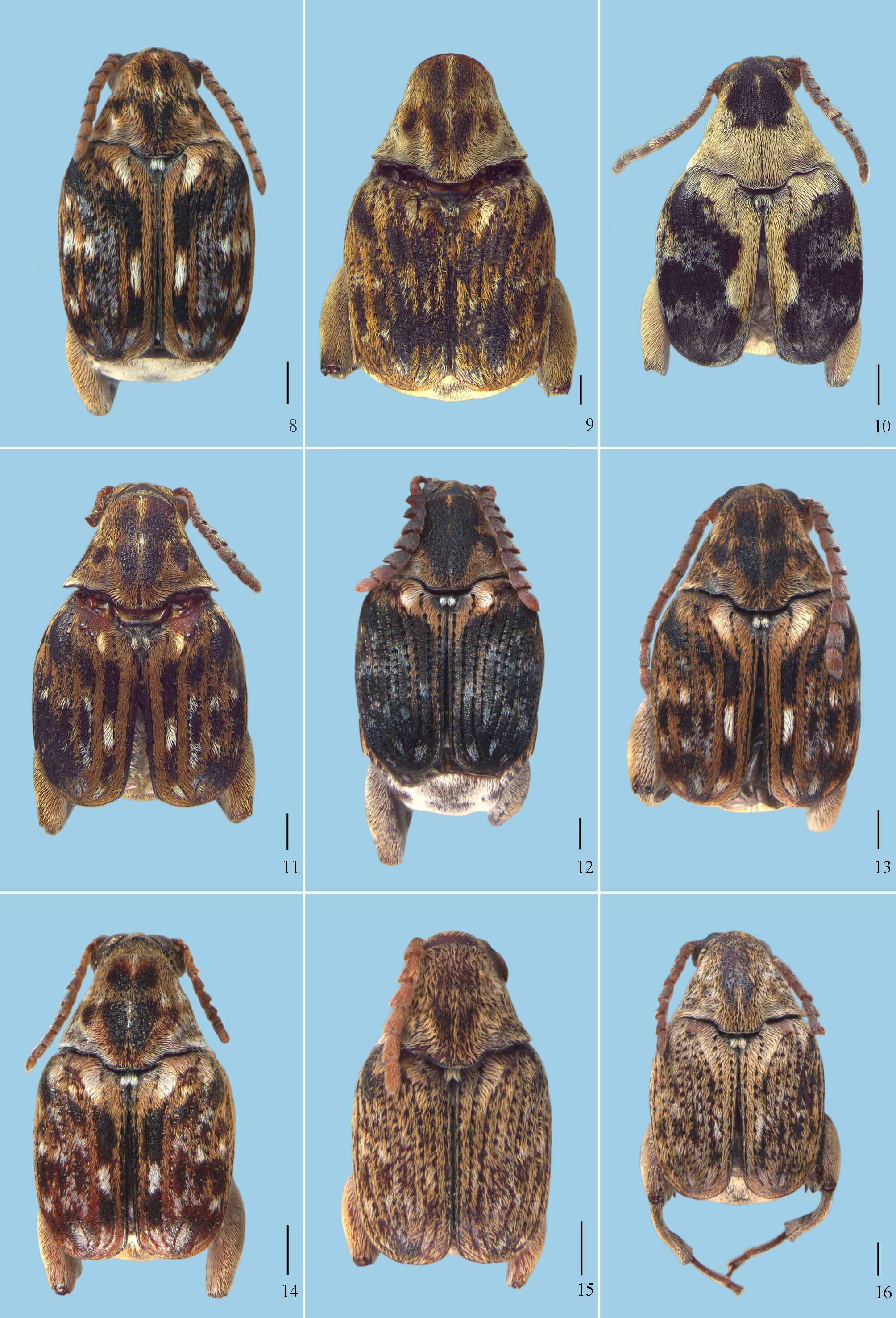
5 (1). Dorsum mostly with yellowish gray setae, forming a “C” pattern on each elytron ( Fig. 10 View FIGURES 8 – 16 ); pronotum with sparse setae that exposes the integument and forms a rounded area on anterior region ( Fig. 10 View FIGURES 8 – 16 ).............................. C. colburni
- Dorsum never mostly yellowish gray; when mostly yellow gray, not forming a conspicuous pattern on each elytron like a “C” ( Figs. 8, 9, 11, 12, 13, 14, 15, 16 View FIGURES 8 – 16 , 17 View FIGURES 17 – 21 ); pronotum with sparse pubescence exposing the integument forming an oval, wide area from anterior to posterior region, divided or not by transversal and longitudinal strip of denser setae ( Figs. 8, 9, 11, 12, 13, 14, 15, 16 View FIGURES 8 – 16 , 17 View FIGURES 17 – 21 )................................................................................. 6
6 (5). Elytral striae with deeply impressed punctures ( Figs. 15, 16 View FIGURES 8 – 16 )................................................... 7
- Elytral striae with moderately impressed punctures ( Figs. 8, 9, 11, 12, 13, 14 View FIGURES 8 – 16 , 17 View FIGURES 17 – 21 )................................ 8
7 (6). Pygidium at median basal region with moderately impressed punctures; tegmen of lateral lobes with “ U” emargination ( Fig. 97 View FIGURES 91 – 99 )................................................... C. punctinotatus Albuquerque & Ribeiro-Costa sp. nov.
- Pygidium at median basal region with deeply impressed punctures; tegmen of lateral lobes with “V” emargination ( Fig. 98 View FIGURES 91 – 99 ).......................................................... C. pygospilotos Albuquerque & Ribeiro-Costa sp. nov.
8 (6). Elytral stria 4 with tooth closer to anterior margin of elytra than base of the tooth of stria 3................ C. biolleyi
- Elytral stria 4 with tooth closer to base of tooth of stria 3 than to anterior margin of elytra...................... 9
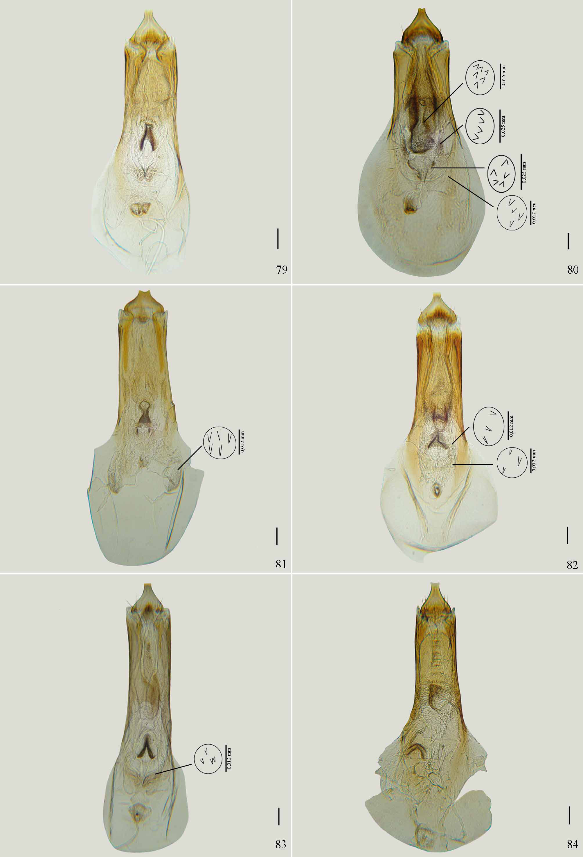
9 (8). Pronotum with median gibbosity divided by transversal sulcus; antenna pale brown or brown with antennomeres 3–11 darker at apex; male genitalia, internal sac with inverted Y-shape sclerite ( Fig. 79 View FIGURES 79 – 84 )................................................................................................. C. aquilus Albuquerque & Ribeiro-Costa sp. nov.
- Pronotum with median gibbosity not divided by transversal sulcus; antenna brown and dark brown or brown to dark brown, never with antennomeres 3–11 darker at apex; male genitalia, internal sac with different forms of sclerites ( Figs. 81–83 View FIGURES 79 – 84 , 86 View FIGURES 85 – 90 )................................................................................................ 10
10 (9). Elytra, striae 3 and 4 with conspicuous teeth at base ( Fig. 11 View FIGURES 8 – 16 ); male genitalia, internal sac at submedian region with squamous hollow sclerite ( Fig. 81 View FIGURES 79 – 84 )............................................................ C. martiale
- Elytra, striae 3 and 4 with less conspicuous teeth at base ( Figs. 13, 14 View FIGURES 8 – 16 , 17 View FIGURES 17 – 21 ); male genitalia, internal sac with different form of sclerite ( Figs. 82, 83 View FIGURES 79 – 84 , 86 View FIGURES 85 – 90 )........................................................................... 11
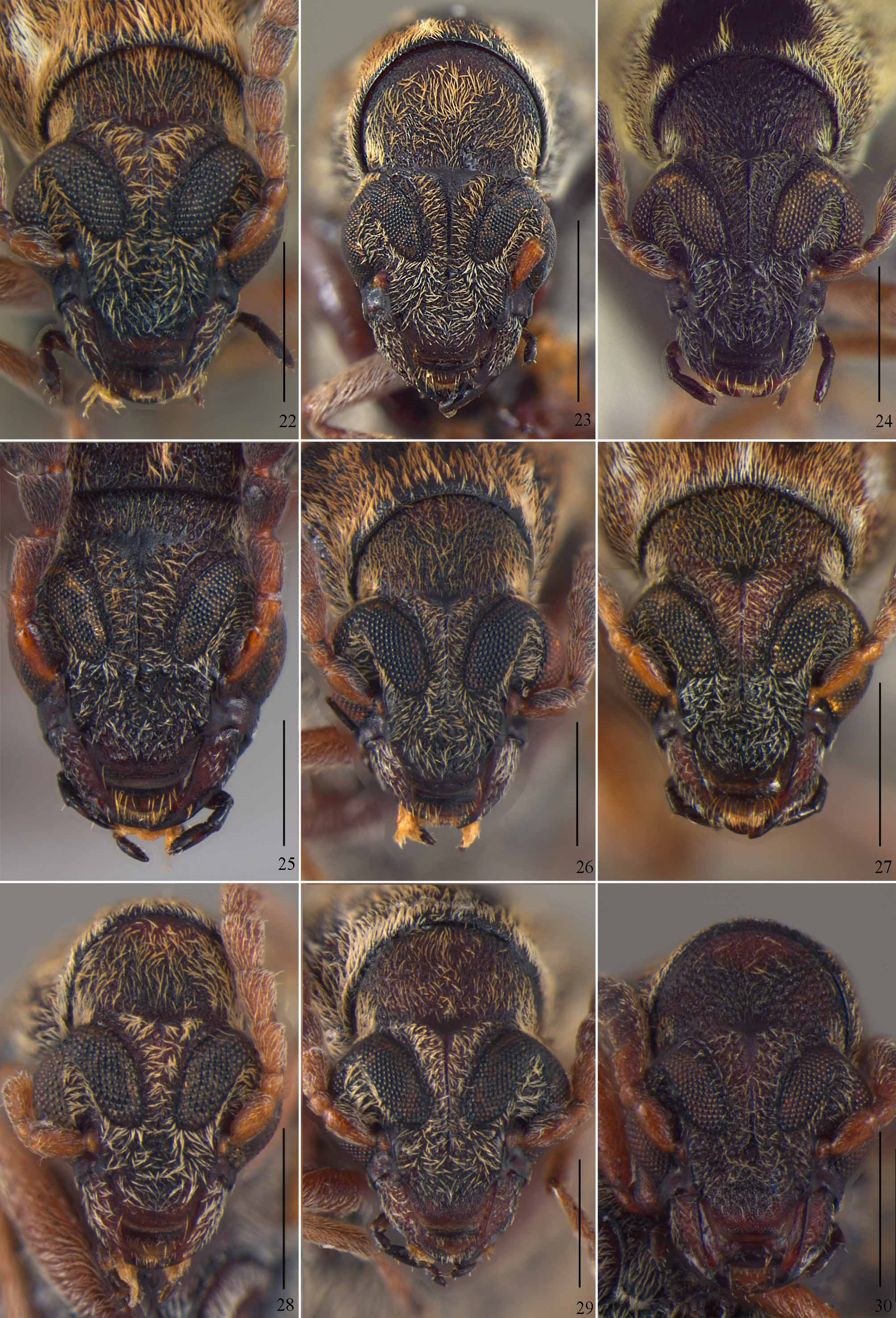
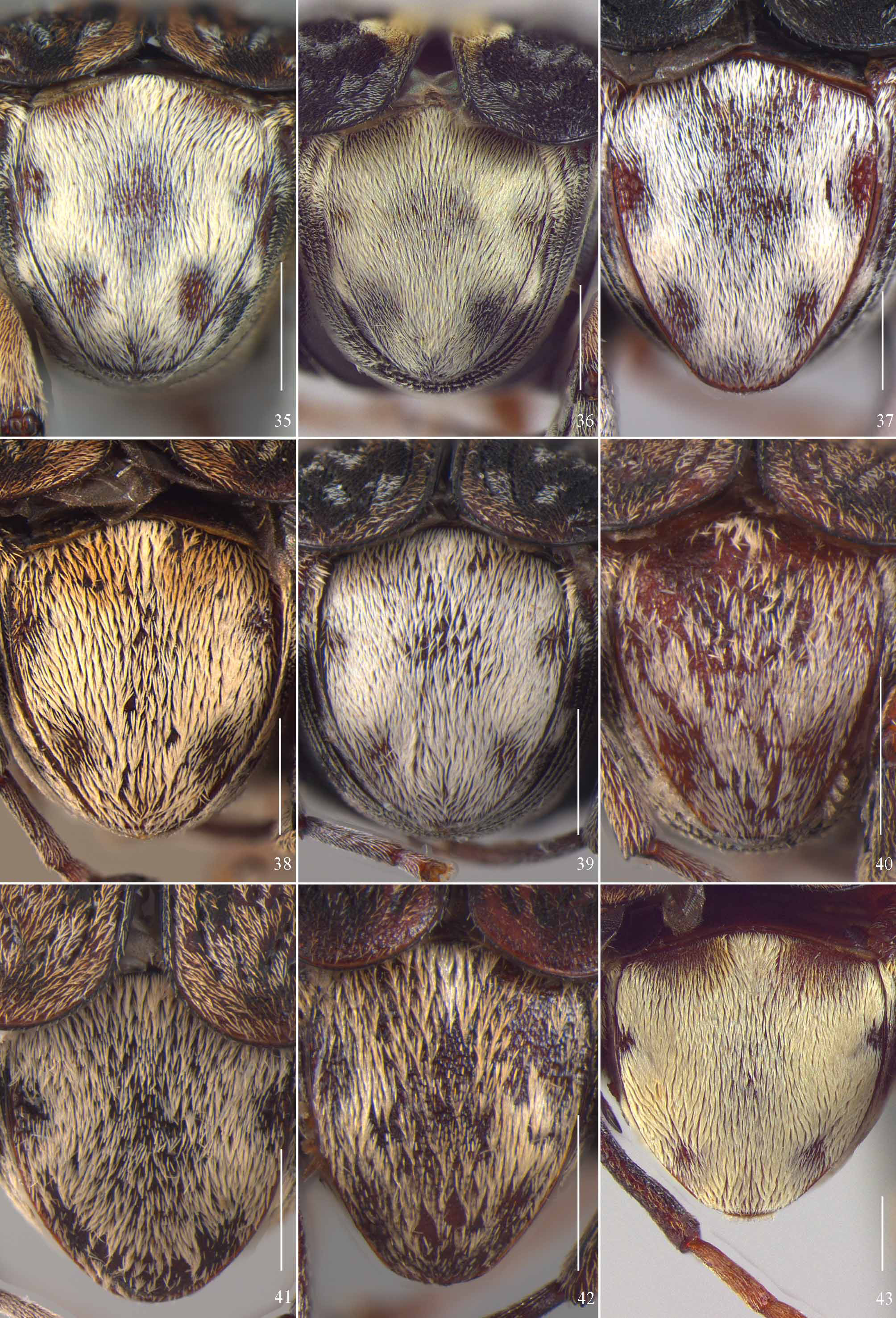
11(10). Ocular index 4.7–7.0 ( Figs. 26, 27 View FIGURES 22 – 30 ); pygidium oval in male ( Figs. 38, 39 View FIGURES 35 – 43 ) and triangular in females ( Figs. 49, 50 View FIGURES 47 – 52 )..... 12
- Ocular index 4.5 ( Fig. 30 View FIGURES 22 – 30 ); pygidium oval in male ( Fig. 42 View FIGURES 35 – 43 ) and female.................................................................................................... C. triangulatus Albuquerque & Ribeiro- Costa sp. nov.
12(11). Male genitalia, lateral lobes of tegmen with internal margin near end of emargination forming a "U" ( Fig. 95 View FIGURES 91 – 99 ).................................................................................................. C. podagricus
- Male genitalia, lateral lobes of tegmen with internal margin near end of emargination forming a “V” ( Fig. 94 View FIGURES 91 – 99 )................................................................... C. milelo Albuquerque & Ribeiro-Costa sp. nov.


No known copyright restrictions apply. See Agosti, D., Egloff, W., 2009. Taxonomic information exchange and copyright: the Plazi approach. BMC Research Notes 2009, 2:53 for further explanation.
|
Kingdom |
|
|
Phylum |
|
|
Class |
|
|
Order |
|
|
Family |
|
|
SubFamily |
Bruchinae |