
Metaperipatus inae, Mayer, Georg, 2007
|
publication ID |
https://doi.org/10.5281/zenodo.175982 |
|
DOI |
https://doi.org/10.5281/zenodo.5690086 |
|
persistent identifier |
https://treatment.plazi.org/id/03FFBE37-D17F-8420-FF5D-FC568FF6F807 |
|
treatment provided by |
Plazi |
|
scientific name |
Metaperipatus inae |
| status |
sp. nov. |
Metaperipatus inae View in CoL sp. nov.
( Figs 2 View FIGURE 1 - 2. 1 –3; 5; 7–9; 11–13; 15; 17–24; 29–32)
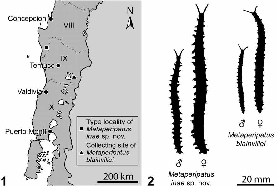
Material examined. Type material: Holotype ɗ ( MZUC – UCCC 31986), Chile, forest near Contulmo, VIII Region del Biobio ( Fig. 1 View FIGURE 1 - 2. 1 ), 38°01’S, 73°11’W, 390 m, 10th July 2004, coll. I. Mayer and G. Mayer. Allotype Ψ ( MZUC – UCCC 31987), collecting data as for holotype. The type specimens are deposited in the Museo Zoológico de la Universidad de Concepción ( MZUC) in Chile. Other material examined: additional specimens were obtained from the same locality as for holotype (n=22) and from laboratory cultures (n=26).
Diagnosis. Body length up to 60 mm (males) or 85 mm (females) when walking ( Fig. 2 View FIGURE 1 - 2. 1 ). Ground color dark greyish-blue, with large irregular orange/red spots formed by numerous orange/red-colored dermal papillae. Number of leg pairs 20 in males and 22 in females. M. inae sp. nov. is distinguished from M. blainvillei (a) by larger body size ( Fig. 2 View FIGURE 1 - 2. 1 ), (b) color pattern of integument characterized by large, irregular orange/red spots (in contrast to body of M. blainvillei which is speckled with single orange/red primary papillae, Figs 3– 6), and (c) by constant number of legs in both sexes (number of leg pairs ranges in M. blainvillei from 19 to 22 in males and from 20 to 22 in females, see review by Ruberg 1985; specimens of M. blainvillei investigated in this study showed the following leg numbers: 13 females and 3 males with 21 leg pairs and one female with 20 leg pairs).
Etymology. The new species is named in honor of the author’s wife, Ina Mayer, who found the first specimen.
Description:
External morphology. Body length: holotype (ɗ) 24 mm, allotype (Ψ) 42 mm (both in contracted condition, preserved in 70% ethanol); live specimens at walking: length of adult males 40–60 mm, length of adult females 65–85 mm ( Fig. 2 View FIGURE 1 - 2. 1 ). Color pattern: dorsal ground color dark greyish-blue, with large irregular orange/ red spots that rarely cross the dorsomedian furrow (Figs 3, 5); dorsomedian furrow unpigmented; ground color of ventrum pale greyish-blue, with similar irregular orange/red spots as on dorsum, spots do not cross ventral midline; ventral pits (= “ventral organs”) white ( Fig. 29 View FIGURES 29 – 32 ). Plicae (= tegumentary folds): number of plicae per segment variable (9–14), with numerous anastomoses. Antennal rings (only those bearing sensory bristles were counted): 49–50 in male holotype, 50–51 in female allotype.
Mouth parts. Tongue with longitudinal row of 6 large sensillae (= “teeth”) of similar size and additional small sensillae sparsely scattered over tongue surface. Jaws: outer blade of jaw with large single tooth and 1– 2 small accessory teeth in adults (Fig. 11), accessory tooth/teeth absent in juveniles (Fig. 12); inner blade of jaw with large single tooth and seven, nine, or ten denticles (Fig. 11 and inset), juveniles initially with only five denticles (Fig. 12), number increases with age; diastema absent.
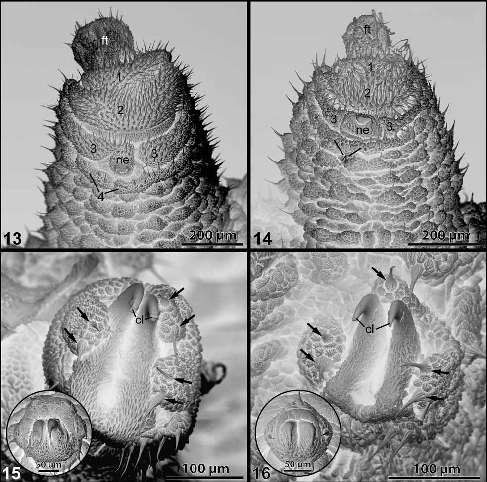
Legs. Number of leg pairs: intraspecifically invariant, but sexually dimorphic, 20 pairs in males (n = 25) and 22 pairs in females (n = 25); last leg pair clawed, oriented posteriorly in both sexes (Figs 7–9), not used in locomotion; length of last pair about 50% length of penultimate leg pair, reduced to same extent as in M. blainvillei (cf. Fig. 10). Spinous footpads: four spinous footpads, fourth pad much narrower than the others and fragmented ( Fig. 13 View FIGURES 13 – 16 ), as in M. blainvillei ( Fig. 14 View FIGURES 13 – 16 ); nephropore in centre of third spinous footpad of leg pairs 4 and 5; no basal foot papillae; distal foot papillae duplicated or triplicated in adult specimens but single in juveniles ( Fig. 15 View FIGURES 13 – 16 ), which resembles the situation in M. blainvillei ( Fig. 16 View FIGURES 13 – 16 ); number and position of (single, duplicated, or triplicated) distal foot papillae variable even within the same individual; there are either (a) two anterior and one posterior, (b) one anterior, one median and one posterior, or (c) two anterior and two posterior papillae ( Figs 15–16 View FIGURES 13 – 16 , and their insets); crural papillae always absent.
Posterior end. Anal cone (area around the anus behind the last leg pair): rounded in both sexes but tapering stronger in females (Figs 7–8). Male accessory gland papillae: anterior papillae absent; paired posterior gland papillae on anal cone (Fig. 7). Genital opening: between last pair of legs in both sexes (Figs 7–9); male gonopore small, cruciform, with four genital pads (Figs 7, 9); female gonopore cruciform, genital pads fragmented (Fig. 8).
FIGURES 3–6. Color patterns of the body in representatives of Metaperipatus . 3, 5, Metaperipatus inae sp. nov. 3, Walking specimen (Ψ). 5, Light micrograph of a dorsal portion of integument ( holotype ɗ: MZUC–UCCC 31986). Large orange/red spots are formed by orange/red-colored dermal papillae. 4, 6, Metaperipatus blainvillei . 4, Walking specimen (Ψ). 6, Light micrograph of dorsal integument speckled with irregularly scattered, orange/red primary papillae. Df = dorsomedian furrow.
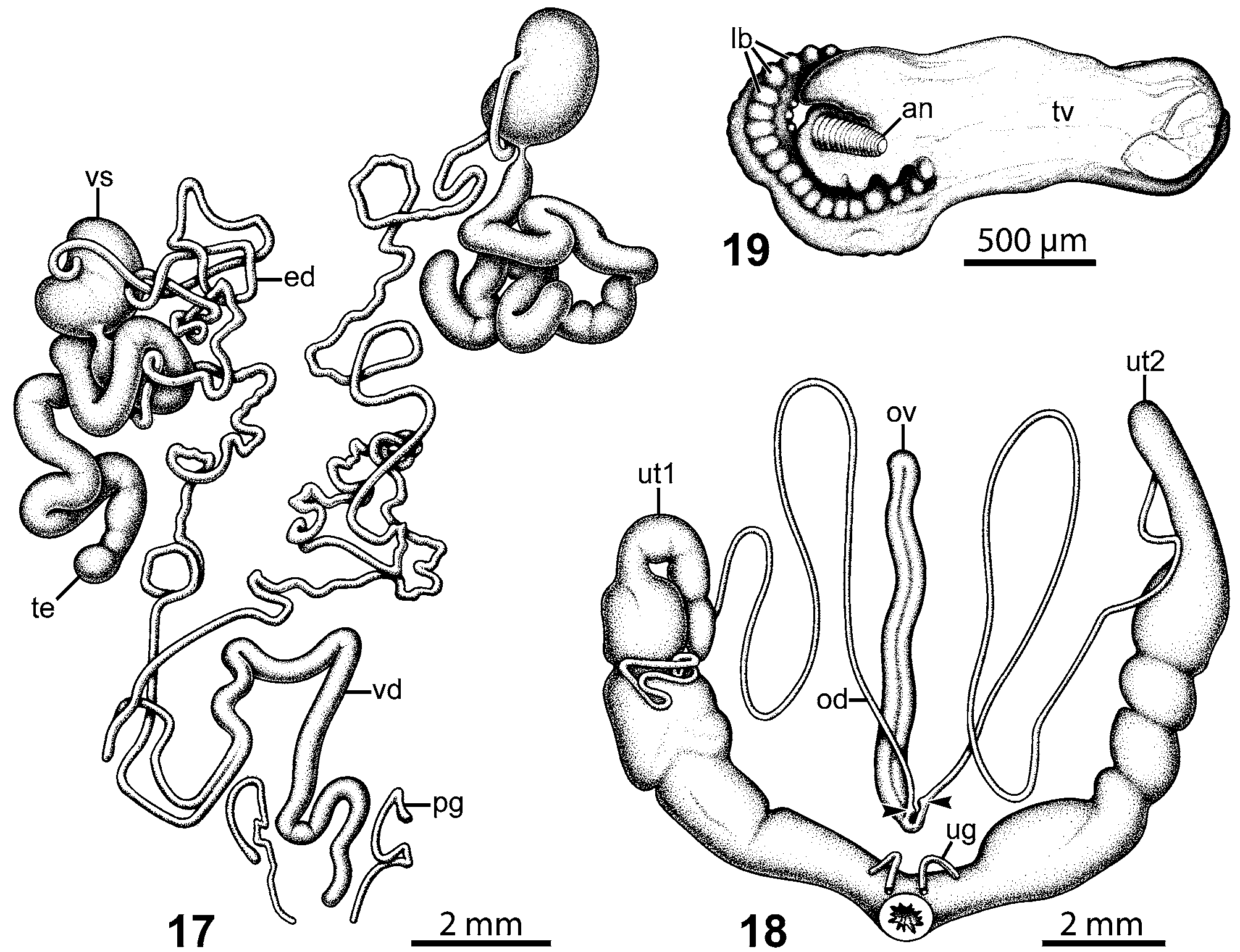
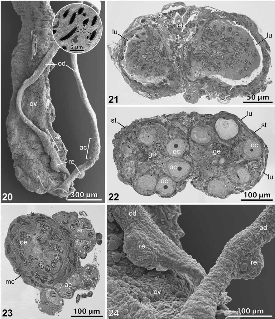
Male genital tract and associated glands. Testes thick, long and twisted ( Fig. 17 View FIGURES 17 – 18 ); seminal vesicles ellipsoid, bean-shaped; efferent ducts twisted and coiled, parallel before fusing to vas deferens; vas deferens continues anteriorly for a short distance before looping posteriorly, then passes beneath nerve cord to gonopore; spermatophore pouch absent; anterior accessory glands absent; posterior accessory glands (= anal glands) paired, tubular, loosely looped, blunt distally; discharge in separate openings on anal segment behind the gonopore (Fig. 7).
Female genital tract and associated structures. Ovary situated between 17th and 21st leg pairs, suspended from pericardial floor (= pericardial septum) along its entire length; distal ovarian ligament absent ( Fig. 18 View FIGURES 17 – 18 ); outer ovarian walls thin; lumens of ovarian tubes separate, crescent-shaped in cross-section, situated laterally ( Fig. 21 View FIGURES 20 – 24 ); ova neither epithelial nor stalked, positioned within a central germinal cell mass; oviducts looped, unite close to ovary ( Fig. 18 View FIGURES 17 – 18 ); receptaculum ovorum absent; seminal receptacles (= spermathecae) positioned on one side of each oviduct almost touching each other ( Figs 18 View FIGURES 17 – 18 , 20, 24 View FIGURES 20 – 24 ), small (80–100 µm), globular; accessory cell groups form irregular swellings of oviduct wall proximal to seminal receptacles ( Figs 20, 23 View FIGURES 20 – 24 ), enclose no lumen which is in contrast to previously described “accessory pouches” in Australian onychophorans (see Walker et al. 2006); oviducts continue into paired uteri with thickened walls; each uterus usually contains 1–2 batches of 2–5 embryos (each batch is characterized by embryos of a similar developmental stage); uteri join to form short muscular vagina leading to exterior; gonopore situated between last, rudimentary pair of legs; ovipositor absent; uterine glands paired, small tubes extending into body cavity, thinner in the proximal portion (towards gland foramen) than in the distal portion (towards hemocoel); gland foramen externally invisible, hidden between genital pads.
Reproduction: Reproductive cycle. Two broods (2–5 young each) were produced within one year (December–January and May–June, see Tab. 1 View TABLE 1 ); females collected in July contained embryos with huge, translucent trophic vesicles associated with embryonic necks (Fig. 19). The embryogenesis of M. inae , thus, resembles that of Peripatopsis sedgwicki from South Africa and Paraperipatus novaebritanniae from Papua New Guinea (see Bouvier 1902; Manton 1949; Willey 1898, 1899).
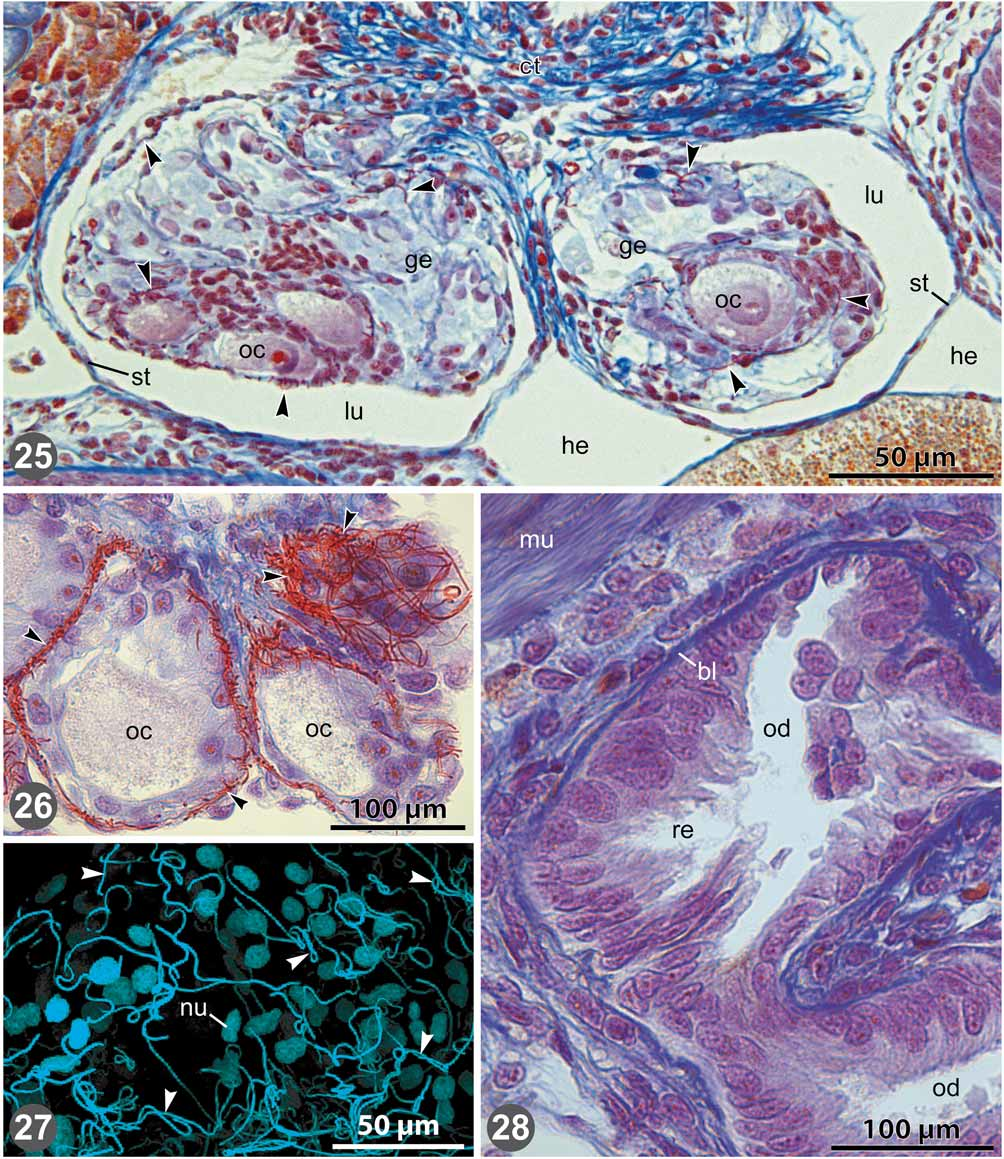
Ovarian structure. The ovarian morphology is similar in M. inae and M. blainvillei . In both species, the ovary is suspended from the pericardial floor along its entire length and bears no distal ligament. Semi-thin and histological sections reveal two medially fused ovarian tubes with separate, crescent-shaped lumens ( Figs 21 View FIGURES 20 – 24 , 25 View FIGURES 25 – 28 ). The ovarian tubes are surrounded by connective tissue towards the hemocoel. Towards the median ovarian portion, each lumen is bordered by a germinal cell mass whereas it is lined by a flattened cellular layer towards the hemocoel ( Figs 22 View FIGURES 20 – 24 , 25 View FIGURES 25 – 28 ). The germinal cell mass is characterized by the presence of maturating oogonia and oocytes, which are lacking in the outer, sterile cell layer.
Mature oocytes have a diameter of 50–100 µm. Within the germinal cell mass, the younger oogonia are usually arranged in the median portion, while the older oogonia and mature oocytes occur towards the exterior in each ovarian tube ( Figs 22 View FIGURES 20 – 24 , 25 View FIGURES 25 – 28 ). There are no follicular stalks associated with oocytes that would grow out of the ovary and project into the hemocoel. Instead, the oocytes retain their position within the germinal layer during maturation. Hence, the external surface of ovaries occurs smooth and not “grape-like” ( Figs 18 View FIGURES 17 – 18 , 20 View FIGURES 20 – 24 ), which is in contrast to other peripatopsids studied thus far.
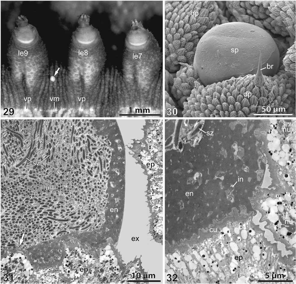
Dermal insemination. In M. inae , white spermatophores are released throughout the year. They are attached to any part of the female’s body, including the ventrum ( Fig. 29 View FIGURES 29 – 32 ). Each male is capable of producing up to 15 spermatophores within 2–3 days. Deposited spermatophores have been observed on both freshly collected and cultured females. After deposition, the spermatophores are spherical in shape ( Fig. 30 View FIGURES 29 – 32 ). Subsequently, they become flattened, and their remains are finally discarded with the molted cuticle during the next ecdysis. Similar spermatophores were also observed on the body surface of female M. blainvillei .
Ultrastructural examination of the spermatophores of M. inae revealed a large lumen densely packed with sperm ( Fig. 31 View FIGURES 29 – 32 ). As in other onychophoran species, sperm cells are characterized by an elongated head containing the nucleus, a mid-piece bearing the mitochondria, and a sperm tail giving rise to a flagellum with a 9 × 2+2 pattern of microtubules and an additional set of accessory microtubules (for details on sperm ultrastructure, see Marotta & Ruhberg 2004). The lumen of the spermatophore is surrounded by an electron-dense envelope, which is 5–10 µm thick. The envelope consists of homogenous material containing various irregular inclusions ( Fig. 32 View FIGURES 29 – 32 ). The basal surface of the spermatophore is closely associated with the cuticle of the female’s body. In some of the contact areas, the envelope is extremely thin or absent. In these areas, the lumen of the spermatophore directly borders the female’s cuticle ( Fig. 31 View FIGURES 29 – 32 ).
Subsequent stages of sperm impregnation into the female’s body have not been studied, but numerous sperm were found on the ovarian surface and within the germinal cell mass in M. inae ( Fig. 20 View FIGURES 20 – 24 , inset) and in M. blainvillei ( Figs 25–27 View FIGURES 25 – 28 ). However, sperm were not detected within the ovarian lumen. Furthermore, sperm were not found within the seminal receptacles (n=4) of M. inae and M. blainvillei , which appeared empty when sectioned ( Fig. 28 View FIGURES 25 – 28 ).
FIGURES 7–10. Characteristics of posterior ends and associated structures in representatives of Metaperipatus (SEM micrographs). 7–9, Posterior ends of Metaperipatus inae sp. nov. in ventral view. 7, Male posterior end showing a cruciform gonopore (go), paired openings of the (posterior) accessory genital glands (po), and small last (20th) pair of legs. 8, Female posterior end showing gonopore and reduced last (22nd) pair of legs. 9, Posterior end of a one week old juvenile (ɗ) showing posteriorly directed last (20th) pair of legs. 10, Two days old juvenile of M. blainvillei (Ψ), posterior end in terminal view showing posteriorly directed last (21st) pair of legs. An = anus, go = gonopore, le = legs or oncopodia (numbered), po = openings of male posterior accessory genital glands. FIGURES 11–12. Characteristics of jaws in Metaperipatus inae sp. nov. (SEM micrographs). 11, Inner (ib) and outer blades (ob) of jaws in adult specimens. Arrow points to an accessory tooth on the outer blade. Denticles (accessory teeth) of the inner blade numbered. Seven denticles are common, but some specimens show ten denticles (inset). 12, Inner and outer blades of jaw in a three months old juvenile. Note that the accessory tooth of the outer blade is lacking and the denticles of the inner blade are lower in number in the juvenile. Ib = inner blade, ob = outer blade.
No known copyright restrictions apply. See Agosti, D., Egloff, W., 2009. Taxonomic information exchange and copyright: the Plazi approach. BMC Research Notes 2009, 2:53 for further explanation.
|
Kingdom |
|
|
Phylum |
|
|
Family |
|
|
Genus |