
Dilemma spectralis, Leal, Jos Ẽ H., 2008
|
publication ID |
https://doi.org/10.5281/zenodo.181996 |
|
DOI |
https://doi.org/10.5281/zenodo.6228965 |
|
persistent identifier |
https://treatment.plazi.org/id/081987A1-A214-BB5A-FF5D-DC6BFF3E8A97 |
|
treatment provided by |
Plazi |
|
scientific name |
Dilemma spectralis |
| status |
sp. nov. |
Dilemma spectralis View in CoL new species
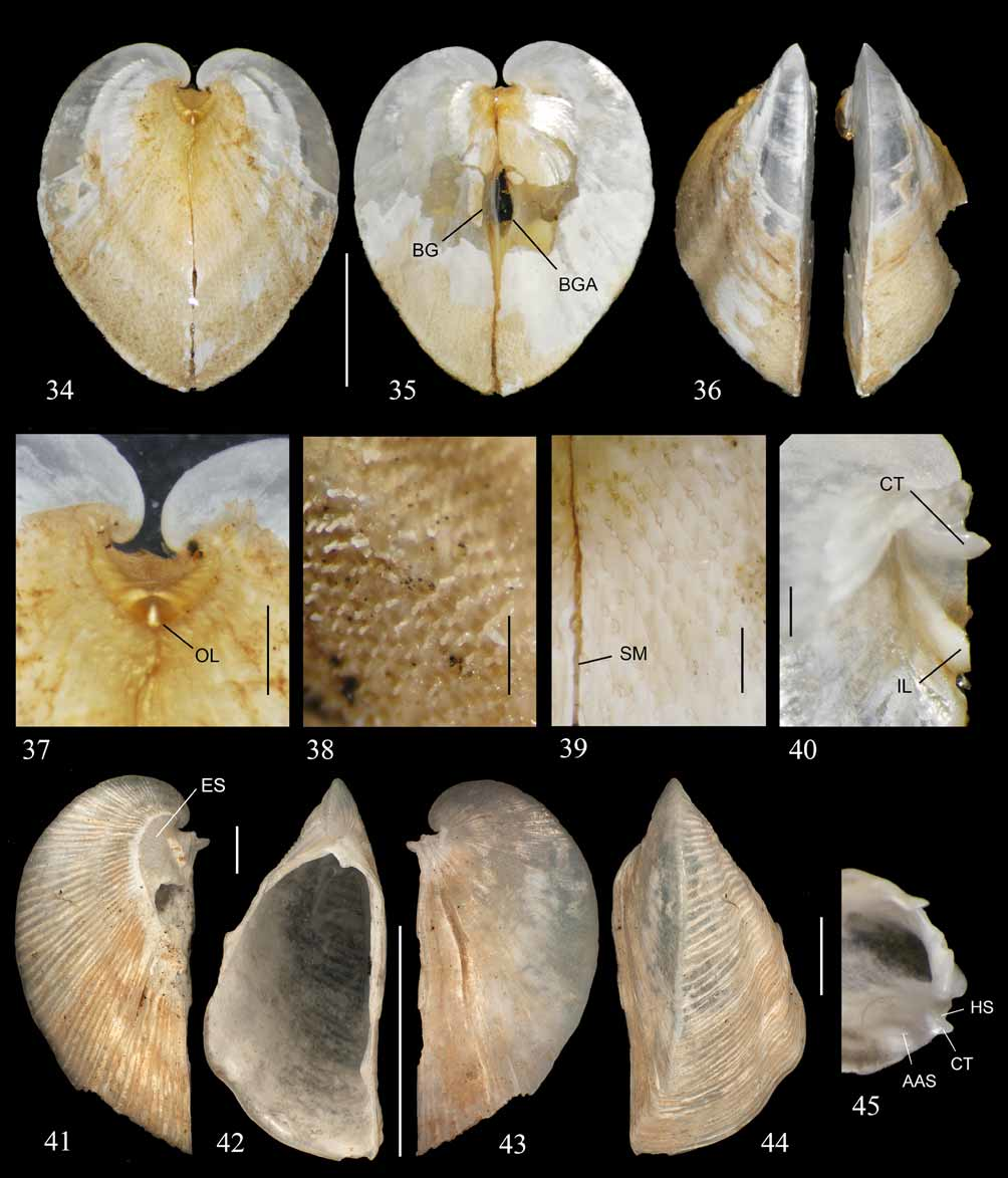
( Figures 34–40 View FIGURES 34 – 45 , 46–58 View FIGURES 46 – 53 View FIGURES 54 – 58 )
Diagnosis. Shell thin, with sculpture of shell pores on external shell layer (and corresponding periostracal spicules); foot digitiform (rather than filiform), large (relative to previous species); byssus narrow; siphonal cowl large, and siphonal tentacles more concentrated ventral to the incurrent siphonal opening; arrangement of siphonal ostia with middle and posterior groups of ostia not converging but forming lines roughly parallel with each other; siphonal ostia distributed as follows: five anterior, four median, and six posterior.
Description. SHELL ( Figures 34–40 View FIGURES 34 – 45 ): General shape and proportions as for genus (above). Shell with minute pores on external layer ( Figure 39 View FIGURES 34 – 45 ) arranged in lines oblique to shell margin ( Figure 39 View FIGURES 34 – 45 , SM). Shell apparently nacreous internally, very thin, equivalve (except for slight anteroposterior overlap at umbo), inequilateral, strongly compressed in anteroposterior direction; lateral outline cardioid ( Figures 34, 35 View FIGURES 34 – 45 ). Moderate byssal gap ( Figure 35 View FIGURES 34 – 45 , BG) in anteroventral position, on central portion of anterior shell surface. Welldeveloped carina separating anterior and posterior shell regions ( Figures 36, 44 View FIGURES 34 – 45 ).
Umbones, ( Figure 37 View FIGURES 34 – 45 ), umbonal cavity, hinge, cardinal-like teeth ( Figure 40 View FIGURES 34 – 45 , CT), hinge sockets, and posterior lateral tooth (present in right valve only) as in previous species.
Ligament, inner ligamental layer ( Figure 40 View FIGURES 34 – 45 , IL), outer ligamental layer ( Figure 37 View FIGURES 34 – 45 , OL) as in previous species, with dorsal periostracum adhering to outer layer.
Anterior adductor muscle scar, posterior adductor muscle scar anterior byssal retractor scar, and posterior byssal retractor scar as in previous species. Pallial line continuous.
Lunule absent. Escutcheon smooth, relatively well-defined, not separated by prominent groove, but distinguishable from remainder of shell by smoother texture and commarginal band of lighter color.
Shell color dull-white. Periostracum yellowish-brown, with hollow spicules ( Figures 38, 39 View FIGURES 34 – 45 ) along lines oblique to the shell margin; spicules coinciding with minute pores on external shell layer.
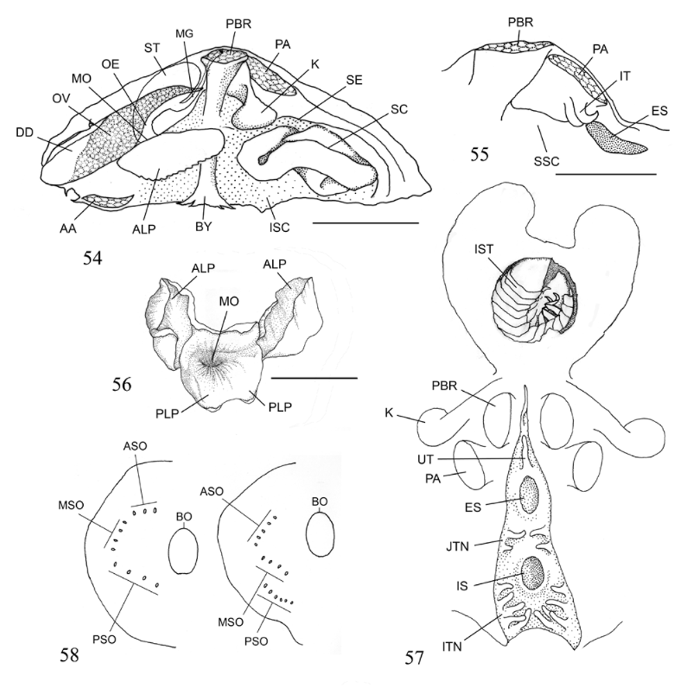
Macroanatomy. MANTLE MARGIN AND SIPHONS: Mantle margins ( Figures 47, 49 View FIGURES 46 – 53 , FM), and siphonal openings as in previous species. Byssal gape ( Figures 35 View FIGURES 34 – 45 , 49 View FIGURES 46 – 53 , BGA) circular to elliptical. Siphons separate; siphonal area well-defined; siphons formed by fusion of inner mantle folds (“ Type A” of Yonge, 1982).
Incurrent siphonal opening ( Figures 46, 47 View FIGURES 46 – 53 , 57 View FIGURES 54 – 58 , IS) and excurrent siphonal opening ( Figures 46, 51 View FIGURES 46 – 53 , 55, 57 View FIGURES 54 – 58 , ES) as in previous species; siphonal cowl ( Figures 47–51 View FIGURES 46 – 53 , 54 View FIGURES 54 – 58 , SC) as in previous species but considerably larger. Base of the incurrent siphon surrounded by 15 simple, tapered tentacles ( Figure 46 View FIGURES 46 – 53 , TN). Large unpaired tentacle ( Figure 46 View FIGURES 46 – 53 , 57 View FIGURES 54 – 58 , UT) as in previous species; following four tentacles (2+2) ( Figure 57 View FIGURES 54 – 58 , JTN) deployed along each side of intersiphonal junction. Remaining ten tentacles (5+5) ( Figure 57 View FIGURES 54 – 58 , ITN) deployed below ventral half of incurrent siphonal opening. Incurrent siphonal opening located almost ventrally, at least twice as wide as excurrent siphonal opening. Relationship between excurrent siphonal opening and angle formed by junction of margins of two valves in posterior direction as in previous species.
Pair of tentacles present internally ( Figures 51 View FIGURES 46 – 53 , 55 View FIGURES 54 – 58 , IT), in supraseptal chamber, between posterior margin of excurrent siphon and posterior adductor muscle.
MANTLE CAVITY: Septum ( Figures 50, 51 View FIGURES 46 – 53 , 54 View FIGURES 54 – 58 , SE), supraseptal (posterior) chamber ( Figure 48 View FIGURES 46 – 53 , 55 View FIGURES 54 – 58 , SSC), and infraseptal (anterior) chamber ( Figures 48 View FIGURES 46 – 53 , 54 View FIGURES 54 – 58 , ISC) as in previous species. Septum attachment to internal shell surface as in previous species.
Septum perforated by byssal opening ( Figure 58 View FIGURES 54 – 58 [right], BO) and by groups of ostia arranged to define three pairs of line segments deployed around byssal gape. Segments containing posterior ( Figures 47, 49, 52 View FIGURES 46 – 53 , 58 View FIGURES 54 – 58 [right], PSO) and middle ( Figures 49, 52 View FIGURES 46 – 53 , 58 View FIGURES 54 – 58 [right], MSO) groups of ostia roughly parallel to one another, each forming ca. 45º angle with sagittal plane; segment defined by anterior group of ostia ( Figures 46, 52 View FIGURES 46 – 53 , 58 View FIGURES 54 – 58 [right], ASO) forming ca. 135º angle with sagittal plane. Ostia distributed as follows: five anterior, four median, and six posterior.
MAJOR SHELL MUSCLES: Relationship between strongly modified shell shape and positioning of adductor muscles as in previous species. Anterior adductor muscles ( Figures 47–50 View FIGURES 46 – 53 , 54 View FIGURES 54 – 58 , AA), posterior adductor muscles ( Figures 46, 48, 50, 51 View FIGURES 46 – 53 , 54, 55, 57 View FIGURES 54 – 58 , PA), posterior byssal retractor muscles ( Figures 46, 48, 50, 51 View FIGURES 46 – 53 , 54, 55, 57 View FIGURES 54 – 58 , PBR), anterior byssal retractor muscles as in previous species. Anterior and lateral septal muscle insertions difficult to observe due to poor specimen preservation. Posterior septal muscle insertions Figure 46 View FIGURES 46 – 53 , PSM) located adjacent to insertion of posterior adductor muscles.
BYSSUS AND FOOT: Byssus ( Figures 47–50 View FIGURES 46 – 53 , 54 View FIGURES 54 – 58 , BY) well developed, elliptical in cross section; byssus of holotype attached to sliver of dense, apparently volcanic, rock. Byssus consisting of consisting of bundle of very fine, fused filaments, originating from collar-like expansion of vestigial foot ( Figures 47, 49, 50, 53 View FIGURES 46 – 53 , F), the latter narrow, digitiform. As in previous species, position and size of vestigial foot indicates that it might never exit infraseptal chamber.
MOUTH AND LABIAL PALPS: Mouth ( Figures 53 View FIGURES 46 – 53 , 54, 56 View FIGURES 54 – 58 , MO) as in previous species; posterior labial palps ( Figure 53 View FIGURES 46 – 53 , 56 View FIGURES 54 – 58 , PLP) reduced to two vestigial projections flush with surrounding surface. Anterior labial palps ( Figure 47, 48, 53 View FIGURES 46 – 53 , 54, 56 View FIGURES 54 – 58 , ALP) very well developed, strongly ridged, pointed distally, potentially expandable in posterodorsal direction.
ALIMENTARY SYSTEM: Esophagus ( Figure 54 View FIGURES 54 – 58 , OE) and stomach ( Figures 46 View FIGURES 46 – 53 , 54 View FIGURES 54 – 58 , ST) generally as in previous species, but stomach more globose. Openings of digestive diverticula situated anteriorly, between esophagus and midgut opening in stomach. Digestive diverticula located in dorsal “horns” ( Figures 46, 48 View FIGURES 46 – 53 , 54 View FIGURES 54 – 58 , DD.) Midgut ( Figure 54 View FIGURES 54 – 58 , MG) and anus as in previous species.
STOMACH CONTENTS: Examination of stomach contents of holotype revealed single specimen, still relatively intact, of possibly unnamed cirolanid isopod (Marilyn Schotte, pers. comm., November 1, 2007); the stomach was greatly distended ( Figure 57 View FIGURES 54 – 58 ). Ms. Schotte could not find any evidence of eyes, ommatidia, or pigment, which indicates that the prey item was an eyeless isopod.
KIDNEYS: Kidneys situated posterodorsally between the remainder of visceral mass and posterior byssal retractor muscle. Kidneys comprising two elongate structures, one on each side of midline of posterior region. Proximal regions of kidney elongate, with distal regions broader, club-shaped ( Figures 46–48 View FIGURES 46 – 53 , 54, 57 View FIGURES 54 – 58 , K).
REPRODUCTIVE SYSTEM: Testes ( Figures 46, 48 View FIGURES 46 – 53 , TE) elongated situated in anteroventral position in relation to ovaries, ovary overlying the testes. Ovaries ( Figures 46, 48 View FIGURES 46 – 53 , 54 View FIGURES 54 – 58 , OV) consisting of pair of elongated sacs symmetrically deployed on each side of midline. Ovaries and testes ventral to digestive diverticula.
Type Material. Holotype, MNHN 20818 (length×height×width [in mm] = 5.20×13.50×11.77), N/ O ALIS, Campagne MUSORSTOM 8, station CP-1112 (beam trawl), P. Bouchet and B. Richer de Forges coll., 0 8 Oct. 1994, from type locality.
Type Locality. Off Republic of Vanuatu, 14°53' S, 167°12' E, 950–961 m, bottom with boulders.
Etymology. From the Latin spectrum, meaning apparition or an evanescent, supernatural image.
Remarks. Dilemma spectralis differs from D. frumarkernorum in having a thinner shell, shell pores (and corresponding periostracal spicules), a filiform (rather than digitiform) foot, a different arrangement of septal ostia, a larger siphonal cowl, and siphonal tentacles more concentrated ventral to the incurrent siphonal opening.

| MNHN |
Museum National d'Histoire Naturelle |
No known copyright restrictions apply. See Agosti, D., Egloff, W., 2009. Taxonomic information exchange and copyright: the Plazi approach. BMC Research Notes 2009, 2:53 for further explanation.
|
Kingdom |
|
|
Phylum |
|
|
Class |
|
|
SuperOrder |
Anomalodesmata |
|
Order |
|
|
Family |
|
|
Genus |