
Placocoris viridis, Mayr, 1864
|
publication ID |
https://doi.org/ 10.11646/zootaxa.4958.1.35 |
|
publication LSID |
lsid:zoobank.org:pub:CC4D6379-ED0B-4166-928F-9416BCB1F5FC |
|
DOI |
https://doi.org/10.5281/zenodo.4716423 |
|
persistent identifier |
https://treatment.plazi.org/id/0A355B65-FF9D-600A-FF08-FBA3FAD7331C |
|
treatment provided by |
Plazi |
|
scientific name |
Placocoris viridis |
| status |
|
Distribution: BRAZIL, ARGENTINA, PERU, PARAGUAY
Placocoris viridis Mayr, 1864:913 .
Mentisa smaragdina Walker, 1868:537 .
Chrysodarecus moneta Breddin, 1903:383 .
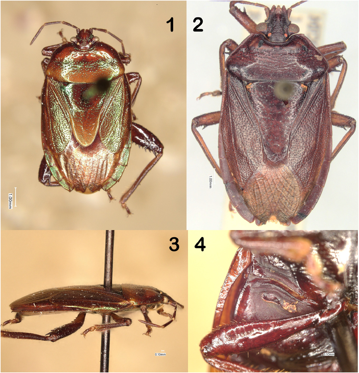
Redescription of Placocoris viridis Mayr. Body elongate-oval, length 10–12 mm, exceedingly dorso-ventrally compressed, flattened ( Fig. 3 View FIGURES 1–4. 1 ). Dorsal surface polished, shiny, coppery with metallic green reflections; venter polished shiny, but without green reflections.
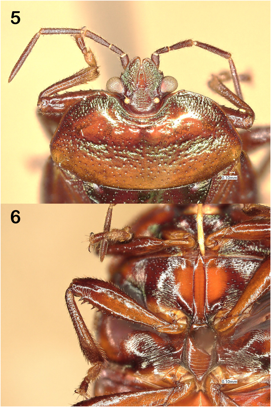
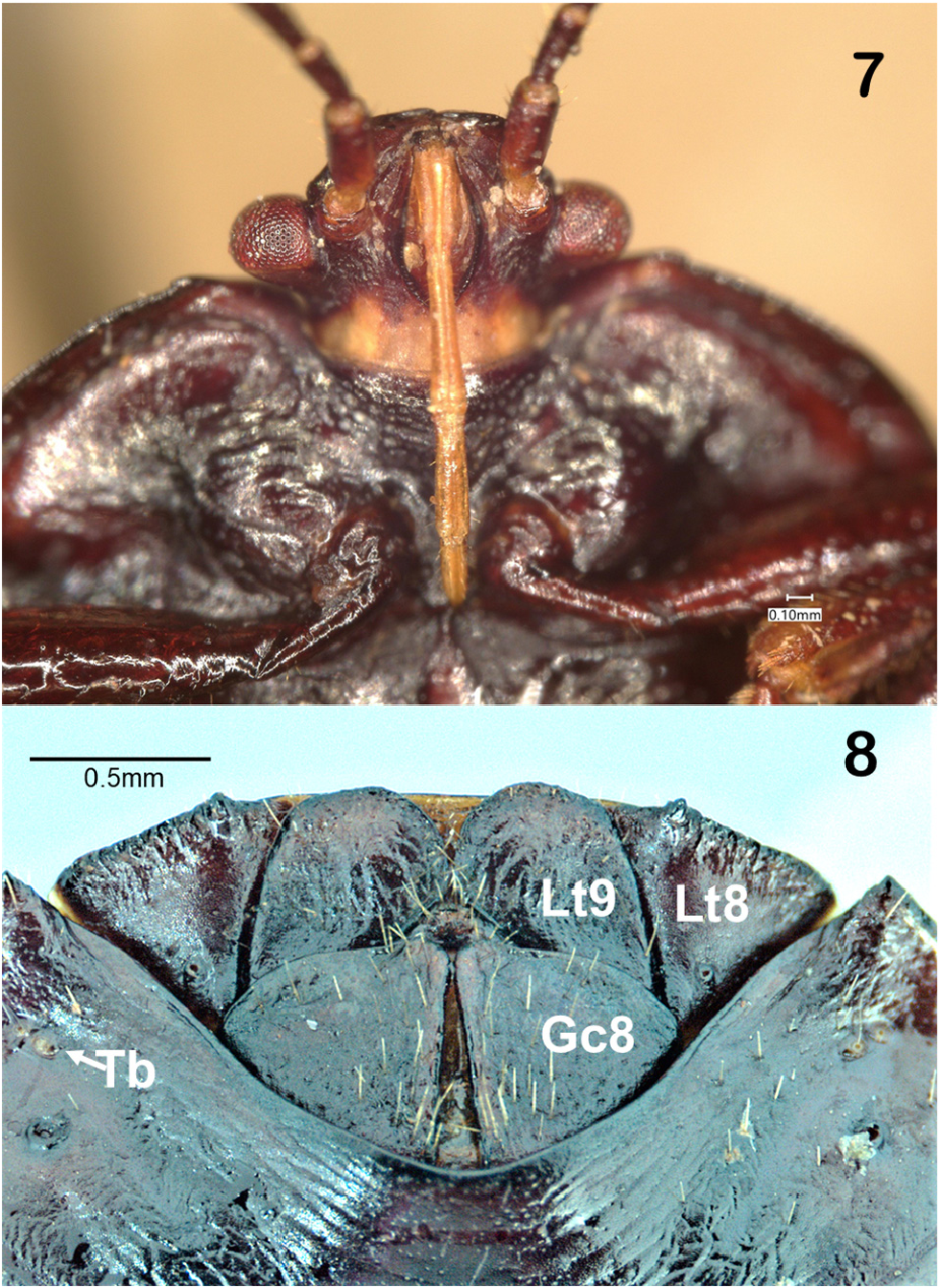
Head short, equal to about 80% of the length of pronotum at midline, trapezoidal in shape; eyes subpedunculate, proximal to cervix ( Fig. 5 View FIGURES 5–6. 5 ). Vertex flat; paraclypei distinctly longer than clypeus and meeting or proximal anteriorly; lateral margins of paraclypei sinuate from dorsal view without anteapical or anteocular processes, apices obtuse. Antennae five-segmented, first segment long, exceeding apex of paraclypeus; antennifers simple. Labium arising anterior to an imaginary line connecting anterior limit of eyes. Rostrum pale, four-segmented without intercalary segment; first segment reposing within and not surpassing the buccula. Buccula low anteriorly, angularly produced medially, low and confluent posteriorly; second rostral segment 2x length of first extending on to prosternum, apex of fourth segment just reaching anterior margin of mesosternum ( Fig. 7 View FIGURES 7–8. 7 ).
Pronotum reniform with anterolateral and posterior margins evenly arcuate, humeri not protruding; anterior margin deeply concave with anterolateral angles weak but situated distinctly laterad of the eyes. Pronotal collar demarcated by an inframarginal groove posterior to the head. Scutellum long, about 1.6 times pronotal length at midline; basal angles devoid of fovea, sides almost straight with constriction at frenal angles nearly obsolete; apex obtuse, broad with width across frenal angles about 70% of basal width; length of postfrenal scutellum subequal to basal portion of scutellum. Corium longer than scutellum, apex arcuate; exocorium broad, about half width of mesocorium; membrane with 9 simple veins, apex coincident with apex of abdomen.
Prosternum weakly tumid anteriorly with elevated keel on posterior half on midline. Mesosternum polished, flat, long and parallel sided, not elevated, coplanar with mesopleura, apex acuminate anteriorly, base truncate posteriorly. Mesosternal evaporatorium extending as a shagreened strip from cleft of mexocoxa along posterior margin to lateralmost angle. Metasternum flat, elongate, ovoid, with evenly curved margins, length about 2.5 x width, coplanar with mesosternum and metapleura; base shallowly, narrowly sulcate for reception of abdominal protuberance ( Fig. 6 View FIGURES 5–6. 5 ). Scent gland orifice situated about half-way along length of an elongated auricle that extends a little more than half-way to the metapleural margin; apex of auricle obtusely truncate and somewhat elevated ( Fig. 4 View FIGURES 1–4. 1 ). Metapleural evaporatorium surrounding the auricle but covering only about a quarter of the metapleural area.
Profemora thickened, apex simple, with a double row of very short, stout, well-spaced, dark tubercles along entire length of inferior surface. Protibia markedly swollen apically with superior surface distinctly, narrowly sulcate. Tubercles on mesofemora similar to arrangement on profemora except those distally are longer, drawn out into curved spines; mesotibia longer than protibiae but not as markedly swollen apically and more broadly sulcate on superior surface. Metafemora long and subclavate, spines arranged in double row on inferior surface, long and curved, becoming longer distally such that length of ultimate pair exceeding width of tibiae. Metatibiae long and distinctly bent at middle. Distal metatarsomere cylindrical on all legs.
Abdominal third urosternite with a small angular protuberance at middle base directed anteriorad in apposition to vertical sulcus on metasternum. Each abdominal sternite is narrowest at the midline except sternite VII which is conspicuously longer at the middle; with IV–VI subequal in width to one another. Spiracular rims darkened. Pair of trichobothria on sternites III–VI situated closer to abdominal margin than to spiracle and somewhat more posterior than spiracle. On segment VII the trichobothrial pair is situated about half-way between the spiracle and the posterolateral angle of the sternite. Four posterior-most connexival segments exposed from above with hemelytra in repose, each connexival angle ending in a minute point. Because of the uniqueness of the specimens the internal reproductive structures were not dissected.
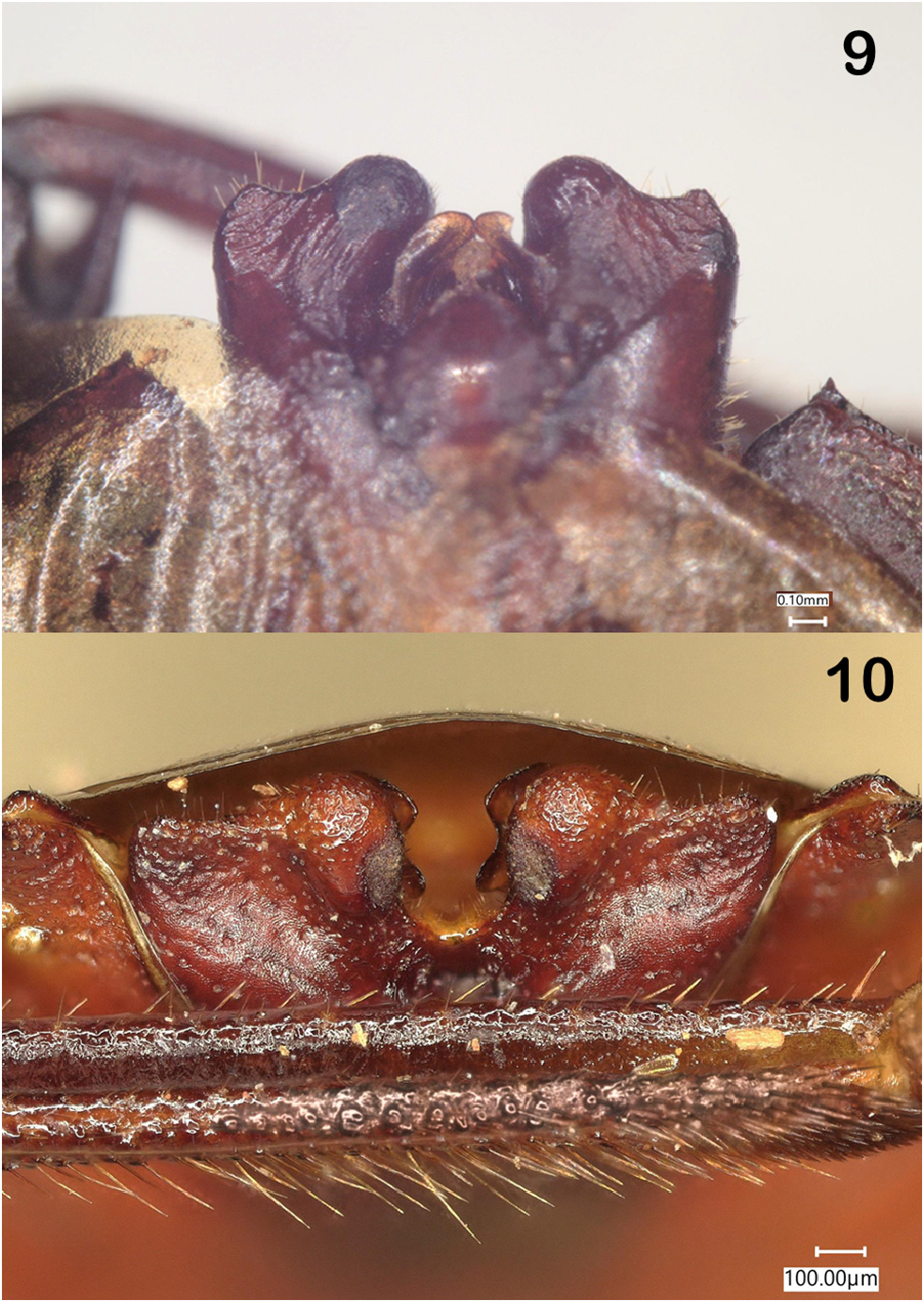
Male Genitalia. The pygophore is heavily sclerotized, dorso-ventrally compressed and seated within abdominal segment VII such that the lumen opens dorsally in reposed position; the posterior surface and the latero-dorsal surfaces are flattened ( Fig. 9 View FIGURES 9–10. 9 ). In ventral view the posterior margin of the pygophore is lobate on each side of the middle, the thickened lobes separated by a deep U-shaped emargination ( Fig. 10 View FIGURES 9–10. 9 ). The proctiger is large, almost filling the pygophoral lumen, suborbicular, about equal in width and length, somewhat angularly produced posteriorly. Parameres foliate, cupped, terminating in two short angular lobes separated by a shallow emargination.
Female Genitalia. The tenth abdominal segment is not visible, covered by the 9 th laterotergites which are broad, rounded in outline posteriorly, contiguous medially, their surface somewhat rough with sparse, coarse, pale setae, the apex only slightly surpassing posterior margin of 8 th tergite. Posterior margin of 8th laterotergite bearing a short tooth-like projection either side of the 9 th laterotergites and with a spiracle near basal angle. Eighth gonocoxites separated at base, contiguous at apex; about as broad as long, posterolateral margin arcuate, medial margin straight, surface flat, bearing scattered coarse, pale, stiff setae ( Fig. 8 View FIGURES 7–8. 7 ).
Material Examined: Male: labeled: (a) “ BRAZIL, Parana, 10 mi. W. Guaraniaçu, 24 FEB 1980, D.B. Thomas Coll. ” (b) “Collected on Bamboo” (c) “ Placocoris viridis Mayr, LHR 80.” Male : labeled: (a) “ PERU: Jajua Prov., Junin Dept. Sani Beni, 840m. ele 16–21 June 1935, Felix Woytkowski.” (b) “ Placocoris viridis Mayr, Det. D.B. Thomas. ” 1 female “ PARAGUAY: Alto Parana Dept., 21–23-X-1989, G. Arriagada. ” Collection of J.E. Eger .
No known copyright restrictions apply. See Agosti, D., Egloff, W., 2009. Taxonomic information exchange and copyright: the Plazi approach. BMC Research Notes 2009, 2:53 for further explanation.
|
Kingdom |
|
|
Phylum |
|
|
Class |
|
|
Order |
|
|
Family |
|
|
Genus |
Placocoris viridis
| Thomas, Donald B. 2021 |
Chrysodarecus moneta
| Breddin, G. 1903: 383 |
Mentisa smaragdina
| Walker, F. 1868: 537 |
Placocoris viridis Mayr, 1864:913
| Mayr, G. L. 1864: 913 |