
Ascidia paulayi, Bonnet, Nadia Yk & Lotufo, Tito Mc, 2015
|
publication ID |
https://doi.org/10.11646/zootaxa.3994.2.8 |
|
publication LSID |
lsid:zoobank.org:pub:6B5BCA21-64BA-4D9B-B8A4-00EE2B7CEE0A |
|
DOI |
https://doi.org/10.5281/zenodo.6120689 |
|
persistent identifier |
https://treatment.plazi.org/id/1C2C805B-0513-4E06-AFEB-F25402438EF7 |
|
taxon LSID |
lsid:zoobank.org:act:1C2C805B-0513-4E06-AFEB-F25402438EF7 |
|
treatment provided by |
Plazi |
|
scientific name |
Ascidia paulayi |
| status |
sp. nov. |
Ascidia paulayi sp. nov.
( Figs. 1–2 View FIGURE 1 View FIGURE 2 )
Ascidia sydneiensis (part): Monniot & Monniot, 1987, morphotype 2, fig. 38.
Material examined. Holotype: FLMNH UF 788-1 ind.; west of Opunohu Bay, Moorea Island, Society Islands, French Polynesia ( 17.487º S; 149.8772º W); 5–7 m depth; fringing reef wall, under rocks and overhangs; 10/xi/ 2009; col.: S. McPherson, T.M.C. Lotufo.
Paratypes: FLMNH UF 787-1 ind.; west of Opunohu Bay, Moorea Island, Society Islands, French Polynesia ( 17.487º S; 149.8772º W); 5–7 m depth; fringing reef wall, under rocks and overhangs; 10/xi/2009; col.: S. McPherson, T.M.C. Lotufo.
FLMNH UF 802-1 ind.; southeast of Moorea Island (near Maatea), Society Islands, French Polynesia ( 17.5749º S; 149.7974º W); 0–3 m depth; sand flat with small rocks on sand, and fringing reef, under and on rocks; 11/xi/2009; col.: S. McPherson, T.M.C. Lotufo, N. Gravier-Bonnet.
Etymology: the species is named after Dr. Gustav Paulay, leader of the team of marine inverebrates of the Moorea Biocode project. Dr. Paulay has coordinated many efforts to study marine invertebrates biodiversity throughout the world’s oceans.
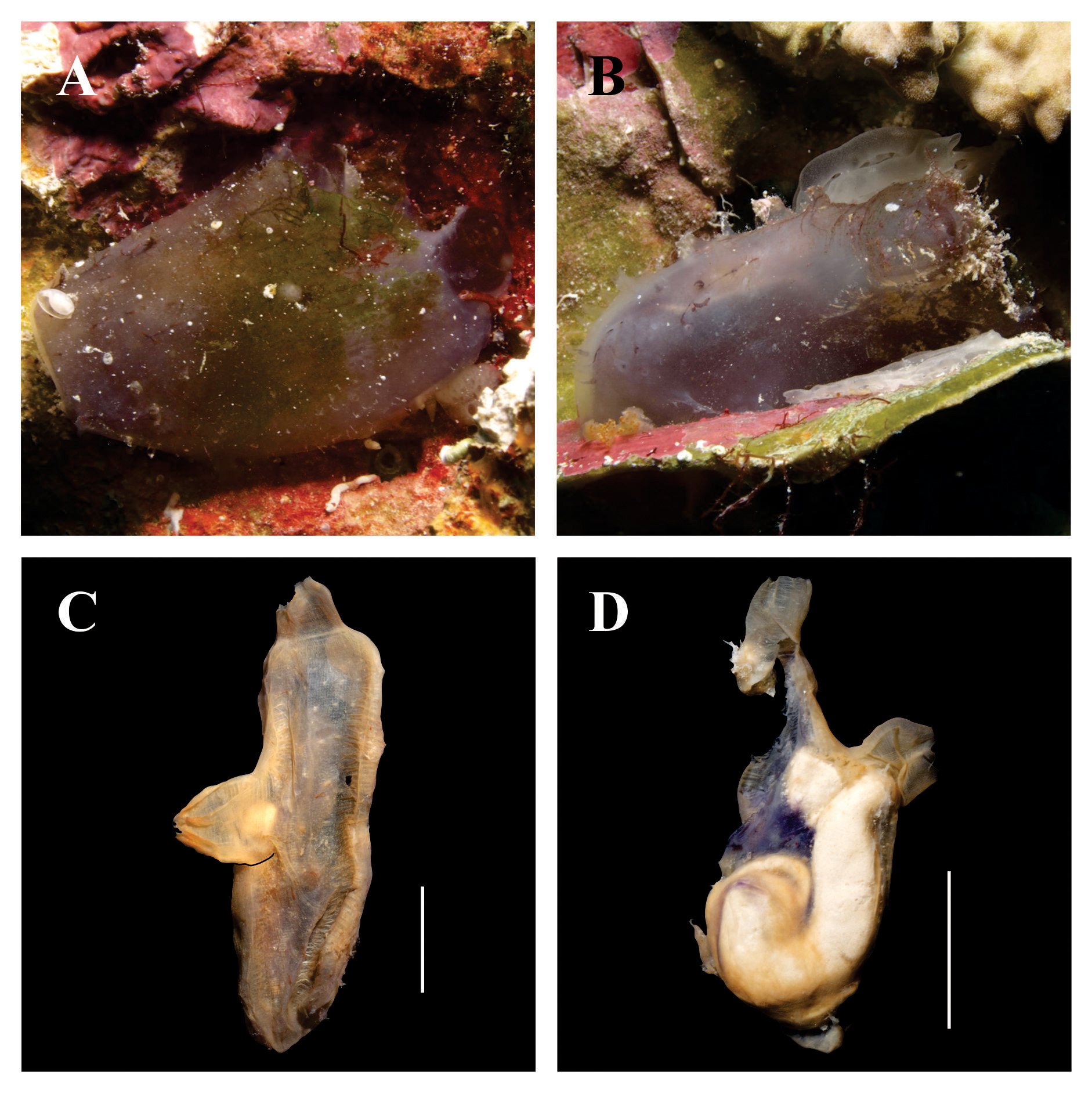
Living animal: individuals up to 4.9 cm in total length, purplish-grey, attached to the substrate by the whole left side of the body. Sand, mud, little calcareous fragments and filamentous algae may be present attached to the free surface ( Figs. 1 View FIGURE 1 A–1B).
Tunic: thin, but firm, 0.7–0.8 mm thick. Translucent and wrinkled after fixation. There are short but thick conical projections, especially on the posterior region of the body ( Figs. 1 View FIGURE 1 A–1B).
Animal without tunic: body oblong or elongated, 17.5–40.3 mm long (from the ring of tentacles to the posterior end) and 11.5–13.0 mm wide. Body wall uncolored. Oral siphon apical, 3.0– 5.2 mm long, and 6–8 fringed lobes on the margin. Atrial siphon displaced posteriorly on the dorsal line, 4.5–16.9 mm from the ring of oral tentacles (approximately ⅓ of the body length); atrial siphon 2.3–8.2 mm long and six fringed lobes on the margin. Ocelli reddish or brownish between the lobes of both siphons.
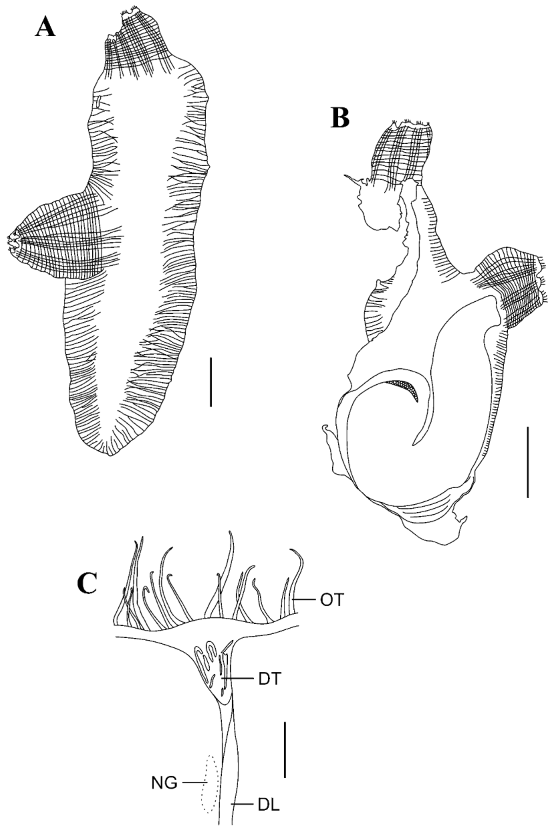
Musculature: bands of parallel fibers along dorsal and ventral margins on the right side of the body are present ( Figs. 1 View FIGURE 1 C–2A); muscle fibers 0.1–0.2 mm thick. On the posterior margin there are few and thinner fibers, almost inconspicuous. On the left side of the body there are short fibers along the dorsal margin ( Figs. 1 View FIGURE 1 D–2B). Longitudinal muscles of the siphons forming bands; these fibers extend for a short distance on both sides of the body. Circular muscle bands of the siphons weak.
Anterior region: oral siphon internal wall usually smooth (only FLMNH UF 787 presents few and sparse papillae). There are 62–92 oral tentacles, with 3–4 different size orders, largest ones with 1.4–2.3 mm in length. Oral tentacles in a thick muscular ring. Prepharyngeal groove double, without projections. Area between the ring of oral tentacles and prepharyngeal groove smooth, 0.3–0.4 mm wide on its whole length. Peritubercular area V– shaped, not extending much farther than the tubercle itself. Aperture of the dorsal tubercle usually convoluted, but in FLMNH UF 802 it is divided into slits ( Fig. 2 View FIGURE 2 C); dorsal tubercle 0.5–2.4 mm anterior-posterior length. Neural ganglion close to the dorsal tubercle (2.6–3.0 mm away).
Pharynx: pharynx wall not pleated. There are 56–61 longitudinal vessels on the right side, 48–60 on the left, 99–146 transverse vessels, and 4–6 stigmata per mesh. Primary papillae simple (straight or curved), but may be bilobed in some parts of the pharynx; papillae with 0.11–0.15 mm in length. Intermediary papillae and parastigmatic vessels are absent. Dorsal lamina doubled anteriorly, on the first 1.2–3.8 mm after the end of the peritubercular area, converging to a single blade with smooth margin ( Fig. 2 View FIGURE 2 C) (FLMNH UF 802 slightly toothed posteriorly due the end of transverse vessels of the pharynx); papillae on the right side of the lamina near to the esophageal aperture are absent. Dorsal lamina passes along the left side of the esophageal aperture and extends to the end of the pharynx. There is a narrow toothed lamina on the right side of the esophageal aperture. Endostyle with smooth margins.
Digestive tract: digestive tube large, situated between the posterior end of the body and the atrial siphon, on the left side of the body ( Figs. 1 View FIGURE 1 D–2B). Stomach elongated with 5–7 internal longitudinal folds. Intestine with both loops (primary and secondary); presence of a sac-like dilation in the descending region; second loop of intestine and rectum also dilated, but not as a sac. Rectum long, in vertical position. Anus with multilobed margin opening anteriorly to the gut loops, close to the atrial siphon. Renal vesicles not observed.
Gonads: ovary ramified, with a central axis inside the primary intestinal loop and branches on the descending region of the intestine and secondary loop. Ovary only conspicuous after removing the pharynx. Oocytes up to 0.23 mm in diameter. Testis not observed. Unfortunately, all samples have some degree of damage in the digestive tube, hindering more accurate details of the gonads.
Distribution. French Polynesia ( Monniot & Monniot 1987, present study).
Remarks. Ascidia paulayi sp. nov. is closely related to Ascidia sydneiensis Stimpson, 1855 due to the presence of finger-like projections in the tunic, the general pattern of body muscles, longitudinal muscle fibers on siphons forming bands, fringed lobes, aperture of the dorsal tubercle convoluted, intestine with a sac-like pouch in the descending region, multilobed anus and ovary ramified ( Kott 1985; Bonnet & Rocha 2011a). However A. sydneiensis has a strongly pleated pharynx, with 6–12 stigmata per mesh, dilation also in the second loop of the intestine, short and narrow rectum, and anus opening posteriorly to the first loop of the intestine ( Kott 1985; Bonnet & Rocha 2011a). Monniot & Monniot (1987) recognized two morphotypes in the samples of A. sydneiensis collected in French Polynesia: 1) similar to those described to Australia ( Kott 1985) and Panama ( Bonnet & Rocha 2011a; Bonnet et al. 2013), and restricted to port zones; 2) similar to the specimens here dissected and found only in natural substrates. Despite Monniot & Monniot (1987) decision of keeping both morphotypes as A. sydneiensis , we think there are characters enough to separate them into two different species. In the study of the ascidians from the western Pacific, A. sydneiensis was reported to Palau, Papua New Guinea and Tonga, with a high intraspecific variability attributed to the different habitats (Monniot & Monniot 2001). Unfortunately there are not descriptions available in the 2001 article, but the plate of the species shows the left side of one individual with the aspect of the digestive tube as the morphotype 1 (p. 308, Fig. 82A), while the figure detailing the gut loop shows the multilobed anus aperture anterior to the top of the intestine (p. 308, Fig. 82D) (Monniot & Monniot 2001). A reexamination of these specimens should be necessary to confirm the identifications.
Three other species of Ascidia have short muscle fibers perpendicular to the margins of the right side of the body, six lobes on the atrial siphon, fringed lobes on both siphons and dilated intestine: A. pacifica Tokioka, 1967 , A. parasamea Kott, 1985 , and A. munda Sluiter, 1898 ( Table 1). Described initially as A. sydneiensis sydneiensis by Tokioka (1953), A. pacifica also presents only 4–5 stigmata per mesh ( Tokioka 1953; Tokioka 1967). However, the tunic covered by minute papillae, aperture of the dorsal tubercle in a simple horseshoe shape, isodiametric intestine, and anus with plain margin and situated on the level of the middle of the digestive tube are characters that distinguish A. pacifica ( Tokioka 1953; Tokioka 1967; Monniot & Monniot 1987).
Similar to Ascidia paulayi sp. nov., A. parasamea also has ramified ovary; the differences are the smooth tunic, up to 12 stigmata per mesh in the pharynx, anus with indented border (not lobed) and opening posteriorly to the first intestinal loop ( Kott 1985). Ascidia munda also presents longitudinal fibers on siphons arranged in bands, dorsal tubercle with a U-shaped aperture with horns irregularly turned in, and around 80 oral tentacles ( Kott 1985; Monniot 1987); the presence of pleats in the pharynx, up to 10 stigmata per mesh, smooth or indented (not lobes) anal border and anus opening posteriorly to the first intestinal loop ( Kott 1985; Monniot 1987) differentiate A. munda and Ascidia paulayi sp. nov.
The peculiar shape of the digestive tube of Ascidia paulayi sp. nov. (descending region of the intestine with sac-like dilation, large rectum and anal aperture anterior to the first intestinal loop) was only described for A. ornata Monniot & Monniot, 2001 , a species from Philippines. However A. ornata may be distinguished from the present species due to the presence of 20 lobes in the oral siphon, a net of muscle fibers on the right side of the body, papillae in the area between the ciliated groove and the ring of oral tentacles, 6–10 stigmata per mesh in the pharynx, stomach almost vertical, anus with smooth border and lobed ovary (Monniot & Monniot 2001).
1. Projections on the surface of the tunic: A. Absent; F. Finger-like; P. Papillae 2. Number of lobes on the oral siphon
3. Lobes on siphons: S. Smooth; F. Fringed
4. Number of oral tentacles
5. Aspect of the dorsal tubercle: U. Horseshoe simple; I. Irregular (convoluted or numerous slits) 6. Number of longitudinal vessels in each side of the pharynx 7. Number of stigmata per mesh
8. Dilatation of the intestine: A. Absent; NS. Not sac-like; S. Sac-like pouch 9. Anus: S. Smooth; I. Indented; B. Bilobed; M. Multilobed 10. Anal position: A. Anterior to the first intestinal loop; P. Posterior to the first intestinal loop 11. Ovary: R. Ramified; L. Lobed
1 2 3 4 5 6 7 8 9 10 11 Species References
A 6–8 S, F 80 I, U 40– 50 5–10 S, NS S, I P R, L A. munda Sluiter, 1898 9, 10, 13
A 7 F 20 + 2 or 3 I? 12 S I P R A. parasamea Kott, 1985 9 smaller
A 8 F 123–340 U 29–80 5–7 A S, B P R A. cf. multitentaculata 8, 14
F?? 400 U? 4–5 A? P? A. multitentaculata Hartmeyer, 4 1912
? 10 S 200 U? 2–3 A S P R A. nerea Kott, 1985 9
F? S 100 U? 4–8 S S P L A. subterranea Kneer et al., 2013 15
F 6–10 F 35–290 I, U 37– 70 6–12 S M P R A. sydneiensis Stimpson, 1855 1, 9, 13, 14
P 6–8 F 62–120 I, U 48– 61 4–6 S M A R A. paulayi sp. nov. 11, 16
P 10? 48–120 U 32– 50 4–5 A S P? A. pacifica Tokioka, 1967 6, 7
P 8 S 60–147 U 34– 56 3–5 NS S P R A. scalariforme Bonnet & Rocha , 14 2011
P?? 60 U?? A B P L A. canaliculata Heller, 1878 * 2
? 8 F 173–283 U 32– 51 6–9 S S P R A. cf. canaliculata 5, 12, pers. obs.
F 8? 60 U? 3 S? P? A. cf. canaliculata 3
References: 1. Stimpson 1855; 2. Heller 1878; 3. Sluiter 1898; 4. Hartmeyer 1912; 5. Michaelsen 1923; 6. Tokioka 1953; 7. Tokioka 1967; 8. Millar 1977; 9. Kott 1985; 10.
Monniot 1987; 11. Monniot & Monniot 1987; 12. Monniot et al. 2001; 13. Bonnet & Rocha 2011a; 14. Bonnet & Rocha 2011b; 15. Kneer et al. 2013; 16. presente study.
Digestive tract and gonads as A. mentula by Ärnbäck (1934) and Millar (1966)
| FLMNH |
Florida Museum of Natural History |
No known copyright restrictions apply. See Agosti, D., Egloff, W., 2009. Taxonomic information exchange and copyright: the Plazi approach. BMC Research Notes 2009, 2:53 for further explanation.
|
Kingdom |
|
|
Phylum |
|
|
Class |
|
|
Order |
|
|
Family |
|
|
Genus |