
Antennoseius ( Antennoseius ) pannonicus Willmann, 1951
|
publication ID |
https://doi.org/10.1051/acarologia/20174159 |
|
DOI |
https://doi.org/10.5281/zenodo.4697138 |
|
persistent identifier |
https://treatment.plazi.org/id/0A7DEE5F-060F-FF87-FF58-FBD5FF304364 |
|
treatment provided by |
Carolina |
|
scientific name |
Antennoseius ( Antennoseius ) pannonicus Willmann, 1951 |
| status |
|
Antennoseius ( Antennoseius) pannonicus Willmann, 1951 View in CoL
( Figures 1-4 View FIGURE View FIGURE View FIGURE View FIGURE , 8A View FIGURE )
Antennoseius pannonicus Willmann, 1951: 109 View in CoL . Antennoseius pannonicus View in CoL . Athias-Henriot, 1961: 461; Ryke, 1962: 662; Karg, 1971: 298; 1977: 4; 1993: 305; Farrier & Hennessey, 1993: 23.
Antennoseius ( Antennoseius) pannonicus View in CoL . Bregetova, 1977: 248; Beaulieu et al., 2008: 47; Lindquist & Moraza, 2009: 34; Moraes et al., 2016: 73 View Cited Treatment .
Female — five specimens measured.
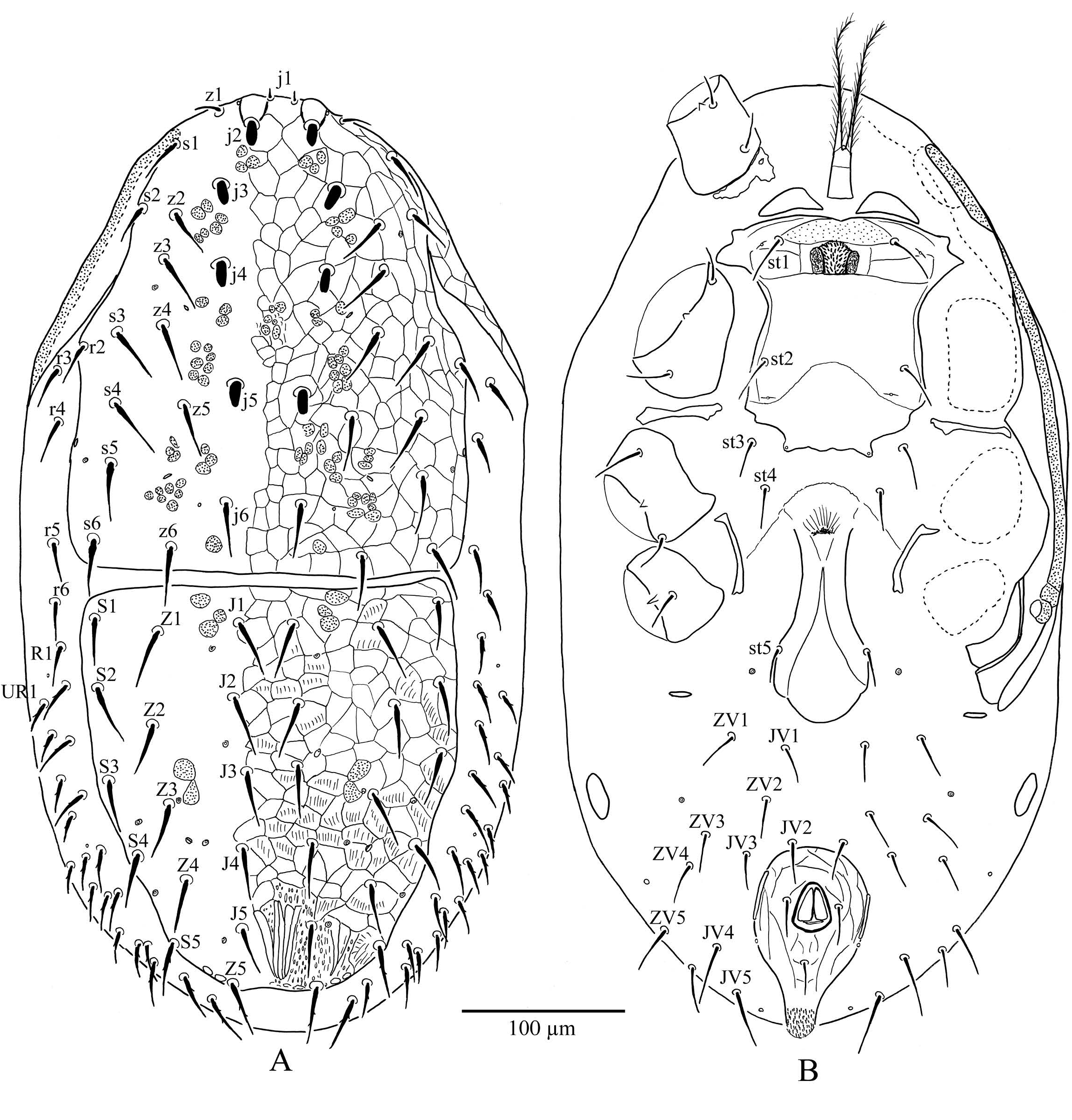
Dorsal idiosoma ( Figure 1A View FIGURE ) — Idiosoma oval, 565 (530 – 590) long; dorsal shield divided and reticulate over entire surface; podonotal shield 294 (288 – 300) long and 240 (238 – 243) wide at j5 level, with 19 pairs of dorsal setae ( r2 seta on the shield) and 7 pairs of pore-like structures (with no distinction made between poroids and glandular pores); j1 short 10 (9-10), j2, j3, j4 and j5 modified, short and spinelike 15 – 18, z1 20 – 21; opisthonotal shield 249 (235 – 263) long and 233 (230 – 238) wide at J2 level, with 15 pairs of dorsal setae and 8 pairs of pore-like structures; J2 35 (34 – 38), Z5 35 (32 – 38); lateral soft integument with 4 pairs of r setae ( r3-6), r3 27 (25 – 28) and 21 – 22 pairs of R – UR setae, dorsal idiosomal setae smooth, except Z5 and most R – UR setae are slightly barbed.
Peritreme — Extending anteriorly to level of seta s1, at venter, anterior to coxa I ( Figures 1A & 1B View FIGURE ). Peritrematal-exopodal plate nearly smooth with three to four longitudinal lines near posterior end of stigma, widened and notched posteriorly; pores on peritrematal-exopodal plate not visible.
Ventral idiosoma ( Figure 1B View FIGURE ) — All ventral setae smooth; tritosternum 108 (105 – 110) long with paired laciniae, free for about three-fourths of total length and pilose, subrectangular base 16 – 18 long and 12 – 13 wide medially; two pairs of subtriangular sclerotized presternal plates; sternal shield 145 (138 – 149) long along midline, 111 (110 – 112) wide at st2 level; with 2 pairs of sternal setae ( st3 off the shield) and three pairs of lyrifissures; st1 35 (33 – 38) long; with a prominent brownish crown-shape configuration between st1 setae, posterior part of sternal shield between lyrifissures iv2 and iv3 weakly sclerotized and laterally eroded; epigynal shield 58 (55 – 60) at greatest width, evenly rounded posteriorly with setae st5 on lateral margins; paragenital poroids iv5 on soft cuticle; endopodal plates weakly developed (except for region between coxae I and II, where fused with sternal shield), posterior section represented by one pair of strips between coxae III and IV parallel to epyginal shield and the other pairs positioned transversely between coxae II and III; anal shield weakly reticulate and pearshaped 109 (102 – 115) long and 69 (65 – 73) of greatest width, with three circumanal setae and gland pores gv3 on shield margin; ten pairs of setae ( JV1- 5 and ZV1-5) on soft cuticle laterad of anal shield; with two pairs of metapodal plates, the smaller pair posteriad of peritrematal plate, the larger pair oval (about 28x10).
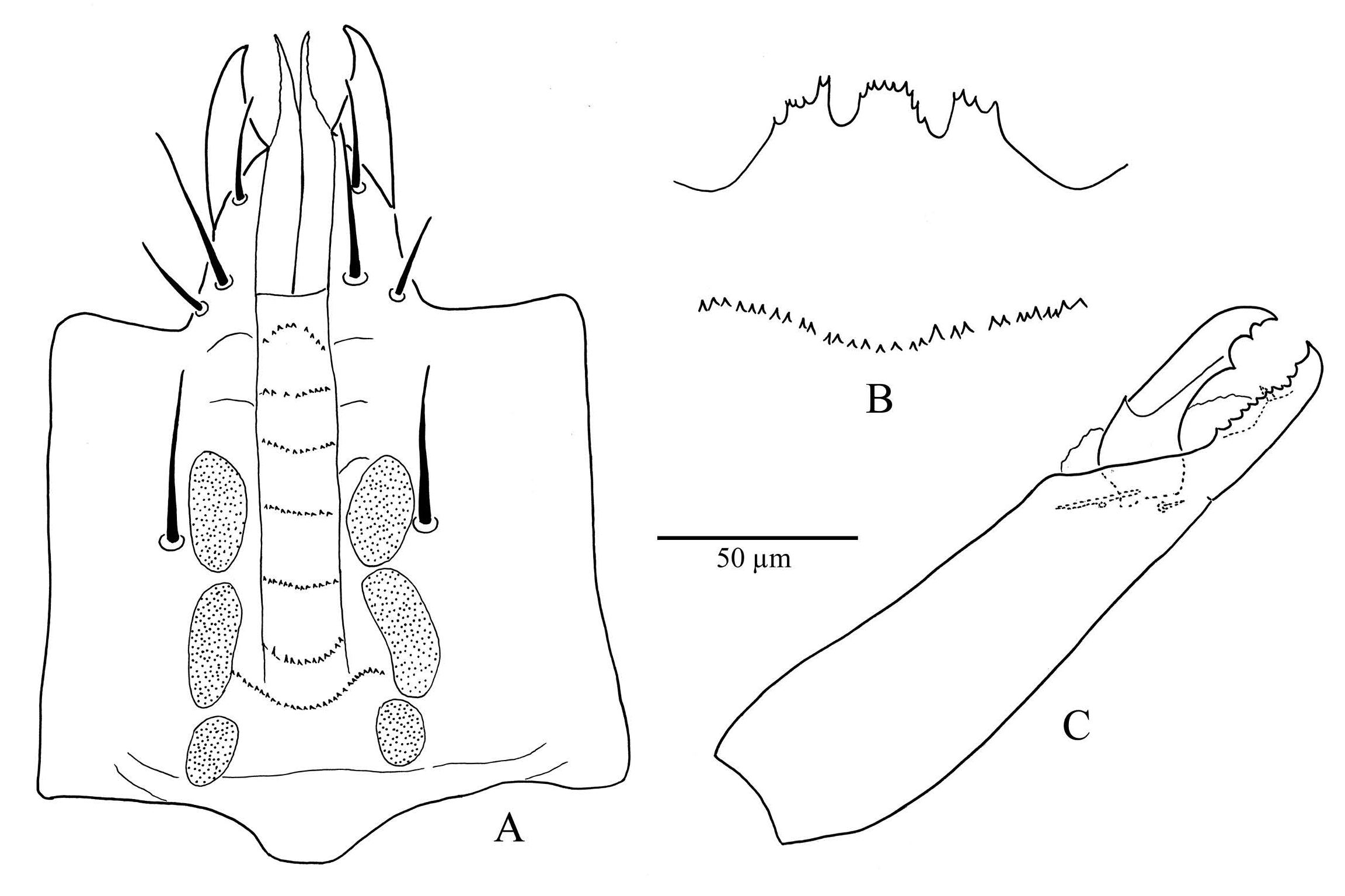
Gnathosoma — Tectum ( Figure 2B View FIGURE ) with anterior margin serrate and with three short projections, dorsal surface with a transverse line of denticles; deutosternum with a smooth transverse line between h3 followed by 7 denticulate transverse lines, denticulate lines 2 – 6 with 12 – 14 denticles, line 7 with 9 – 10 denticles and curved, proximalmost line longer with 25 – 30 denticles ( Figure 2A View FIGURE ); palpcoxal setae ( pc) 27 – 29, hypostomal setae h1 25 – 26, h2 10 – 12, and h3 26 – 27, all smooth; corniculi horn-like; internal malae slightly fimbriated laterally; fixed cheliceral digit with setiform pilus dentilis and a row of about 10 teeth, movable digit tridentate ( Figure 2C View FIGURE ); palp apotele two-tined.
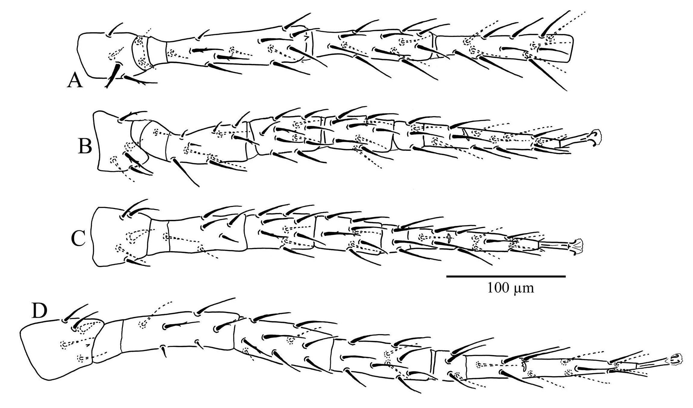
Legs ( Figure 3 View FIGURE ) — Lengths, Leg I 607 (600 – 610), leg II 420 (410 – 430), leg III 410 (400 – 420), leg IV 557 (550 – 560); most of leg setae smooth except some setae lightly barbed, dorsal seta of trochanter I slightly thickened and barbed, ventral setae of trochanters I-IV slightly thickened; setation of legs I – II – III – IV: coxae 2 – 2 – 2 – 1, trochanters 6 – 5 – 5 – 5, femora 12 – 11 – 6 – 6, genua 13 (2 3/1 3/2 2) – 11(2 3/1 2/1 2) – 9 (2 2/1 2/1 1) – 9 (2 2/1 3/0 1), tibiae 13 (2 3/1 3/2 2) – 10 (2 3/1 2/1 1) – 8 (2 2/1 1/1 1) – 10 (2 2/1 2/1 2), tarsus II-IV 3 3/2 3/2 3+ mv,md.
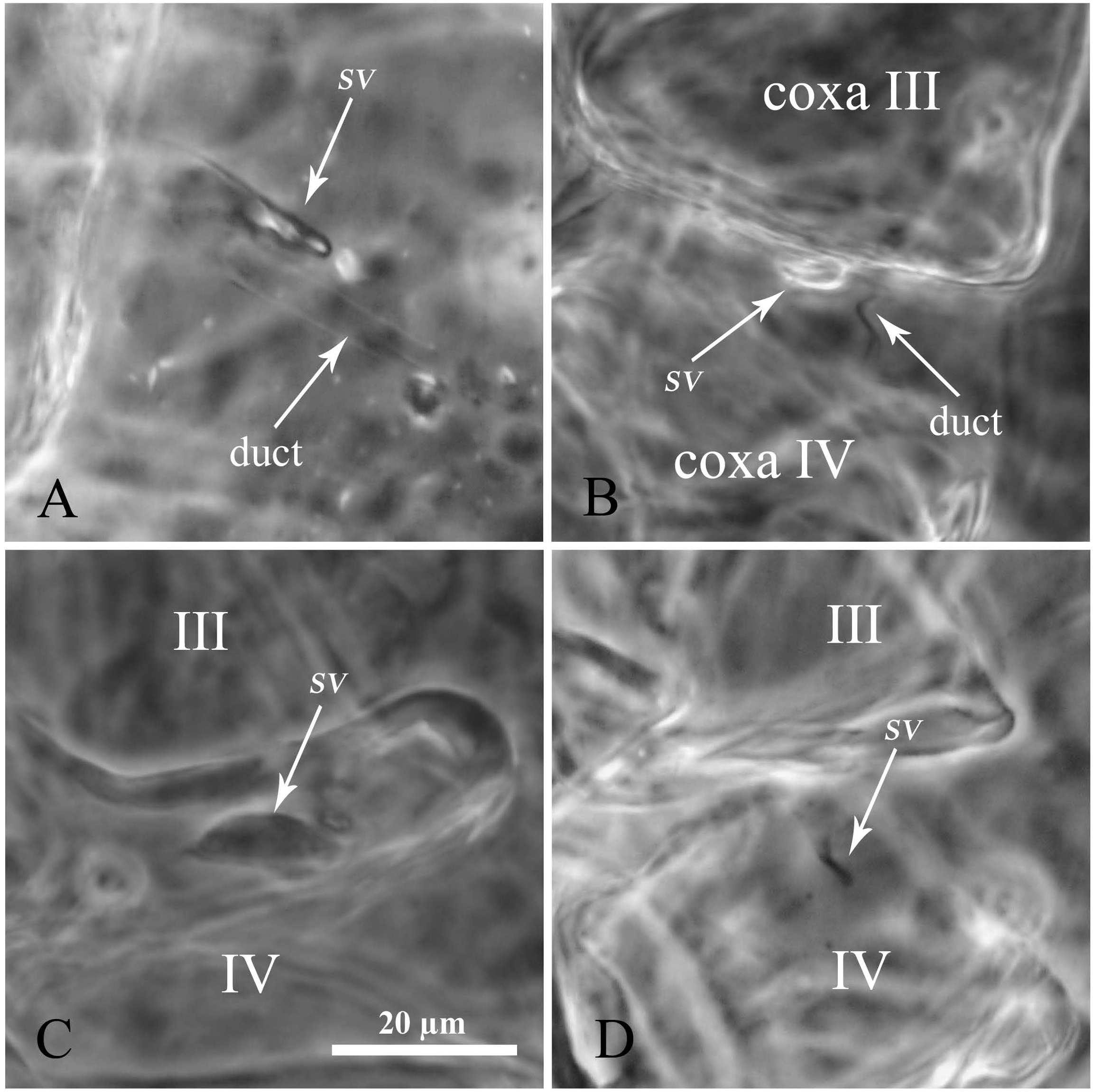
Spermathecal Apparatus — This species has apparently type A complex (laelapid) of sperm access system ( Figure 8A View FIGURE ); sacculus vestibules fingershaped with relatively wider duct, situated between coxae III and IV .
Male — Not phoretic and not found.
Specimens examined — 23 June 2015: 8♀♀ on Harpalus rufipes (Degeer) ; 20♀♀ on Poecilus cupreus (Linne) ; 3♀♀ on Pterostichus madidus (Fabricius) ; 23 Sep. 2015: 3♀♀ on Calathus fuscipes (Goeze) ; 12♀♀ on Harpalus calceatus (Duftschmid) ; 1♀ on Nebria salina Fairmaire et Laboulbene ; 18♀♀ on Pterostichus madidus (Fabricius) ; 3♀♀ on Scybalicus oblongiusculus (Dejean) ; 04 Aug. 2015: 3♀♀ on Harpalus distinguendus (Duftschmid) ; 1♀ on Pterostichus melanarius (Illiger) , all collected by Bogdan Dehelean in Villeneuve de Mezin, Lot-et-Garonne, South West France .
Remarks — The prominent brownish crownshape configuration on the sternal shield of A. pannonicus is a unique feature that allows identification of this species by just using a dissecting microscope. Descriptions of legs, tectum and hypostome, which were lacking in the original description by Willmann (1951) are provided here. Figure 4 View FIGURE shows the position of mites under the elytra of a carabid beetle.

No known copyright restrictions apply. See Agosti, D., Egloff, W., 2009. Taxonomic information exchange and copyright: the Plazi approach. BMC Research Notes 2009, 2:53 for further explanation.
|
Kingdom |
|
|
Phylum |
|
|
Class |
|
|
Order |
|
|
Family |
|
|
Genus |
Antennoseius ( Antennoseius ) pannonicus Willmann, 1951
| Faraji, F., Dehelean, S. - B., Vuyk, M. & Bakker, F. 2017 |
Antennoseius ( Antennoseius ) pannonicus
| Moraes G. J. & Britto E. P. J. & Mineiro J. L. de & Halliday B. 2016: 73 |
| Lindquist E. E. & Moraza M. L. 2009: 34 |
| Beaulieu F. & DechOEne A. D. & Walter D. E. 2008: 47 |
| Bregetova N. G. 1977: 248 |
Antennoseius pannonicus
| Karg W. 1993: 305 |
| Farrier M. H. & Hennessey M. K. 1993: 23 |
| Karg W. 1977: 4 |
| Karg W. 1971: 298 |
| Ryke P. A. J. 1962: 662 |
| Athias-Henriot C. 1961: 461 |
| Willmann C. 1951: 109 |