
Pseudosinella argentea Folsom, 1902
|
publication ID |
https://doi.org/10.5281/zenodo.275457 |
|
DOI |
https://doi.org/10.5281/zenodo.6208548 |
|
persistent identifier |
https://treatment.plazi.org/id/0B21878A-636B-FD1F-F7EF-C970FB6B8EC2 |
|
treatment provided by |
Plazi (2016-04-08 05:23:07, last updated 2022-02-21 18:04:21) |
|
scientific name |
Pseudosinella argentea Folsom, 1902 |
| status |
|
Pseudosinella argentea Folsom, 1902
Figs 35–54 View FIGURES 35 – 44 View FIGURES 45 – 54 , Table 5 View TABLE 5
Material Examined. PENDLETON Co., Quarry Cave, 20 May 2006, 2 on one slide and 3 in alcohol; Cedar Cave, 15 May 2006, 1 on slide; Hoffman Pit, 20 May 2006, 1 on slide, 3 in alcohol; MONOGALIA Co., Maiden Run # 1 Cave, 12 June 2006, 1 on slide.
Descriptive notes. Length to 2.5mm; color in alcohol white, without trace of pigment, or just a wash of light blue pigment near base of antennae. Ant. 4 subapical sense organ clavate, or appearing capitate in some angles ( Fig. 35 View FIGURES 35 – 44 ). Ant. 3 sense organ formed by two enlarged sensilla ( Fig. 36 View FIGURES 35 – 44 ). Dorsal head chaetotaxy ( Fig. 37 View FIGURES 35 – 44 ) with 7–9 macrosetae along base of antennae; A0, A 2, and A 3 macrosetae; Pa 5 a microseta; A 1 and M0 ciliate microsetae, all other dorsal microsetae smooth; seta S0 anterior to S 3; M 1 /S 1 near M 2. Eyes absent. Prelabral setae ciliate, all labral setae smooth. Labral intrusion and papillae as in Fig. 39 View FIGURES 35 – 44 . Pleural fold setae ciliate; peristomal setae pss0 normal ciliate, pss 1–2 ciliate and bothriotricha-like. Ungulum of maxilla with 4 teeth. Lateral process of labial papilla E slightly curved dorsally and barely reaching papillar tip. Proximal seta of labial palp with seta Y subequal to somewhat longer than seta Z. Ventral head chaetotaxy as Fig. 38 View FIGURES 35 – 44 : labial chaetotaxy M 1 M 2 rEL 1 L 2 A 1–5, microseta r smooth and thin walled; post-labial chaetotaxy includes 4 + 4 ciliate setae along ventral groove, and columns C, E, L and O with 2, 2, 3 and 2 setae respectively; L 2 and O 1 modified into short, ciliate and sharply acuminate microsetae. Inner macrochaetotaxy of the body 00/ 0100+ 2.
argentea sensu Christiansen & Bellinger (1998) .
1 Original description (Christiansen & Bellinger 1986) mentions 6–7 setae but Figure 46 View FIGURES 45 – 54 shows 9.
Abd. 1 with setae in linear arrangement, a 6 present. Chaetotaxy of Abd. 2 as in Fig. 42 View FIGURES 35 – 44 ; a 2, a 6 and all supplementary setae of bothriotrichal complexes fan-shaped; a 2 p and a 3 more strongly ciliate than others; a 3 external and reaching tip of as; as reaches the socket of m 3 in some individuals, but not in others; m 3 e not reaching socket of m 3; supplementary seta m 4 i ciliate, to weakly fan-shaped (the presence of microsetae m 4 i is often difficult to ascertain because the surrounding fan-shape setae tend to obscure m 4 i ’s socket); m 5 a normal macroseta; p 5 p a short, ciliate mesoseta. The male from Quarry Cave has supernumerary setae associated with seta p 5 p on Abd. 2 and m 4 i appears to be displaced anteriorly towards a 5, but only on one side of the body (arrows on Fig. 44 View FIGURES 35 – 44 ). Abd. 3 ( Fig. 45 View FIGURES 45 – 54 ) with a 2, a 6, am 6 and all supplementary setae associated with bothriotrichal complexes fan-shaped, all other setae weakly ciliate, but appearing smooth at low magnification; a 3 not reaching as; as about 0.5x the length of m 3 and reaching base of m 3; d 2 present; a 7, m 7 and p 7 normal microsetae, m 7 a a long and slender acuminate macroseta, a 7 inserted anterior and close to am 6, m 7 posterior to p 6. Abd. 4 bothriotrichal complex as in Fig. 46 View FIGURES 45 – 54 : all supplementary setae anterior to T 2, including seta s, fan-shaped; C 1 p and T 3 weakly ciliate (appearing smooth at low magnifications); tip of T 3 reaching base of D 1 p; D 1 p posterior to T 3 and reaching the base of Pe; Pe and Pi fan-shaped; the male from Quarry Cave carries an asymmetric supplementary seta between Pe and T 5 (arrow in Fig. 46 View FIGURES 45 – 54 ). General chaetotaxy of Abd. 4 ( Fig. 47 View FIGURES 45 – 54 ) with macrosetae B 5, B 6, T 6, T 7, D 2 D 3, E 2, E 3, F 1, F 3; E 1 and F 2, microsetae; macroseta B 5 on line drawn between A 5 and C 2; F 2 closer to E 3 than E 2; microseta posterior to E 3 absent. Abd. 4 usually with 1 + 1 posterior setae, but varying among some caves: specimens from Quarry Cave, Hoffman’s Pit and Cedar Cave, and Maiden Cave have 2 + 2, 3 + 3 and 4 + 4 posterior setae, respectively. Trochanteral organ with 14–15 setae. Metathoracic femora with two outstanding acuminate macrosetae inserted near basal third of the segment ( Fig. 48 View FIGURES 45 – 54 ). Tenet hair as long as unguiculus, acuminate or truncate, varying according to locality. The two individuals from Quarry Cave, and the individual from Hoffman’s Pit have acuminate tenet hairs on the pro- and mesothoracic legs ( Fig. 49 View FIGURES 45 – 54 ), but truncate on metathoracic legs ( Fig. 52 View FIGURES 45 – 54 ); the individual from Cedar Cave has all tenent hairs truncate, and the individual from Maiden Run Cave has all tenent hairs acuminate. Unguiculus lanceolate with indistinct serrations on posterior lamella, all other lamellae smooth. Unguis with three inner teeth ( Figs. 49, 52 View FIGURES 45 – 54 ): basal pair unequal in size ( Fig. 51 View FIGURES 45 – 54 ), the difference in size between teeth being more prominent on metathoracic legs; unpaired tooth prominent and closer to ungual tip than base. Outer ungual teeth ( Fig. 50 View FIGURES 45 – 54 ) short, closer to ungual base on third pair than on first two pairs of legs (cf. Figs. 49, 52 View FIGURES 45 – 54 ). Collophore with 8 + 8 disto-lateral setae, four ciliate and four smooth; anterior face with variable number of setae, but generally arranged as in Fig. 54 View FIGURES 45 – 54 ; posterior face ( Fig. 53 View FIGURES 45 – 54 ) with 5 + 5 proximal and nine distal setae. Manubrial plate ( Fig. 43 View FIGURES 35 – 44 ) with six outer and two inner setae separated by two pseudopores. Mucro ( Fig. 41 View FIGURES 35 – 44 ) normal, with teeth subequal; mucronal spine with basal swelling.
Remarks. These specimens belong to the P. argentea complex, although specific determination remains ambiguous and is based on the preponderance of similarity with that apparently very variable species. The material from West Virginia keys out to P. flatua Christiansen & Bellinger, 1996 in Christiansen & Bellinger (1998) by the presence of a ciliate seta a 2 p on Abd. 2, but differs from that species in many chaetotacic characters of the labrum, labium, postlabium and ventral tube ( Table 5 View TABLE 5 ). The variation in tenent hair shape and number of posterior setae on Abd. 4 suggest that these samples may include more than one species, but as with other cave forms, the limited material available makes a final determination difficult.
Christiansen K. & Bellinger P. (1996) Cave Pseudosinella and Oncopodura new to science. Journal of Caves and Karst Studies, 58, 38 - 53.
Christiansen K. & Bellinger P. (1998) The Collembola of North America north of the Rio Grande; A taxonomic analysis. Grinnell College, Grinnell, Iowa, 1518 pp.
Folsom, J. W. (1902) Collembola from the grave. Psyche, 9, 363 - 367.
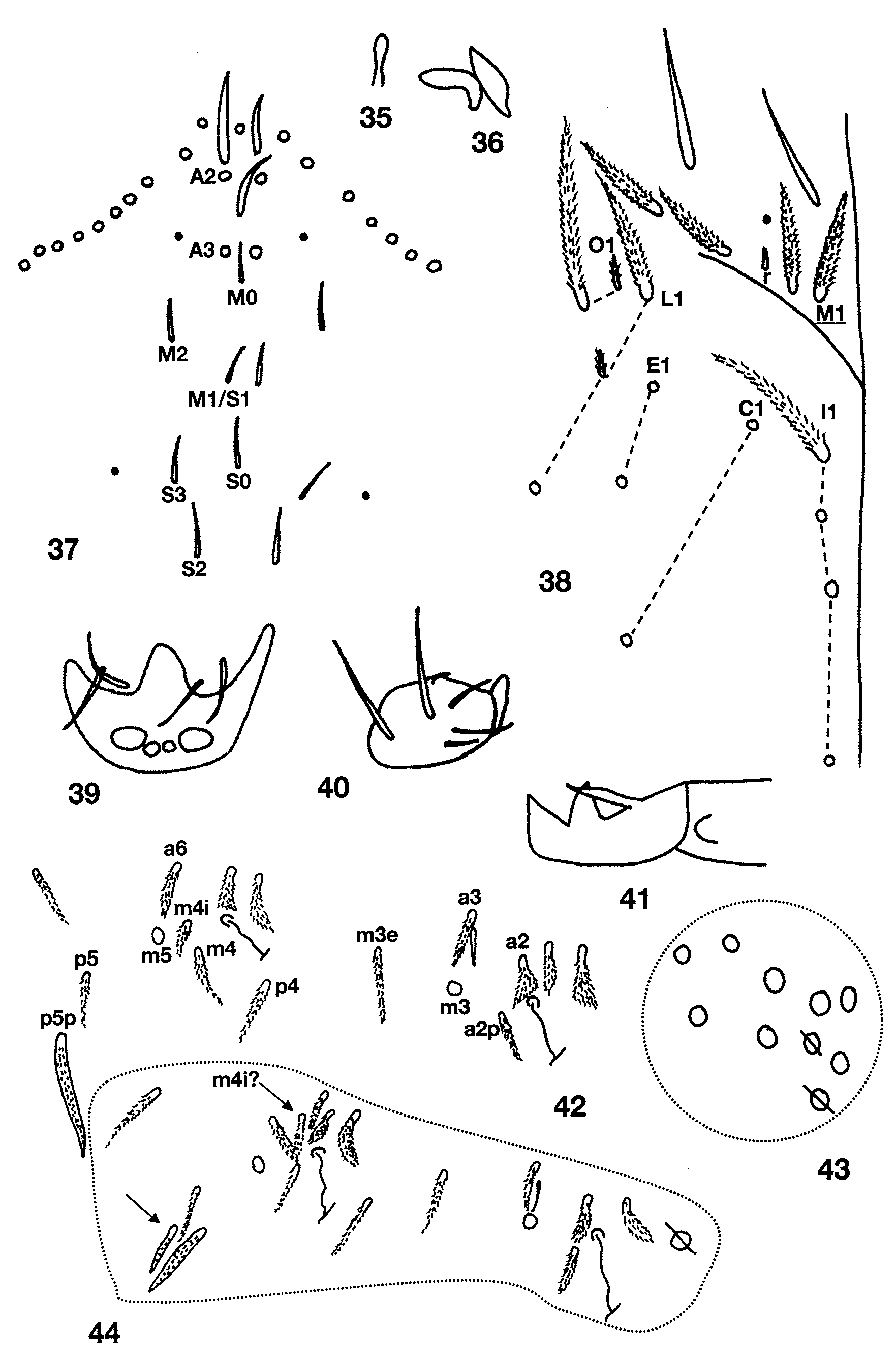
FIGURES 35 – 44. Pseudosinella argentea: 35, Subapical sensilla of forth antennal segment; 36, Sense organ of third antennal segment; 37, Dorsal head chaetotaxy; 38, Labial and postlabial chaetotaxy; 39, Anterior margin of labrum; 40, Sublobular plate of outer maxillary lobe; 41, Mucro; 42, Chaetotaxy of second abdominal segment; 43, Distribution of setae on distal manubrial plate, left side; 44, Atypical chaetotaxy of second abdominal segment in another individual.
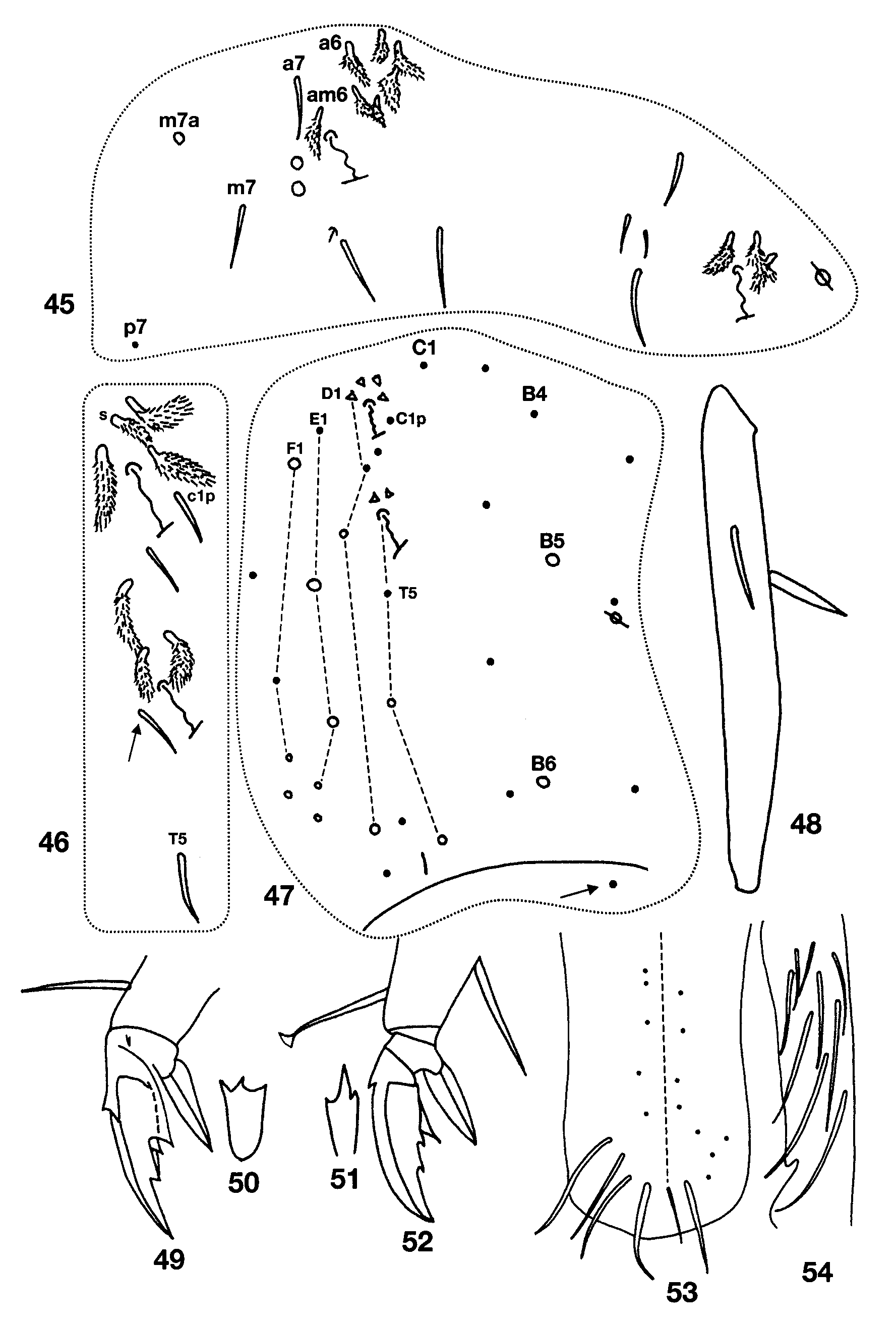
FIGURES 45 – 54. Pseudosinella argentea: 45, Chaetotaxy of third abdominal segment; 46, Bothriotrichal complex of fourth abdominal segment; 47, Complete chaetotaxy of fourth abdominal segment; 48, Metatibiotarsus showing enlarged setae; 49, Prothoracic claw complex; 50, Anterior view of outer ungual teeth on metathoracic leg; 51, Posterior view of inner ungual teeth of prothoracic leg; 52, Metathoracic claw complex; 53, Posterior chaetotaxy of collophore; 54, Anterior chaetotaxy of collophore, left side.
TABLE 5. Comparison between Pseudosinella from West Virginia here identified as argentea, and flatua, granda and
| Species | Head macroseta A0 | Prelabra l setae | Sense organ Ant. 3 | Labial setae m1 & m2 | Labial seta r | Head ventral groove setae | Abd. 2 seta a3 | Abd. 2 seta a2p | Ventral Tube anterior chaetotaxy | Ventral Tube posterior chaetotaxy (total/distal margin) | Ventral Tube vesicle base chaetotaxy |
|---|---|---|---|---|---|---|---|---|---|---|---|
| West Virginia ‘ argentea ’ | 1 | ciliate | swollen | M1≈M2 | smooth, thin walled blunt microseta | 4 ciliate | ciliate | ciliate & ≈ a2 | 9 | 19/9 | 7–8; 4 ciliate |
| argentea | 1 | ? | swollen | M1≈M2 or M1≈M2 | smooth or truncate microseta or absent | 4 ciliate | ? | smooth | ? | ? | ? |
| granda | 2 | ciliate | swollen | M1≈M2 | Absent or conic reduced | 4 ciliate | smooth | ciliate &> a2 | 9 1 | 13/7 | 8–9; 4–5 ciliate |
| flatua | 1 | smooth | slightly swollen | m1<M2 | smooth acuminate microseta | 3 smooth 1 ciliate | smooth | ciliate &> a2 | 11–13 | 14/? | 11–13 |
No known copyright restrictions apply. See Agosti, D., Egloff, W., 2009. Taxonomic information exchange and copyright: the Plazi approach. BMC Research Notes 2009, 2:53 for further explanation.
|
Kingdom |
|
|
Phylum |
|
|
Class |
|
|
Order |
|
|
Family |
|
|
Genus |
Pseudosinella argentea Folsom, 1902
| Soto-Adames, Felipe N. 2010 |
argentea sensu
| Christiansen & Bellinger 1998 |
1 (by plazi, 2016-04-08 05:23:07)
2 (by ImsDioSync, 2017-06-17 15:55:21)
3 (by ImsDioSync, 2017-06-17 20:09:27)
4 (by ExternalLinkService, 2019-09-26 07:03:05)
5 (by ExternalLinkService, 2022-01-30 17:01:26)
6 (by ExternalLinkService, 2022-02-21 18:04:21)
7 (by plazi, 2023-10-25 11:52:45)