
Spinonychiurus nazguli, Pomorski, Romuald J. & Kapruś, Igor J., 2015
|
publication ID |
https://doi.org/10.11646/zootaxa.3914.2.1 |
|
publication LSID |
lsid:zoobank.org:pub:84210442-8F95-440B-8D50-9EDD4C75BA8D |
|
DOI |
https://doi.org/10.5281/zenodo.6118366 |
|
persistent identifier |
https://treatment.plazi.org/id/10737675-FFD4-8F53-BDB0-21576735AD0A |
|
treatment provided by |
Plazi |
|
scientific name |
Spinonychiurus nazguli |
| status |
sp. nov. |
Spinonychiurus nazguli sp. nov.
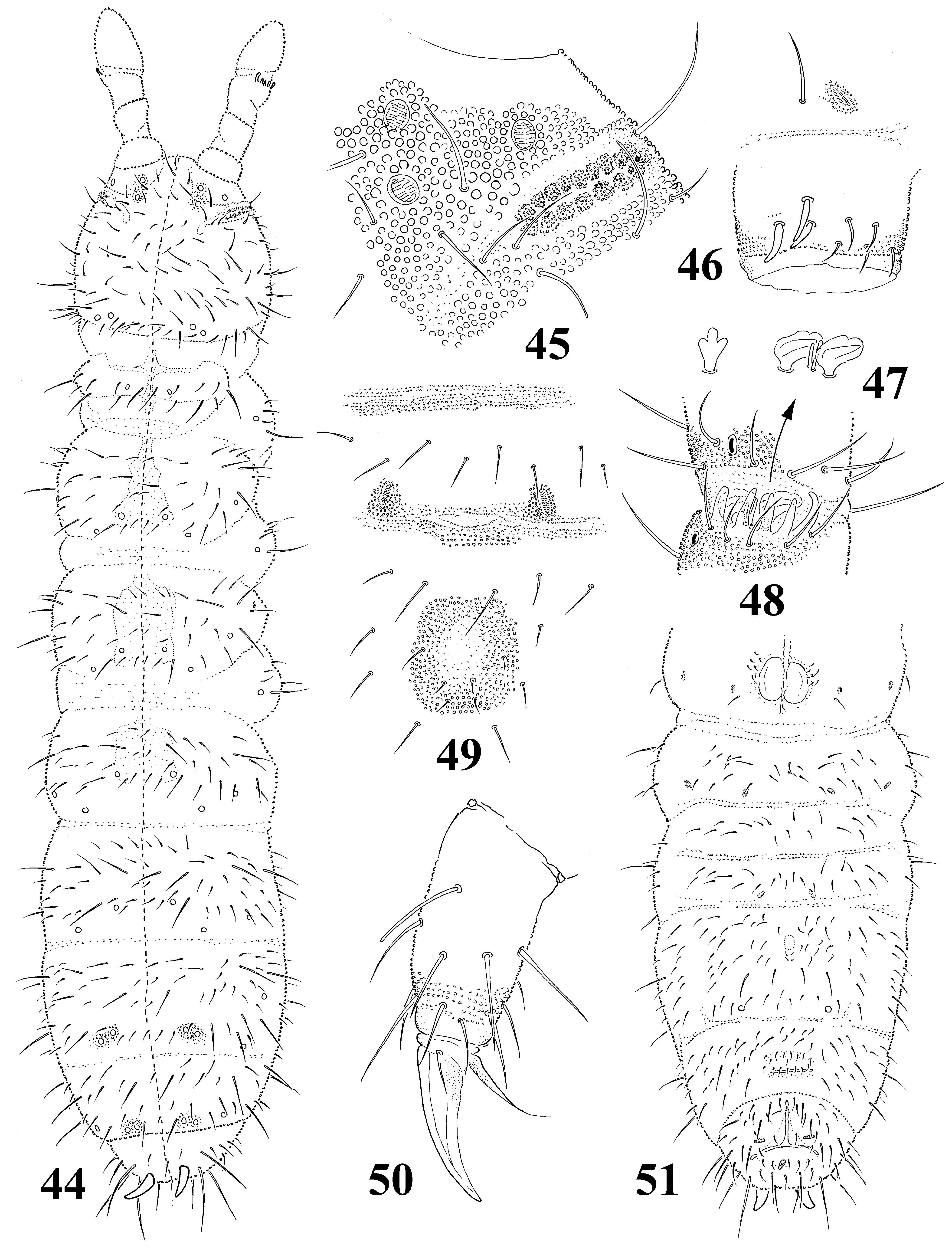
( Figs 44–51 View FIGURES 44 – 51 )
Type material. Holotype reproductive male (mounted on slide) and 12 paratypes (5 unreproductive males, 6 females, 1 juv., mounted on 6 slides); moss near Kok Zhaiyk waterfall, litter under “archa” ( Juniperus turkestanica ) bushes and roots of grasses and herbs on mountain meadow, ca. 2600 m alt.; 11.vi.2006; Dzhety Oguz, Karakol area, Issyk–Kul district, Kyrgyzstan, leg. R.J. Pomorski (preserved in the collection the Department of Biodiversity and Evolutionary Taxonomy, Zoological Institute, Wrocław University, Wrocław).
Etymology. The species is dedicated to Mrs. Nazgul Turdubekova from Helsinian Foundation of Human Rights. Thanks her hospitality and friendly help I had a possibility to investigate of Kyrgyzian springtails.
Description. Color white. Body length: females 1.25–1.5 mm, males 1.0– 1.1 mm. Body shape cylindrical with curved anal spines, longer than inner edge of hind claw (in relation 1.25) ( Fig. 44 View FIGURES 44 – 51 ). Granulation of body surface fine and rather uniform, stronger around pseudocelli on front of head and in middle part of thoracic terga II and III. Antennal bases well marked.
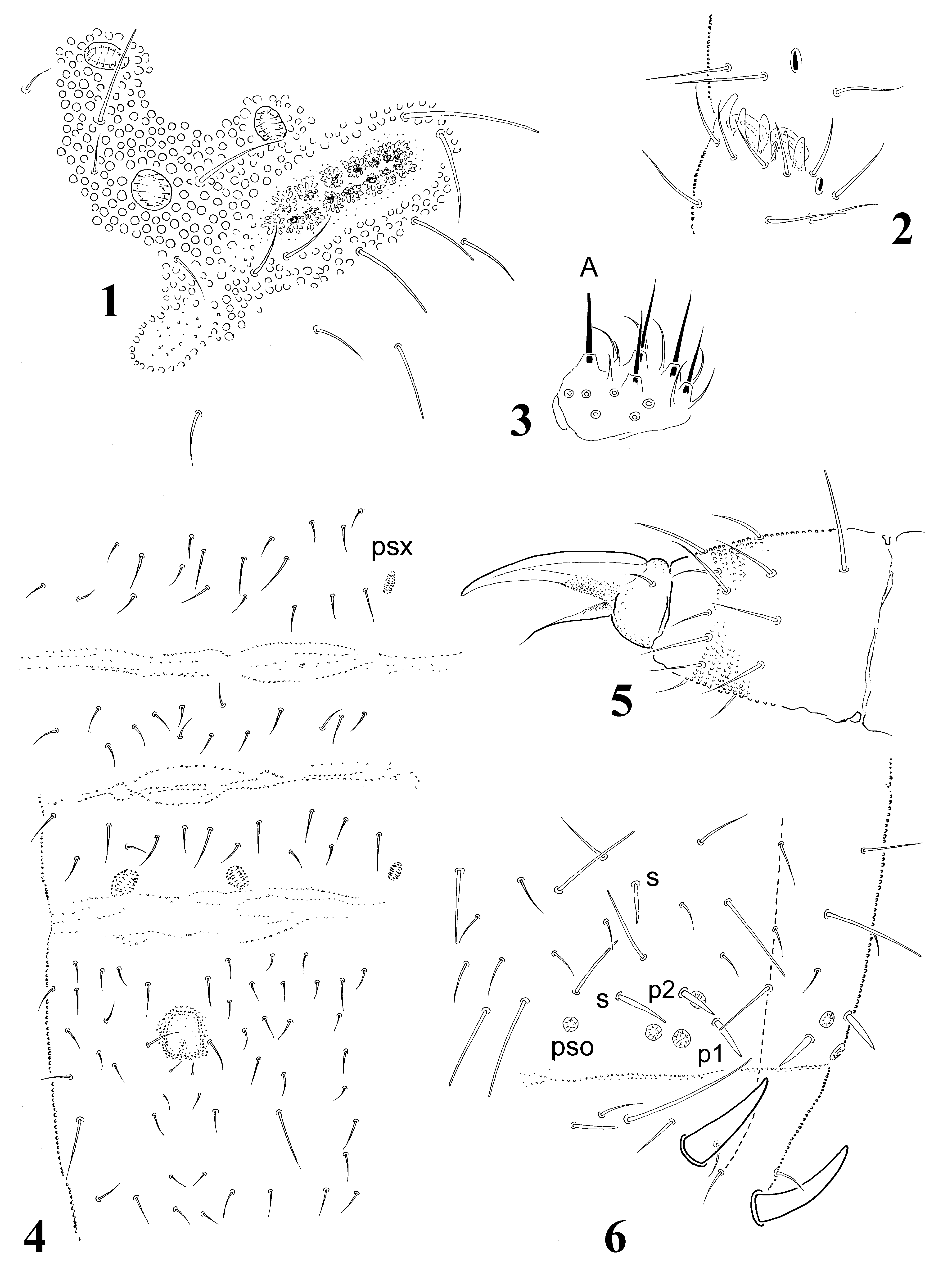
Antennae shorter than head (ratio 0.8). Antennal segment IV with typical subapical organite and two poorly marked sensilla (dorsal-subapical and internal-subbasal). Microsensillum on antennal segment IV in latero-external position, in proximal whorl of chaetae. Antennal III sensory organ with 5 guard chaetae, 5 papillae, 2 small sensory rods and 2 bent, smooth with longitudinal ribs sensory clubs, and microsensillum located slightly below antennal III sensory organ ( Figs 47, 48 View FIGURES 44 – 51 ).
Postantennal organ consists of 15–18 granulated vesicles ( Fig. 45 View FIGURES 44 – 51 ). Labial palp of A type.
Pseudocellar formula dorsally 33/133/33343, ventrally 1/000/00001. Subcoxae1 of I–III legs with 1 pseudocellus each. Parapseudocellar formula: 1/000/ 221003 (each anal valve with parapseudocellus). Localization of parapseudocelli on abdominal sterna I–VI as in Fig. 51 View FIGURES 44 – 51 . Subcoxae1 of I–III legs with 1parapseudocellus each.
Dorsal chaetotaxy as in Fig. 44 View FIGURES 44 – 51 , nearly symmetrical, well differentiated into meso- and microchaetae. Thoracic terga II and III with microsensilla laterally. Body sensory chaetae s not differentiated. Thoracic tergum I with 8+8 chaetae. Abdominal tergum IV with p0 chaeta. Abdominal tergum V with a0 and p0 chaetae. Abdominal tergum VI with 2 axial chaetae and 1+1 prespinal chaetae. Subcoxae1 of I–III legs with 4, 5, 5 chaetae respectively.
Thoracic sterna I–III with 0+0, 1+1, 1+1 chaetae respectively. Ventral tube of females with 6+6 chaetae, and 1+1 chaetae at base. Furca reduced to small area of fine granulation and three rows of manubrial chaetae behind its posterior edge ( Fig. 49, 51 View FIGURES 44 – 51 ). Claws without teeth. Empodial appendage without basal lamella, shorter than inner edge of claw (ratio 0.9). Tibiotarsi I–III with 9 chaetae in distal whorl ( Fig. 5 View FIGURES 1 – 6 ). Males with ventral organ, it consists of 3+3 thickened chaetae, located on ventral tube in posterolateral position ( Fig. 46 View FIGURES 44 – 51 ).
Remarks. Because of presence of 9 chaetae in distal whorl of tibiotarsi S. nazguli sp. nov. seems to be similar to S. issykkulensis sp. nov. and S. natashae sp. nov. (described below). It differs from these species in the structure of labial palp and the presence of 1+1 pseudocelli on thoracic tergum I and abdominal sternum IV. S. issykkulensis sp. nov. and S. natashae sp. nov. have labial palp of AB type, 2+2 pseudocelli on thoracic tergum I and they have no pseudocelli on abdominal sternum IV.
No known copyright restrictions apply. See Agosti, D., Egloff, W., 2009. Taxonomic information exchange and copyright: the Plazi approach. BMC Research Notes 2009, 2:53 for further explanation.
|
Kingdom |
|
|
Phylum |
|
|
Class |
|
|
Order |
|
|
Family |
|
|
Genus |