
Onychiurus gulinensis, Sun & Zhang, 2012
|
publication ID |
https://doi.org/10.1080/00222933.2012.707236 |
|
persistent identifier |
https://treatment.plazi.org/id/131087DD-EB7D-F014-FE63-58C4FEEDFA30 |
|
treatment provided by |
Felipe |
|
scientific name |
Onychiurus gulinensis |
| status |
sp. nov. |
Onychiurus gulinensis sp. nov.
( Figures 1–3 View Figure 1 View Figure 2 View Figure 3 ; Table 1)
Type material
Holotype male, one male paratype and three female paratypes. China: Jiangsu Province: Nanjing: Gulin Park : 7 November 2007, litter, Berlese extraction, Jigang Jiang and Xin Sun leg. (C9557). –ibid: Qingliangshan Park: seven paratypes on slide, 26 February 2006, litter, Berlese extraction, Yongzheng Luo and Li Liu leg. (C9314).
Holotype and eight paratypes on slide in NJU, three paratypes in MNHN.
Description
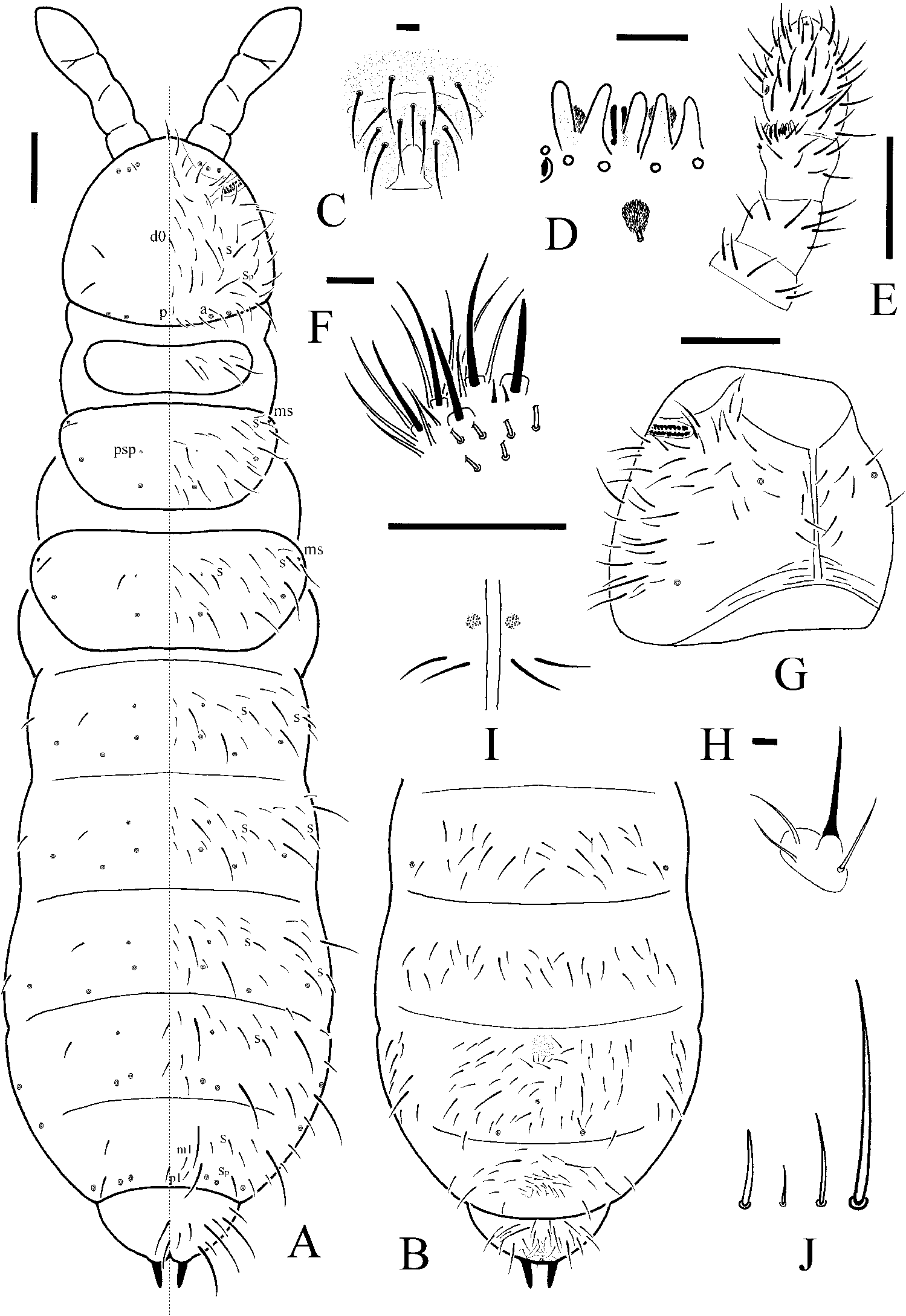
Body length: female 1.7–2.0 mm, male 1.6 mm; holotype 1.6 mm. Body shape: cylindrical, Abd. III –IV more or less broadened. Body colour: white in alcohol .
Pseudocelli formula 32 / 022 / 33343 dorsally, 11 / 000 / 01010 ventrally, subcoxa 1 of legs I–III with one pso each ( Figure 1A,B View Figure 1 ). Parapseudocelli: subcoxa 1 of legs I–III with one psx each, 1+1 on Abd. I sternum, near base of ventral tube. Pseudopore formula 00 / 011 / 11110 dorsally, 00 / 111 / 000x0 ventrally.
Formula for s-chaeta 11 / 012 / 222120 dorsally. Sp present on head. The s-microchaetae tiny and blunt, present on Th. II and III dorsally ( Figure 1A,B View Figure 1 ).
Head: antennae short and distinctly segmented, as long as head. Length ratio of antennal segments I: II: III: IV = 1: 1.5: 1.5: 2.3–2.5 ( Figure 1E View Figure 1 ). Ant. I with 10 chaetae. Ant. II with 16 chaetae. Ant. III sensory organ composed of five papillae, five guard chaetae, two small rods and two weakly granulated sensory clubs, both morel-like; lateral ms just behind sensory organ ( Figure 1D View Figure 1 ). Ant. IV subapical organite with apex globular; basolateral ms at about one-third length from base. Antennal base with smaller granulation. PAO composed of 20–21 compound vesicles arranged in two rows along axis of organ. Dorsal cephalic chaeta d 0 present. 4+4 p-chaetae between posterior a-pso on head, p 1 anterior to others ( Figure 1A View Figure 1 ). Mandible with strong molar plate and four apical teeth. Maxilla bearing three teeth and six lamellae. Maxillary palp simple with one basal chaeta and two sublobal hairs ( Figure 1H View Figure 1 ). Labral chaetae 4 / 342 ( Figure 1C View Figure 1 ). Labial papillae of AC type, papillae A–E respectively with one, four, zero, three and four guard chaetae ( Figure 1F View Figure 1 ). Labium with six proximal, four basomedian (E, F, G, and f) and six basolateral (a, b, c, d, e, e’) chaetae. Postlabial chaetae 4+4 along ventral groove ( Figure 1G View Figure 1 ).
Body chaetotaxy: ordinary chaetae differentiated into mesochaetae and macrochaetae, ratio sp: m1: p1 on Abd. V = 1: 0.7: 0.6. Th. I tergum with 7–8+7–8 chaetae. Th. II–Abd. III terga respectively with 3+3 chaetae along axial line, without axial chaetae. Abd. IV tergum with two axial chaetae (m 0 and p 0), Abd. V tergum with one axial chaeta (p 0), Abd. VI tergum with two axial chaetae (a 0 and m 0) ( Figure 1A View Figure 1 ). Th. I, II and III sterna with 0+0, 2+2 and 2+2 (3) chaetae respectively between legs ( Figure 1I View Figure 1 ). Subcoxa 1 of legs I–III with four or five chaetae each, subcoxa 2 with one, four and four chaetae respectively.
Appendages: tibiotarsi I, II and III with 22 (11, 8, 3), 21 (11, 8, 2) and 21 (11, 8, 2) chaetae. Unguis without teeth. Unguiculus slender and pointed, 0.7 times as long as inner edge of unguis, with very narrow inner basal lamella ( Figure 2D View Figure 2 ). Ventral tube with 8–9+8–9 distal chaetae, without anterior and basal chaetae ( Figure 2A View Figure 2 ). Furca reduced to a finely granulated area, with four small dental chaetae in one row posterior to furcal rudiment; three manubrial rows of chaetae present ( Figure 2B View Figure 2 ).
Genital plate with 18–19 circumgenital chaetae and two genital chaetae in female, 32–40 circumgenital chaetae and eight genital chaetae in male ( Figure 2F View Figure 2 ). Anal valves with numerous acuminate chaetae; each lateral valve with chaetae a 0 and 2a 1; upper valve with chaetae a 0, 2a 1, b 0, 2b 1, 2b 2, c 0, 2c 1, 2c 2 ( Figure 2C View Figure 2 ). Anal spines set on distinct papillae, as long as inner edge of hind unguis ( Figures 1A,B View Figure 1 , 2E View Figure 2 ).
Ecology
In broad-leaved litter of a park in town.
Etymology
Named for the type locality.
Remarks
Onychiurus gulinensis sp. nov. can be easily distinguished by its dorsal pseudocellar formula as 32 / 022 / 33343, the presence of 11 chaetae in distal whorl of tibiotarsi and the granulated sensory clubs of AIIIO.
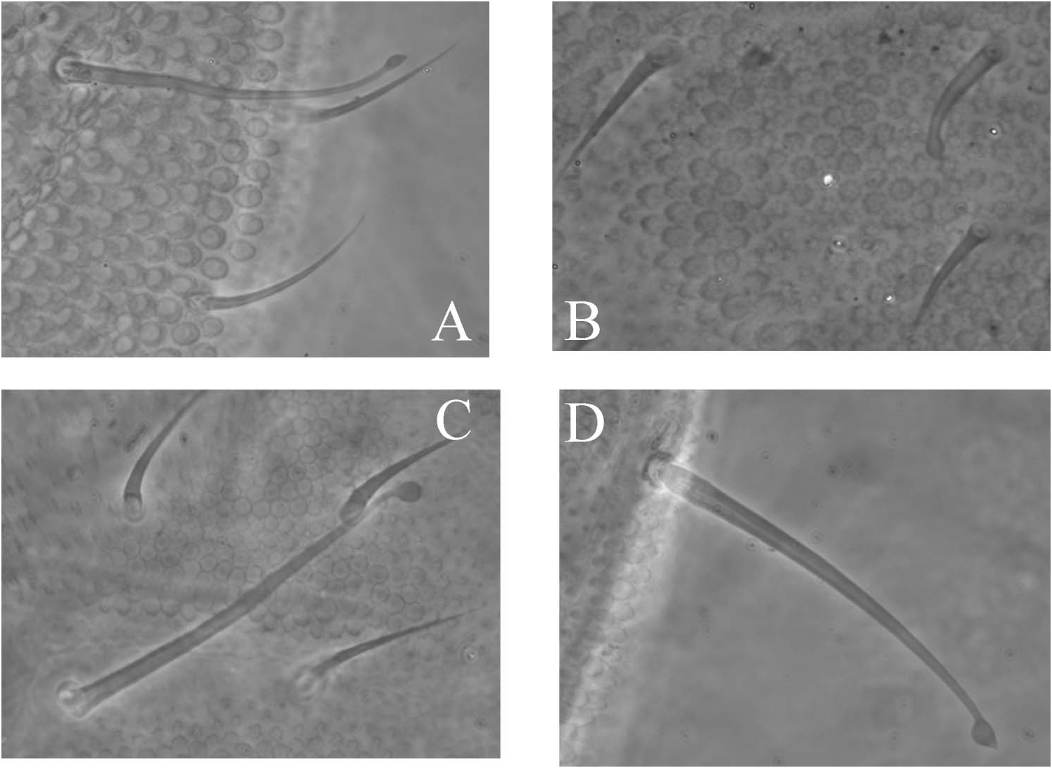
Five paratypes ( two males and three females) have a large number of clavate chaetae on body and appendages ( Figures 2E View Figure 2 , 3A–D View Figure 3 ). Chaetotaxy of these specimens is still identical to that of “normal” specimens. The clavate chaetae are differentiated dorsally (s-chaetae, macrochaetae and some mesochaetae), ventrally (a few mesochaetae), on subcoxae (two or three chaetae) and in the basal whorl of tibiotarsus (two chaetae Y), but absent on antennae. This modification of chaetal morphology is similar to that already described for Onychiurus stillicidii and sporadically recorded in other Poduromorpha ( Deharveng 1983) , except that antennal chaetae are not modified in our specimens.

| MNHN |
Museum National d'Histoire Naturelle |
No known copyright restrictions apply. See Agosti, D., Egloff, W., 2009. Taxonomic information exchange and copyright: the Plazi approach. BMC Research Notes 2009, 2:53 for further explanation.