
Docosia
|
publication ID |
https://doi.org/ 10.5281/zenodo.213192 |
|
DOI |
https://doi.org/10.5281/zenodo.6180627 |
|
persistent identifier |
https://treatment.plazi.org/id/15369321-6459-FF90-33AE-FDB6FA47F857 |
|
treatment provided by |
Plazi |
|
scientific name |
Docosia |
| status |
|
Key to males of Docosia View in CoL View at ENA species in Central Asia
1. Sc setose and ending free; terminalia as figured by Laštovka & Ševčík (2006: fig. 1) and Kurina (2008b: figs. 16–21). Uzbekistan................................................................... D. gilvipes View in CoL (Haliday in Walker 1856)
- Sc bare, ending in R1................................................................................... 2
2. Laterotergite setose.....................................................................................3
- Laterotergite bare..................................................................................... 5
3. All coxae entirely blackish, only forecoxa occasionally somewhat paler to brownish................................. 4
- Coxae yellow with only bases infuscated. Ventroapical margin of gonocoxa emarginated with a bare finger-like medial process ( Fig. 7 View FIGURE 7 b). Gonostylus with two subapical spines, more basal one much smaller and with clearly delimited basal body ( Figs. 7 View FIGURE 7 a–c). Tergite 9 apically widening with slightly convex apical margin ( Fig. 7 View FIGURE 7 d). Cercus with 10 combs of retinacula ( Fig. 7 View FIGURE 7 e). Turkmenistan......................................................................... D. koyentagi sp. nov.
4. Ventroapical margin of gonocoxite wavy with a sclerotized, bare, finger-like medial process and with lateral processes bearing four subapical spines each. Gonostylus bifid: ventral lobe bent posteriorly, without apical spine; dorsal lobe bent medially with subapical tubercle on medial margin. Tergite 9 subquadrate, basal margin with shallow medial incision. Cercus with 5 combs of retinacula ( Kurina 2006: fig. 4). Kazakhstan......................................... D. sogetensis Kurina, 2006 View in CoL
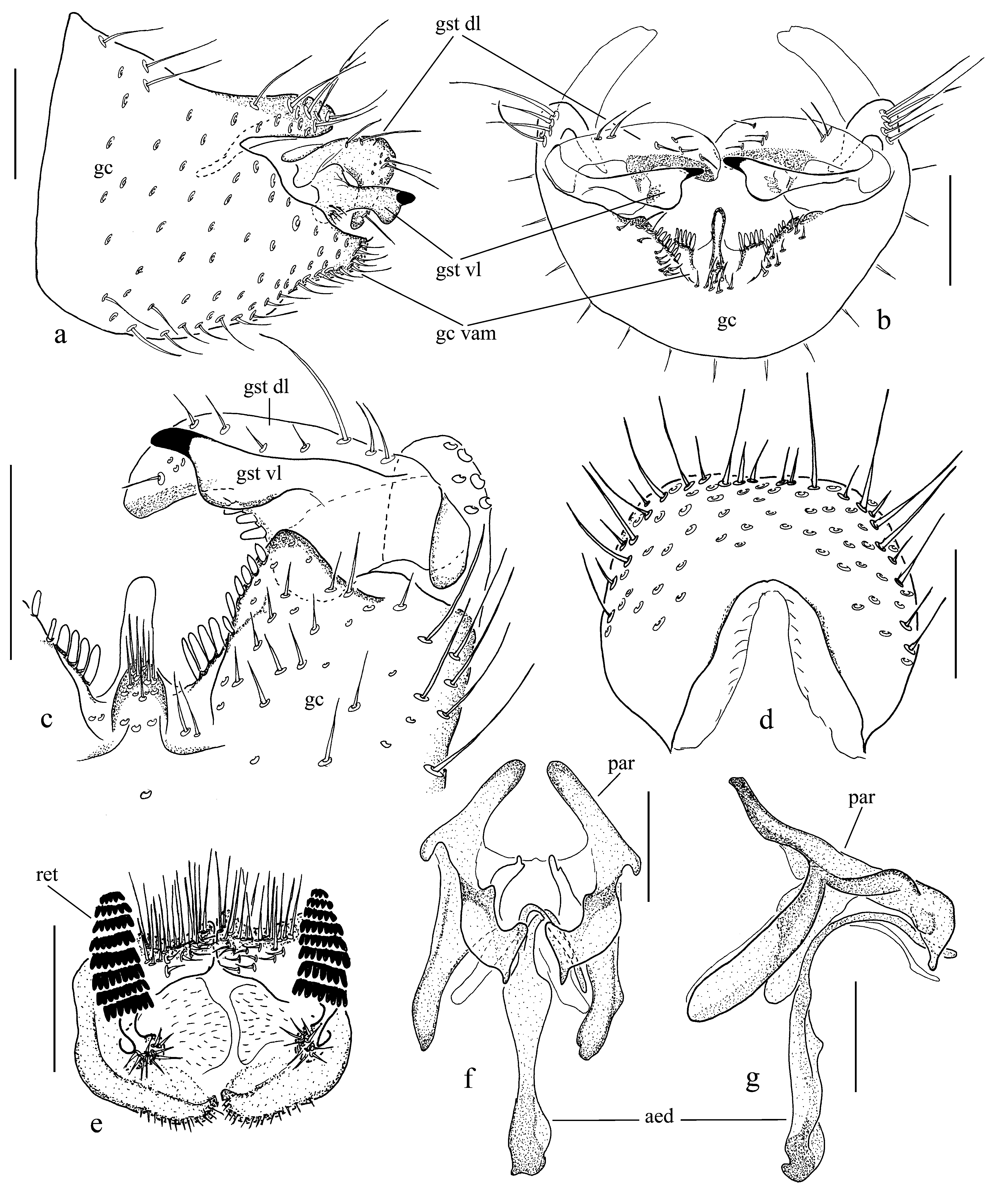
- Ventroapical margin of gonocoxite deeply emarginated with a basally setose, finger-like medial process ( Fig. 3 View FIGURE 3 b). Ventroapical margin of gonocoxite with gradually shortening spines from medial process laterally ( Fig. 3 View FIGURE 3 c). Gonostylus bifid: ventral lobe medially swollen and with apical spine; dorsal lobe bent medially with three short basal spines on medial margin ( Fig. 3 View FIGURE 3 a–c). Basal margin of tergite 9 with a deep incision ( Fig. 3 View FIGURE 3 d). Cercus with 8 combs of retinacula ( Fig. 3 View FIGURE 3 e). Uzbekistan................................................................................................. D. bartaki sp. nov.
5. Ventroapical margin of gonocoxite without medial process but with a flange bearing short strong setae ( Kurina 2006: fig. 3). Kazakhstan..................................................................... D. agnesiana Kurina, 2006
- Ventroapical margin of gonocoxite with medial process (e.g. Figs. 5 View FIGURE 5 b, 6b) and with (e.g. Fig. 6 View FIGURE 6 c) or without (e.g. Fig. 5 View FIGURE 5 c) an additional internal flange................................................................................ 6
6. Ventroapical margin of gonocoxite with short, apically flattened or concave medial process bearing an aggregation of short setae ( Fig. 5 View FIGURE 5 c)........................................................................................ 7
- Ventroapical margin of gonocoxite with protruding medial process bearing modified setae or with setae located on internal flange ( Figs. 4 View FIGURE 4 c, 6c, 8c)................................................................................. 8
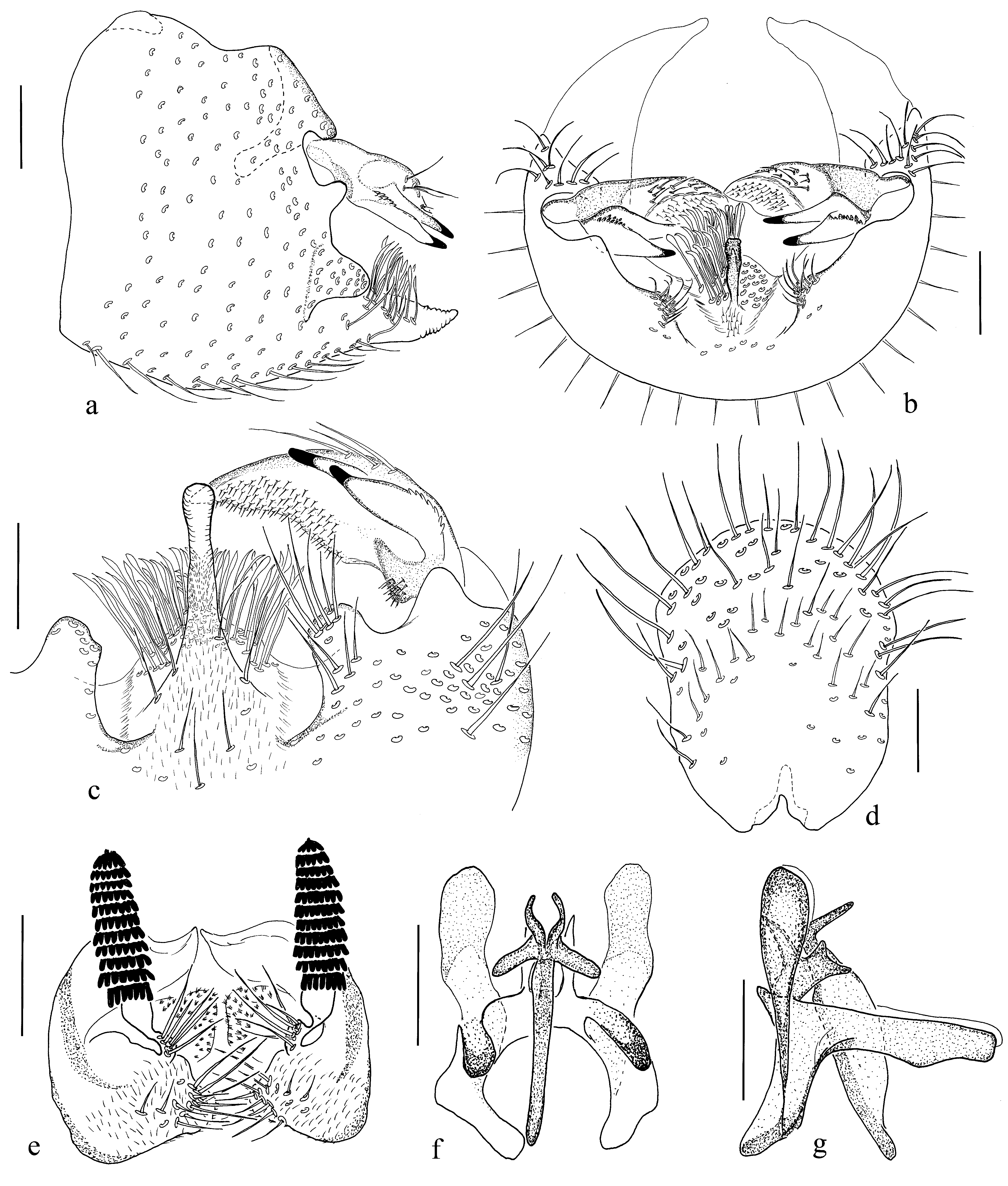
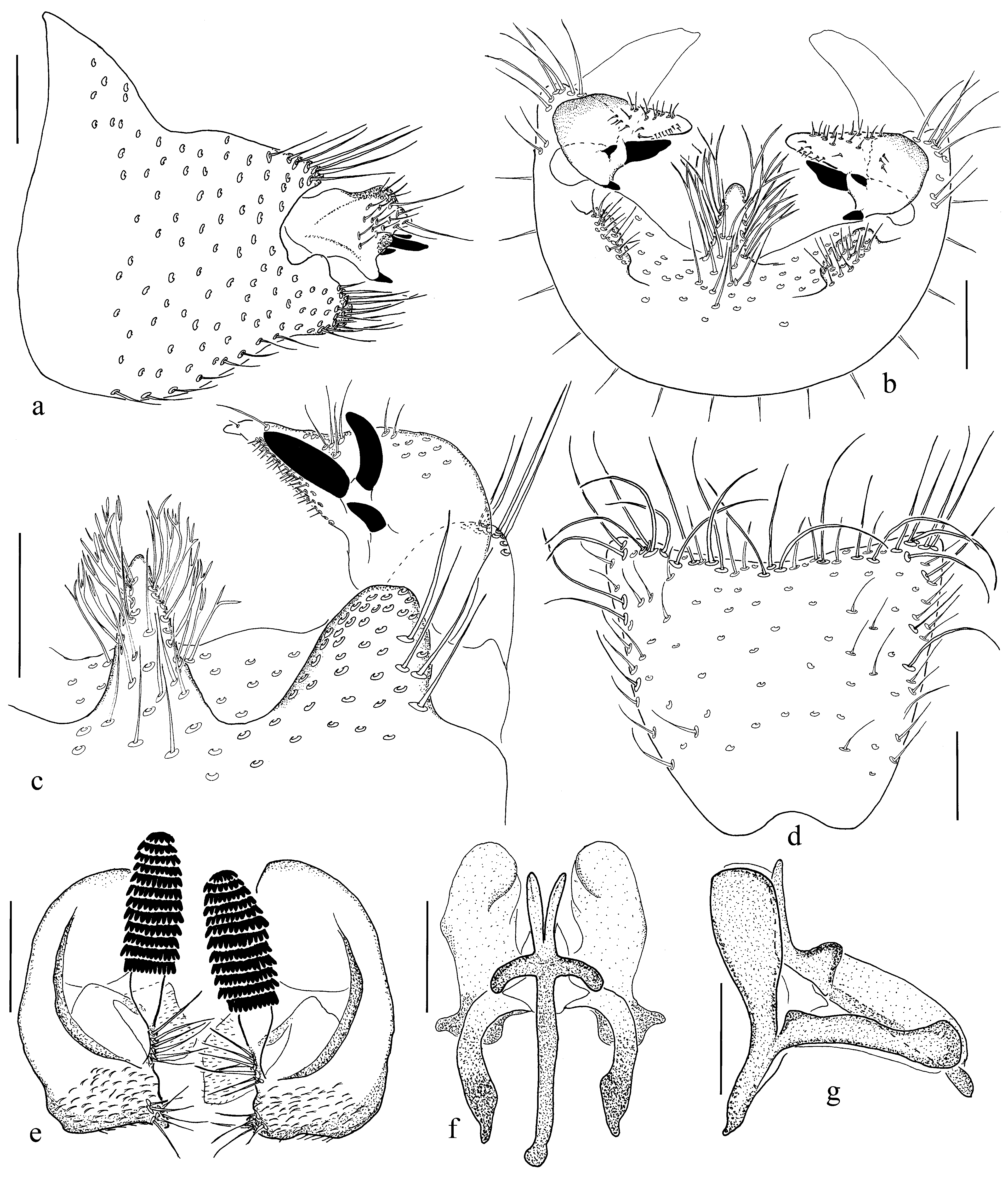
7. Medial process of ventroapical margin of gonocoxite apically convex ( Figs. 5 View FIGURE 5 b–c). Gonostylus with 6 subequal spines on apical half and with basal and medial extensions, the latter bearing 6 fine setae ( Figs. 5 View FIGURE 5 a–c). Tergite 9 apically widening, without basal incision ( Fig. 5 View FIGURE 5 d). Cercus with 10 combs of retinacula ( Fig. 5 View FIGURE 5 e). Uzbekistan................ D. chimganica sp. nov.
- Medial process of ventroapical margin of gonocoxite apically emarginate. Gonostylus with three spines: the apical twice as long as the subapical which is longer than the basal. Gonostylus with only basal extension bearing short setulae. Tergite 9 apically widening and with basal incision. Cercus with 11 combs of retinacula ( Kurina 2006: fig. 2). Kazakhstan, Uzbekistan...................................................................................... D. selini Kurina, 2006 View in CoL
8. Ventromedial projection on gonocoxite evenly tapering, bearing an aggregation of apically split megasetae ( Figs. 4 View FIGURE 4 c, 8c)... 9
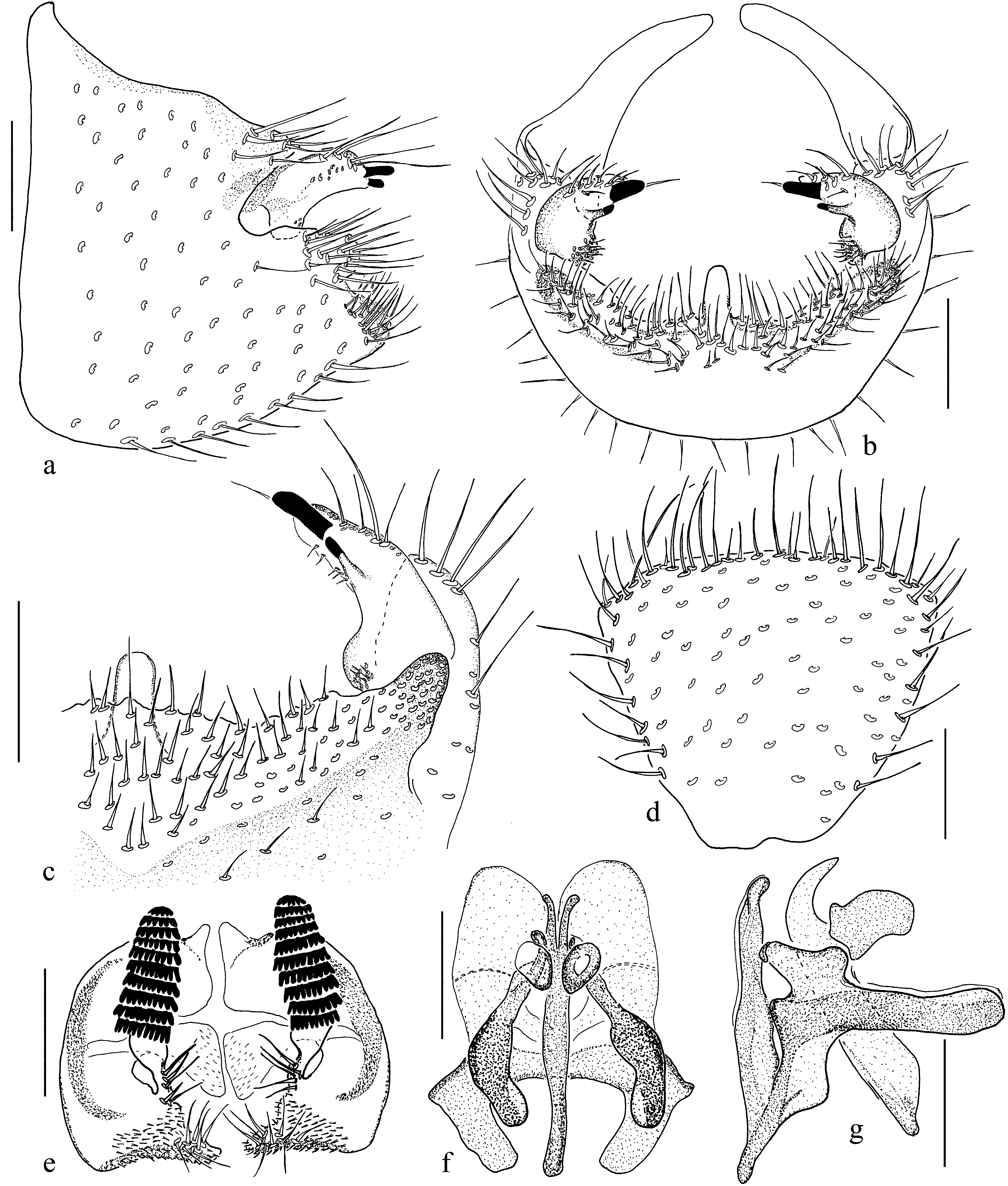
- Ventromedial projection on gonocoxite fin-like ( Figs. 6 View FIGURE 6 b–c). Megasetae on internal flange of ventroapical margin of gonocoxite lanceolate ( Fig. 6 View FIGURE 6 c). Gonostylus crescent, apically tapering with two narrow lobes, both with apical spines, ventrally ( Figs. 6 View FIGURE 6 a–c). Tergite 9 suboval, with basal incision ( Fig. 6 View FIGURE 6 d). Cercus with 11 combs of retinacula ( Fig. 6 View FIGURE 6 e). Kazakhstan, Turkmenistan................................................................................... D. distributa sp. nov.
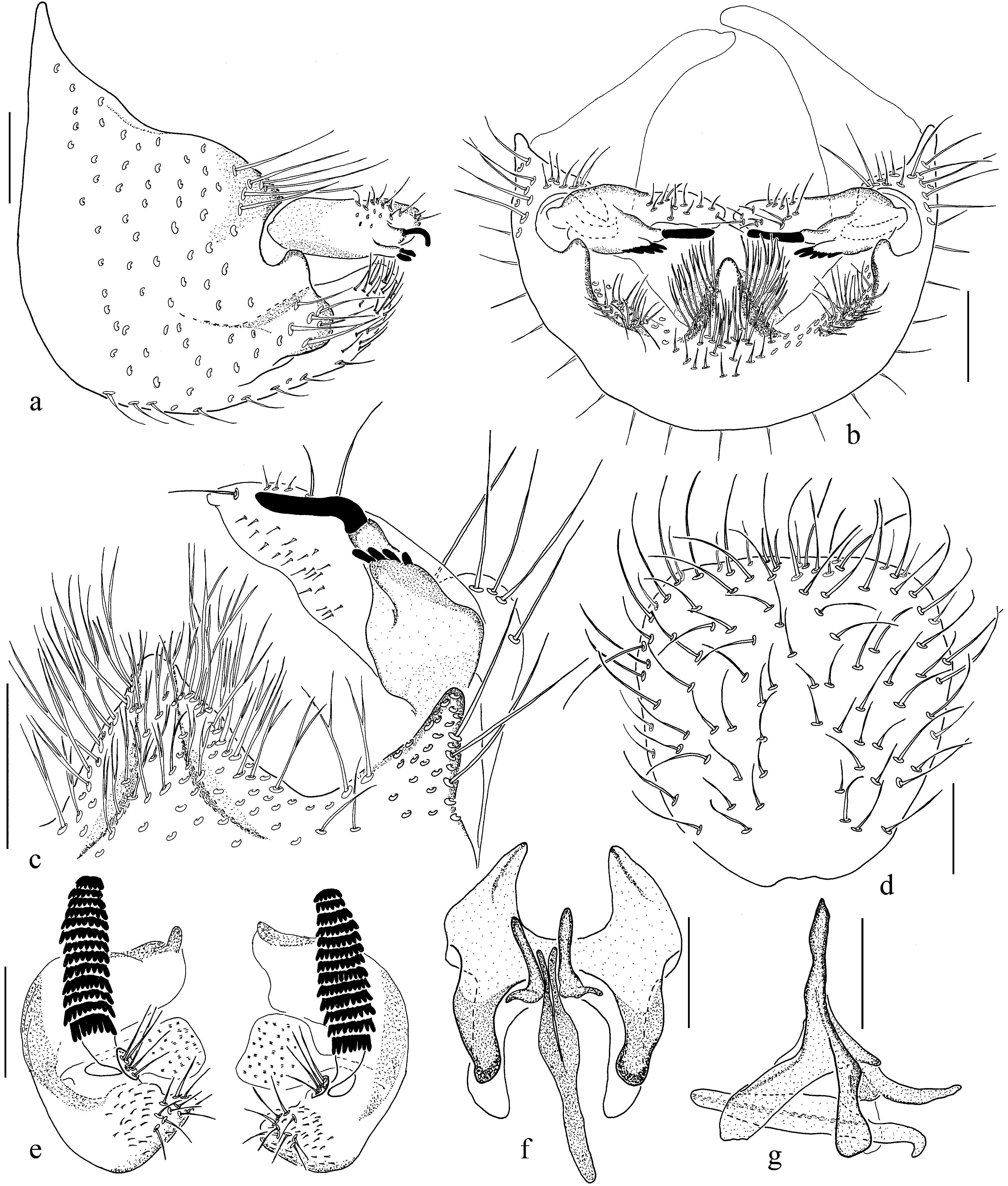
9. Ventromedial projection of gonocoxite wide, ventrolateral extensions narrow ( Fig. 4 View FIGURE 4 c). Gonostylus suboval, apically somewhat tapering, ventrally with a geniculate spine on clearly delimited basal body and four short spines on a common basal body ( Figs. 4 View FIGURE 4 a–c). Tergite 9 subquadrate with straight apical margin ( Fig. 4 View FIGURE 4 d). Cercus with 13 combs of retinacula ( Fig. 4 View FIGURE 4 e). Turkmenistan................................................................................... D. blagoderovi sp. nov.
- Ventromedial projection of gonocoxite narrow, ventrolateral extensions wide ( Fig. 8 View FIGURE 8 c). Gonostylus humpbacked, apically clearly tapering, bearing three spines on ventral side medially: apical two spines more prominent than basal one ( Figs. 8 View FIGURE 8 a–c). Tergite 9 apically widening with concave apical margin ( Fig. 8 View FIGURE 8 d). Cercus with 12 combs of retinacula ( Fig. 8 View FIGURE 8 e). Turkmenistan.................................................................................... D. turkmenica View in CoL sp. nov.

No known copyright restrictions apply. See Agosti, D., Egloff, W., 2009. Taxonomic information exchange and copyright: the Plazi approach. BMC Research Notes 2009, 2:53 for further explanation.
|
Kingdom |
|
|
Phylum |
|
|
Class |
|
|
Order |
|
|
Family |