
Puto brom Powell & Miller, 2024
|
publication ID |
https://doi.org/ 10.11646/zootaxa.5443.3.1 |
|
publication LSID |
lsid:zoobank.org:pub:44123F53-17F2-4205-8B14-1155D41FCEBA |
|
DOI |
https://doi.org/10.5281/zenodo.11045269 |
|
persistent identifier |
https://treatment.plazi.org/id/184087D2-FFD4-FFF7-E1ED-FCBAD74FFE08 |
|
treatment provided by |
Plazi |
|
scientific name |
Puto brom Powell & Miller |
| status |
sp. nov. |
Puto brom Powell & Miller , sp. n.
Suggested common name: bromeliad giant mealybug
Material examined
Holotype adult female mounted singly on slide. Left label “ Puto / Mexico / ex Bromeliad leaf / IX-23-74 / Nogales 2962 / J. Kaiser, R. Duke / Balsam.” Right label “ Puto brom / Powell & Miller / n.sp.”, USNM.
Paratype: Same data as holotype (1 adult ♀ on 1 slide) USNM .
Other material examined: MEXICO: Veracruz, taken in quarantine at Brownsville, Texas , September 17, 1975, coll. Burgess, Chace, Arsego, ex. bromeliad leaf. (1 ad. ♂ on 1 slide) USNM .
Etymology: The species epithet “ brom ” is a noun in apposition based on an abbreviation of the scientific name of the host plant, an unknown species of Bromeliaceae .
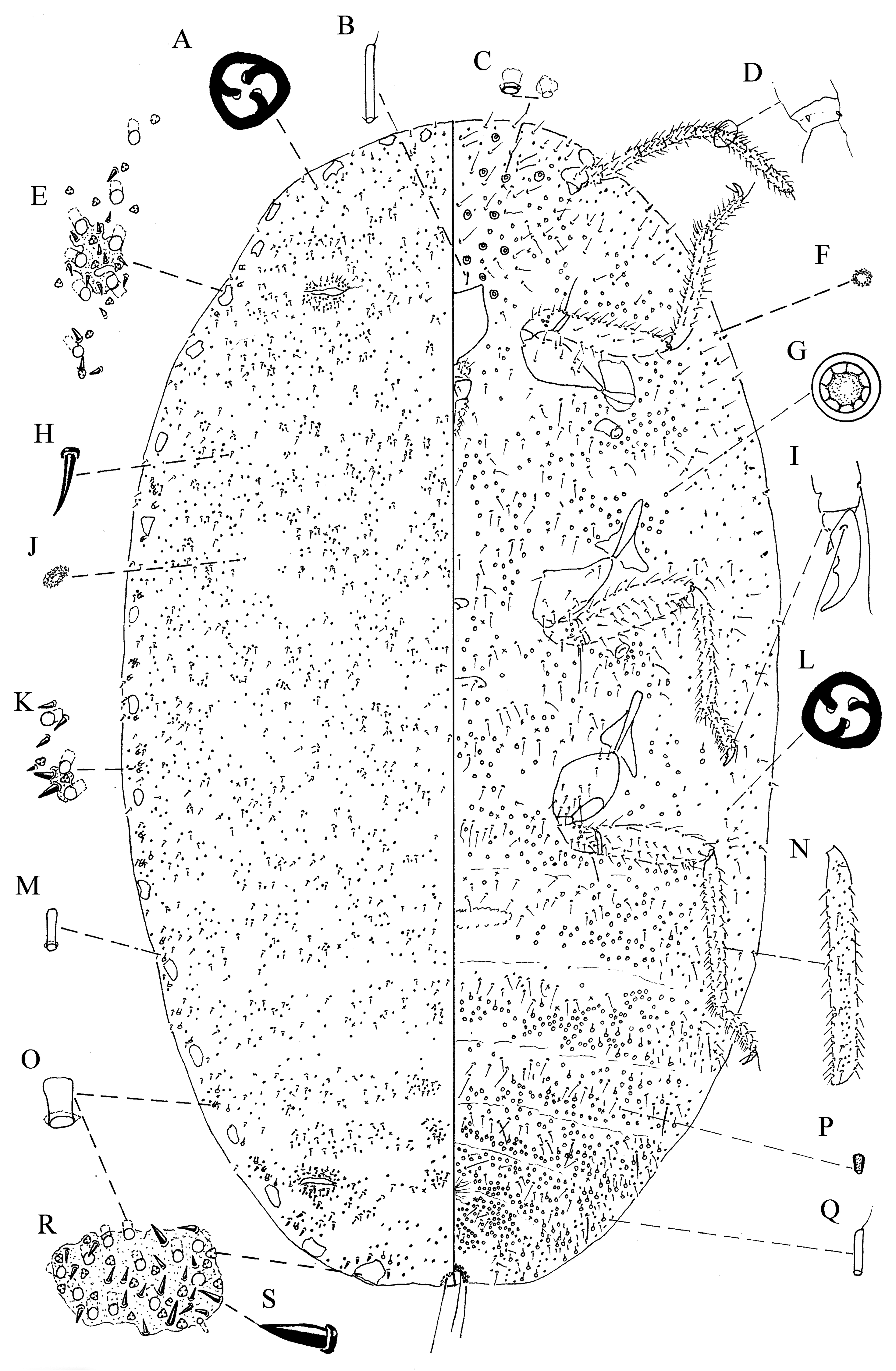
Adult female
( Fig. 12 View FIGURE 12 )
Description: Slide-mounted holotype 8.65 mm long, 5.4 mm wide; paratype 8.7 mm long, 5.15 mm wide; body elongate oval.
Dorsum with 18 or 19 pairs of cerarii; anal-lobe cerarii each with 15–17 lanceolate setae (paratype 16–19), 14 or 15 trilocular pores (paratype 12–13), 10–12 large-sized oral-rim type tubular ducts (paratype 11–12), each duct 18–19 µm in diameter, and 1 or 2 smaller-sized oral-rim type tubular ducts also in sclerotized area, each about 11 µm in diameter (paratype 8 µm); additional areas of large-sized oral-rim type tubular ducts, cerarian setae with basal sclerotization, and trilocular pores, present marginally outside of sclerotized cerarii; frontal cerarii each with 9–11 lanceolate setae (paratype 10 or 11), 4 or 5 trilocular pores (paratype 2–3), and 4 oral-rim type tubular ducts (paratype 4); each cerarius without associated discoidal pores; marginal cerarii with basal sclerotization. Anterior and posterior ostioles present. Multilocular disc-pores absent; trilocular pores of 1 size, numerous, concentrated in medial and submedial clusters with dorsal setae on abdominal segments, scattered over thorax and head, abundant on lips of ostioles; discoidal pores scattered over body. Small oral-rim type tubular ducts only present marginally, interspersed with large oral-rim type tubular ducts. Dorsal setae lanceolate, forming concentrated medial and submedial clusters on abdominal segments II–VII and scattered elsewhere, clustered on lips of ostioles, scattered on head and thorax, and present on margin between cerarii on head and thorax. Longest seta on abdomen about 26 µm (paratype 22 µm) long, thinner than cerarian setae, longest seta in abdominal cerarii 32 µm (paratype 34 µm) long.
Anal ring bent around abdomen apex, bearing 6 anal-ring setae with apices acute; posterior anal-ring setae each 428 µm (paratype 408 µm) long; 2 times (paratype 1.83 times) as long as greatest diameter of anal ring.
Venter with multilocular disc-pores abundant on abdominal segments II–VIII, especially abundant on posterior margin of V and on VI–VIII, also on thorax and head; multilocular disc-pores on abdomen predominantly 9-locular, with some 10-locular. Trilocular pores of 1 size, numerous, scattered over body; discoidal pores scattered over body. Oral-collar tubular ducts of 4 sizes; with 2 sizes of shorter oral-collar tubular ducts abundant on segments IV–VIII, and scattered on thorax and head; 1 or 2 long oral-collar tubular ducts present anterior to mouthparts; and fourth type of oral-collar tubular duct scattered, numbering 10 (paratype 12) anterior to mouthparts, poorly sclerotized, wide and short, unlike typical elongate oral-collar tubular ducts in cluster anterior to mouthparts found in other species of Puto . Long hair-like setae forming rows across abdomen, longest ventral seta 226 µm (paratype 201 µm) long, longest seta on segment V 169 µm (paratype 151 µm) long, longest seta on head 226 µm (paratype 200 µm) long. Circulus elongate oval, 890 µm long by 127 µm wide (paratype 818 µm long by 147 µm wide). Labium 440 µm (paratype 448 µm) long. Antennae each 9-segmented, total length 1.99 mm (paratype with both antennae broken); apical segment 238 µm long, segment III 323 µm (paratype 340 µm) long; antennal intersegmental sensilla present between segments III and IV, IV and V, and VI and VII; coeloconic sensilla present on segment IX. Hind tibiae each with small translucent pores. Femora each 1.03 µm (paratype 1.07 mm) long, tibiae each 1.21 mm (paratype 1.31 mm) long, tibia/tarsus 3.16 (paratype 3.00). Hind trochanters each with 4 campaniform sensilla on each surface. Tarsal digitules acute, claw digitules acute, as long as claw; claw denticle and basal spurs present.
Remarks: Coeloconic sensilla were difficult to observe on these specimens due to the stain. We only observed them on segment IX on this species but anticipate that they could be present on other segments, given what we have seen in other species of Puto .
Diagnosis: Puto brom is most similar to P. lasiorum ( Cockerell, 1901) by having: (i) oral-rim type tubular ducts in the cerarii but not in the medial areas of the dorsum, and (ii) clusters of lanceolate setae dorsomedially on the posterior abdominal segments. It differs from P. lasiorum as follows (characters of P. lasiorum are presented in parentheses): (i) small cerarii, with the anal-lobe cerarius about half the size of the hind coxa (anal-lobe cerarius about equal in size to the hind coxa); (ii) dorsomedial clusters of lanceolate setae without conspicuous sclerotization (with conspicuous sclerotization); and (iii) two sizes of oral-rim type tubular ducts outside of abdominal cerarii on dorsum (with one size of oral-rim type tubular duct outside of the abdominal cerarii on dorsum).
Adult male (macropterous)
( Figs 13 View FIGURE 13 , 14b View FIGURE 14 )
Description: Slide-mounted; body elongate, with abdominal segment VIII produced laterally.
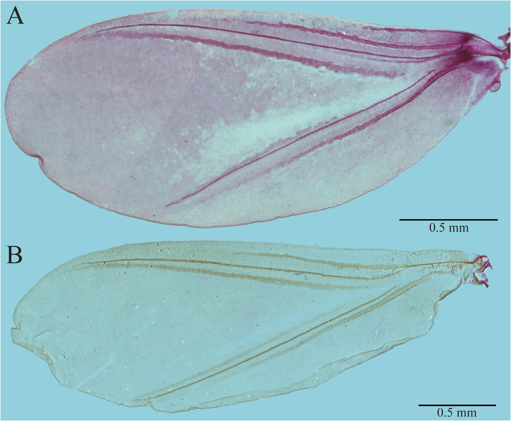
Dorsum with 1 pair of glandular pouches (gp); each glandular pouch with 2 setae, each apically acute, setae approximately same size, about 280 μm long. Multilocular disc-pores in glandular pouches, each glandular pouch with about 263 tightly clustered pores, each multilocular disc-pore with predominantly 5 loculi with some 4 loculi; discoidal pores absent, setae around margin of glandular pouches; tubular ducts (gpt) present at base of glandular pouch setae (gls). Multilocular disc-pores in cluster on prothorax, segmental row one pore wide on metathorax and abdominal segments I–VII, predominantly with 4 loculi. Hair-like setae (hs) slender, apically acute, shorter than those on venter, in segmental rows, with marginal clusters on each side of each segment; setae on pro-, meta-, and mesothorax; head setae anterior to postoccipital ridge (por), on ocular sclerite (ocs), along dorsal arm of midcranial ridge (dmcr). Microtrichia (not illustrated) on all abdominal segments. Posterior ostioles (po) present (not illustrated), poorly developed. Abdominal tergites (at) with 2 areas of sclerotization on posterior margin of each segment from abdominal segments II–V; segment VIII with heavily sclerotized yoke-shaped medial tergal plate (mtp) on anterior margin; metapostnotal sclerite present (not illustrated); metapostnotal ridge absent. Scutellum (scl) well developed, with 38 setae; scutellar ridge (sclr) well-developed. Scutum (sct) sclerotized throughout, except for medial triangular membranous area (mta) anterior to scutellum. Prescutum, prescutal suture, and prescutual setae absent. Pronotal ridges (prnr) well developed (not illustrated); pronotal sclerites (prn) present (not illustrated). Hamulohalteres each with 3 or 4 setae (not illustrated). Wings each 3.05 mm long, covered in microtrichia, each with alar lobe (al), alar sclerites (alsc), and veins (wv) including subcosta (sc) arising from alar sclerites; radius (r) joining subcosta; possible media (m) represented by sclerotized area arising 1⁄4 distance from alar sclerite, cubitus anterior (cua) present, not joined with other veins; cubitus posterior (cup) arising from alar sclerites, additional band of sclerotization anterior to cubitus anterior; without setae and sensoria. Tegula (teg) each with 20 setae. Postoccipital ridge (por) well developed, fused with postocular ridge (pocr). Dorsal arm of midcranial ridge (dmcr) narrow, touching postoccipital ridge posteriorly; with anterior swelling, connected to lateral (lmcr) arms anteriorly; surrounded by median crest (mc) with irregular sclerotization with about 2 setae, with membranous area between ocular sclerite and sclerotization surrounding dorsal arm of midcranial ridge; ocular sclerite with setae not in distinct clusters, with 18 setae on each side. With 3 dorsal simple eyes (dse) on each side, those nearest dorsal arm of midcranial ridge largest, each 53 μm long, on lateral edge smallest, each 38 μm long. Lateral ocellus (lo) about same size as lateral simple eye, each 43 μm in diameter.
Genitalia on side, anus not visible. Penial sheath (ps) 625 μm long; elongate, apically with several small papillae (pap); anterior portion with 17 long setae, each about 90 μm long, posterior portion with about 10 small setae, each about 12 μm long. Aedeagus (ae) u-shaped, attached on membranous surface, about 1.43 mm long, apex bifurcate, one side with three small denticles, other with five.
Venter with multilocular disc-pores present near anterior and posterior spiracles (not illustrated), in marginal clusters on abdominal segments II–VII, medially between mesothoracic legs (not illustrated), anterior to mesosternum (stn 2), in cluster laterad to proepisternum + cervical sclerite (pepcv) (not illustrated), in cluster (10) on unsclerotized area anterior to ocular sclerite and preocular ridge (pocr). with predominantly 4 loculi on abdomen, with predominantly five-loculi around spiracles, with both 4 and 5 loculi on head. Discoidal pores present around spiracles (not illustrated). With hair-like setae, apically acute, longer than those on dorsum, in lateral clusters and in segmental rows on abdomen, in clusters on metathorax, mesothorax, and prothorax; head setae abundant in cluster, about 35 on each side, on medial area of ocular sclerite (ocs) posterior to ventral simple eyes (vse), present (about 12) (specimen rolled) near midline of head between antennal base and ventral eye (ve) anterior to preocular ridge. Microtrichia on II–VIII (not illustrated). Abdominal sternites without sclerotization, abdominal segment VIII with sclerotization mediolaterally. Metapleural ridge well developed, with two precoxal ridge extensions, the anterior one longer, reaching the metasternal apophysis (not illustrated). Mesosternum (stn 2) well-developed, with large furca (f); lateropleurites on mesothorax triangular, with membranous area between lateropleurite and mesepisternum (not illustrated). Mesopleural ridge with precoxal ridge of mesothorax and additional posterior ridge extension anterior to mesocoxal articulation (not illustrated). Prosternum (stn 1) with sclerotized prosternal ridge that ends with a prosternal apophysis, surrounded by well-developed sclerotized triangular sclerite (not illustrated). Proepisternum and cervical sclerite (pepcv) well developed (not illustrated). Preoral ridge (pror) well developed, fused with postocular ridge (pocr). Ventral midcranial ridge (vmcr) well developed anteriorly, extending to lateral arms (lmcr) between antennae, fused with preocular ridge and preoral ridge. Ocular sclerite (ocs) sclerotized throughout, area anterior to preocular ridge unsclerotized. Mouth displaced, but apparently present (not illustrated). Cranial apophysis not clearly visible. With 4 ventral simple eyes (vse) on each side, those nearest dorsal arm of midcranial ridge largest, each 55 μm long, those on lateral edge smallest, each 41 μm long. Totaling seven pairs of simple eyes and one pair of ocelli wrapping around head. Metathoracic legs longest, coxa 303 μm long, trochanter 177 μm long, femur 647 μm long, tibia 1.00 mm long, tarsus 284 μm long, claw (c) 72 μm long, tibia/tarsus 3.5. Hind trochanter each with 4 campaniform sensilla (camp) on each surface. Claw with denticle (dt) and pair of basal spurs (bs); digitules on tarsus (tdt) acute, shorter than claw; digitules on claw (cdt) acute, shorter than claw. Spine-like setae on tarsus, tibia; long hair-like setae on tibia, femur, trochanter, and coxa; many hair-like setae on trochanter, femur, and tibia with satellite setae, short hair-like setae on all segments. Antenna likely 10-segmented; third antennal segment about 1.6 times longer than apical segment, last segment apically rounded or pointed. Elongate hair-like setae (hs) on segments II–X, many with a very small satellite seta (sats); short setae (shs) on first 3 segments, antennal bristles (not illustrated) on apical 3 segments; a pair of antennal intersegmental sensilla present at least between segments III and IV; coeloconic sensilla present at least on segments II and X.
Remarks: The male was not collected in association with the females, but both were taken in quarantine from Mexico on the same host, within the span of a year. Thus, we cannot be sure that the male described above is P. brom and the specimen is therefore not included in the type series. The description is based on a single specimen that was dissected into many pieces. Parts of the description (e.g., total body length or width of penial sheath) are missing where measurements were not able to be taken and the illustration reflects the condition of the specimen. Nineteen antennal segments were broken into ten separate pieces on the slide, making it difficult to ascertain which segments were which. Segments I–IV from the right side of the head were together and intact and the apical three segments of an unknown side were also still together. See the “Remarks” section of the P. philo adult male for comments about wing venation.
Diagnosis: The male of presumed P. brom keys out separately from any other known Puto male. It is most similar to P. simmondsiae McKenzie, 1961 but differs in that the glandular pouch of P. brom is larger, each with about 230 pores (160 pores in P. simmondsiae ). Only two other species of Puto are known from Guatemala: P. mexicanus ( Cockerell, 1893) and P. ulter Ferris, 1950 . The male of P. brom (?) has more setae on each tegula (20) than that of P. mexicanus (12) or P. ulter (8). Only two species of Puto are presently known on bromeliads, P. ulter and P. barberi . In addition to having fewer setae on the tegula, P. ulter also has clubbed tarsal digitules while P. brom (?) has simple tarsal digitules. Puto barberi is apparently parthenogenetic, no males are known ( Villegas García et al. 2013).

| USNM |
Smithsonian Institution, National Museum of Natural History |
No known copyright restrictions apply. See Agosti, D., Egloff, W., 2009. Taxonomic information exchange and copyright: the Plazi approach. BMC Research Notes 2009, 2:53 for further explanation.
|
Kingdom |
|
|
Phylum |
|
|
Class |
|
|
Order |
|
|
Family |
|
|
Genus |