
Scalibregma lanai, Mendes & Rizzo & Paiva, 2023
|
publication ID |
https://doi.org/ 10.11646/zootaxa.5353.5.3 |
|
publication LSID |
lsid:zoobank.org:pub:7E9D426F-D88A-4F70-9067-683A4C9A653A |
|
DOI |
https://doi.org/10.5281/zenodo.10010293 |
|
persistent identifier |
https://treatment.plazi.org/id/C29BD4BF-0121-43B1-8B1B-B041040BCFE8 |
|
taxon LSID |
lsid:zoobank.org:act:C29BD4BF-0121-43B1-8B1B-B041040BCFE8 |
|
treatment provided by |
Plazi |
|
scientific name |
Scalibregma lanai |
| status |
sp. nov. |
Scalibregma lanai sp. nov.
urn:lsid:zoobank.org:act:C29BD4BF-0121-43B1-8B1B-B041040BCFE8
Figures 3–4 View FIGURE 3 View FIGURE 4 , Figure 5 View FIGURE 5 (A–C)
Type material. UERJ4662 (Holotype) : AMBES7 A1 R1 , Lat: -21.046058 Long: -40.541447, 25 m, 22 Jan 2012 ; UERJ4617 (Paratypes) : AMBES14 E2 R3 , Lat: -19.301700 Long: -39.389819, 38 m, 15 Jul 2013, 2 specimens . MNRJP007674 (Paratype): SANSED5H3 R3 , Lat: -23.0353 Long: -42.2260, 75m, 01 Nov 2011 ; MNRJP007675 (Paratype): SANSED5E4 R1 , Lat: -24.20626491 Long: -44.8077, 100m, 09 Nov 2019, 1 specimen .
Additional material. Ambes Project: UERJ4649 : AMBES7 A2 R2 , Lat: -21.057539 Long: -40.383225, 40 m, 22 Jan 2012, 1 specimen ; UERJ 8746 : FOZ14 R1 , Lat: -19.708947 Long: -39.649267, 38 m, 14 Dec 2010, 1 specimen mounted to SEM. UERJ9009 : FOZ13, Lat: -19.792453 Long: -39.720856, 41 m, 15 Dec 2010, 1 specimen .
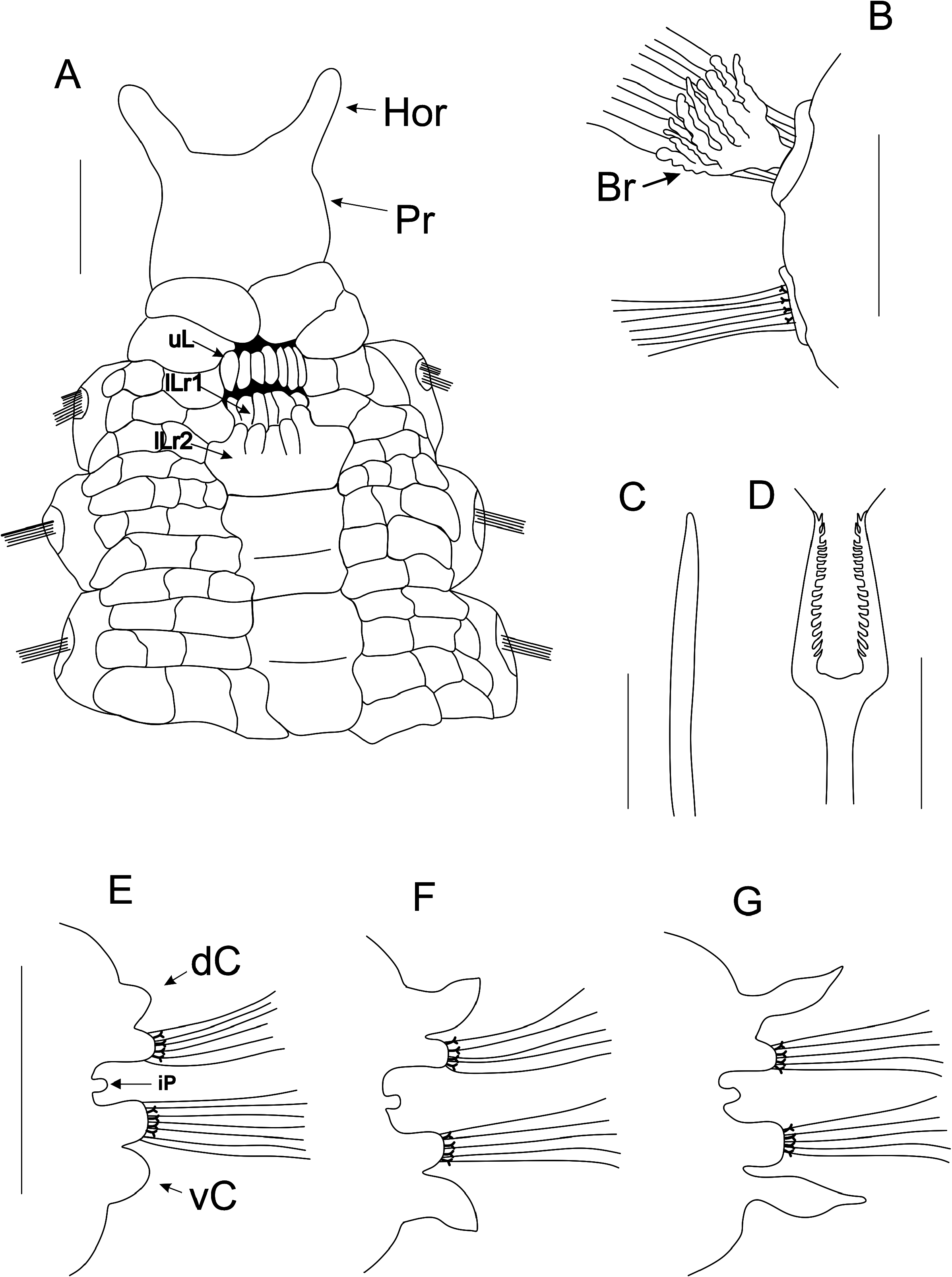
Diagnosis. Trapezoidal prostomium, T-shaped, with two short lateral horns. Eyes present as two dorsal patches. Nuchal organs present as a sausage-like everted structure, with internal grooves. Mouth formed by a single row of paired lobes on upper lip and two rows of paired lobes on lower lip. Branched branchiae present from chaetiger 3–5. Dorsally and ventrally, chaetiger 1–2 triannulate becoming quadriannulate to pentannulate on posterior chaetigers. Blunt, curved apically spinous chaetae present on chaetiger 1–2 anterior to capillaries and numbering 8–9, being replaced by lyrate chaetae with equal tynes on chaetiger 3.
Description. Holotype 14 mm long and 1–4 mm wide for 34 chaetigers, pygidium absent. Paratypes measuring 10–15 mm long for 0.1–3.5 mm wide for 28–30 chaetigers. Large species, adults 13–20 mm long to 1–5 mm wide across 35–43 chaetigers. Body arenicoliform, expanded on chaetiger 4–5 up to mid-body segments. Color in alcohol, pale tan. Body surface covered by annulated secondary rings, each one formed by numerous rectangular pads anteriorly and quadrangular to rounded pads posteriorly, giving a complex areolate appearance. These pads may have small individual dark blue or black glands inside, consisted of entangled thin cellular tubules.
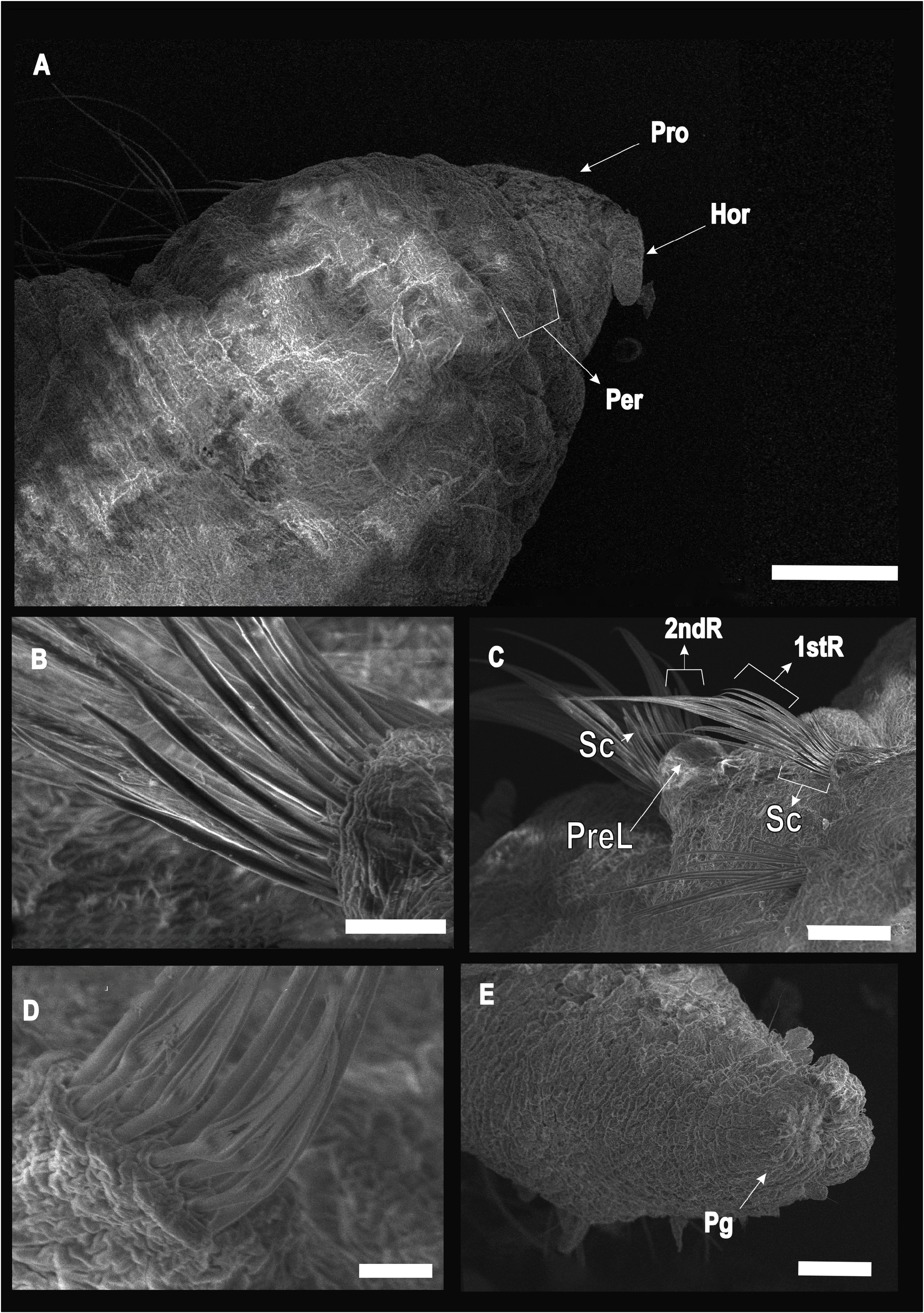
Trapezoidal prostomium, T-shaped, with two short lateral horns ( Figs 3A View FIGURE 3 ; 4A View FIGURE 4 ). Eyes present as two dorsal patches ( Fig. 5B View FIGURE 5 ). Nuchal organs present as a sausage-like everted structure, with internal grooves. Peristomium achaetous, biannulate dorsally and ventrally ( Figs 3A View FIGURE 3 ; 5A–B View FIGURE 5 ). Peristomium may have small rectangular pads around each annulus ( Figs 3A View FIGURE 3 ; 5A–B View FIGURE 5 ). Mouth formed by a single row of paired lobes on upper lip and two rows of paired lobes on lower lips ( Figs 3A View FIGURE 3 ; 5A View FIGURE 5 ). Upper lip row formed by up to seven paired lobes. Lower pair of rows on inferior lip with 4–6 paired lobes on superior most row and 5–7 on inferior most row, formed by contribution of superior portion of ventral groove’s first pad ( Figs 3A View FIGURE 3 ; 5A View FIGURE 5 ). Ventral groove starts from inferior-most row of lower lip with a large single pad paired with chaetiger 1 ( Figs 3A View FIGURE 3 ; 5A View FIGURE 5 ). Proboscis smooth.
Dorsally and ventrally, chaetigers 1–2 triannulate, then quadriannulate to pentannulate on far posterior chaetigers ( Figs 3A View FIGURE 3 ; 5A–B View FIGURE 5 ). Ventral groove from chaetiger 1 with each body segment having a single pad merging with following one, forming a mid-ventral ridge line continuing to end of body. First pad trapezoidal, fused to mouth’s lower lip, sometimes subdivided two times ( Figs 3A View FIGURE 3 ; 5A–B View FIGURE 5 ); following pads may be subdivided two or three times, forming bi- to quadriannulate quadrangular to rectangular slender blocks ( Figs 3A View FIGURE 3 ; 5A–B View FIGURE 5 ).
Branched branchiae present from chaetiger 3–5, posterior to capillary fascicles on notopodia ( Figs 3B View FIGURE 3 ; 5B View FIGURE 5 ). First and second pair similar in size but smaller than third one. First chaetiger small, conspicuous, formed by a main strong rhomboid pad sustaining three smaller rounded pads ( Figs 3A View FIGURE 3 ; 5A–B View FIGURE 5 ). From middle pad emerges a small interramal papillate organ; from superior and inferior pads noto and neuropodial lobes emerge as parapodia each with two rows of capillary chaetae ( Figs 3A View FIGURE 3 ; 5A–B View FIGURE 5 ). Capillaries present in two rows anteriorly, with fine bristles sparsely distributed ( Fig. 4C View FIGURE 4 ); 8–9 short spinous chaetae present from chaetiger 1–2 anterior to capillaries and near their basis ( Figs 3C View FIGURE 3 ; 4B–C View FIGURE 4 ), being replaced by lyrate chaetae with equal tynes on chaetiger 3 ( Figs. 3D View FIGURE 3 ; 4D View FIGURE 4 ). Posterior chaetigers following same parapodial pattern, but larger, with a conspicuous knob-like interramal papilla and slender parapodial lobes ( Fig. 3E–G View FIGURE 3 ). Posterior region with a single row of capillaries and 4–6 lyrate chaetae. Ventral and dorsal cirri present from chaetigers 14–15. Parapodial cirri with a gradation in size and shape, with the first pair small, rounded and distally pointed, becoming larger and lanceolate in posterior parapodia ( Fig. 3E–G View FIGURE 3 ).All annuli pads with a single patch of entangled dark blue or black glands dorsally and ventrally ( Fig. 5A–B View FIGURE 5 ). Glands consisting of robust entangled cellular patches, covering inner portion of each parapodial cirrus entirely, without dorsal or ventral differentiation ( Fig. 5A–C View FIGURE 5 ). Last 2–3 segments cirriferous and achaetous. Pygidium with a long crenulated margin formed by several lobes ( Fig. 4E View FIGURE 4 ), with three long anal cirri, two ventral and one mid-ventral.

No known copyright restrictions apply. See Agosti, D., Egloff, W., 2009. Taxonomic information exchange and copyright: the Plazi approach. BMC Research Notes 2009, 2:53 for further explanation.
|
Kingdom |
|
|
Phylum |
|
|
Class |
|
|
Family |
|
|
Genus |