
Skeletonema pseudocostatum Medlin emend . Zingone et Sarno
|
publication ID |
https://doi.org/10.11646/phytotaxa.607.3.2 |
|
DOI |
https://doi.org/10.5281/zenodo.8243243 |
|
persistent identifier |
https://treatment.plazi.org/id/1F6E031E-FFD6-5675-BCDB-0ED5DB05F95C |
|
treatment provided by |
Plazi |
|
scientific name |
Skeletonema pseudocostatum Medlin emend . Zingone et Sarno |
| status |
|
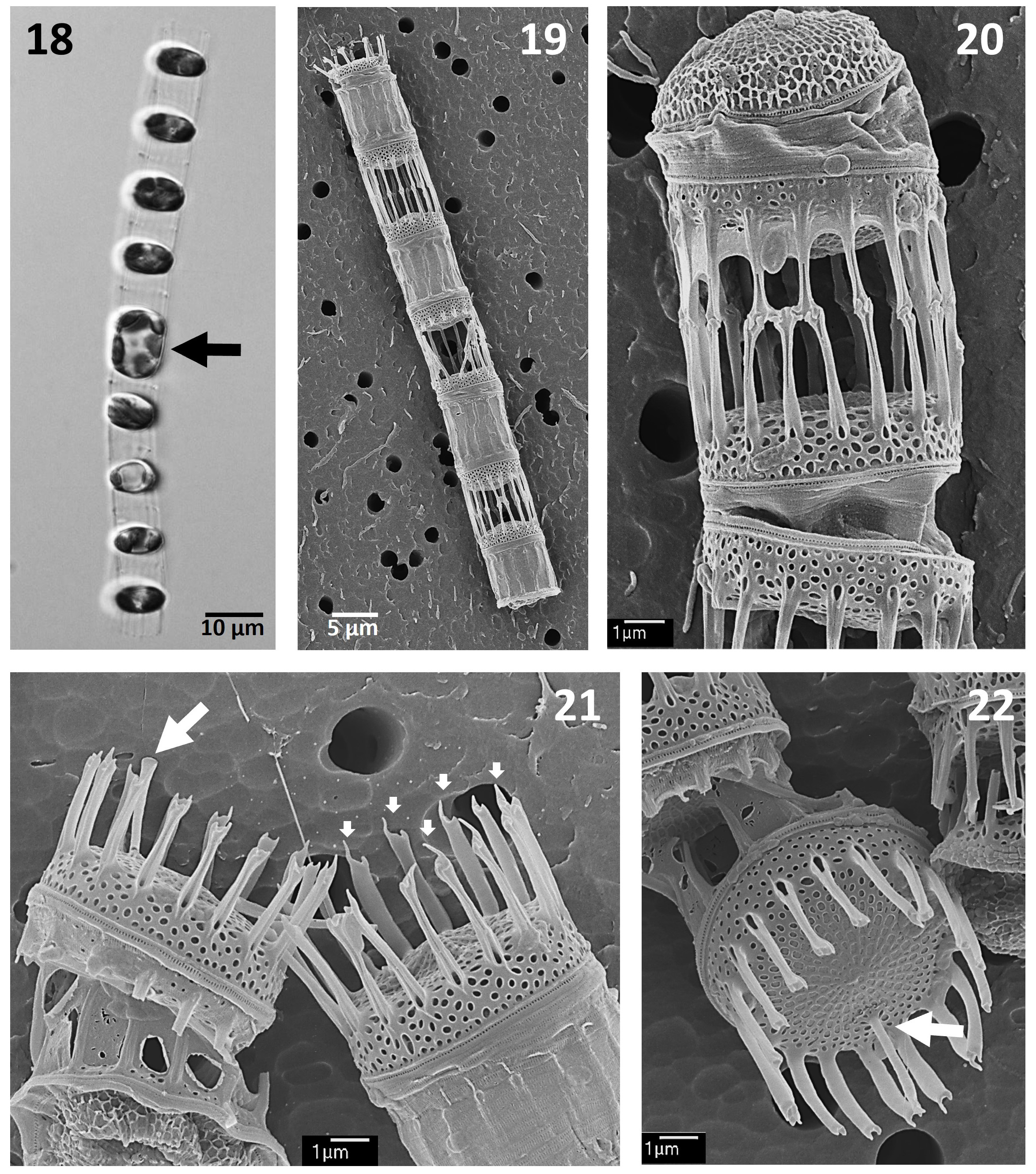
Skeletonema pseudocostatum Medlin emend. Zingone et Sarno
Figs 18–22 View FIGURES 18–22
References: Medlin et al. 1991, p. 522; Sarno et al. 2005, p. 162, figs 7, A–I; Gu et al. 2012, p. 254, figs 21–23; Hernández-Becerril et al.
2013, p. 81, figs 16–25.
Description: Chains are relatively long and straight (slightly curved), with 4–9 cells per chain ( Figs 18, 19 View FIGURES 18–22 ). Cells have two chloroplasts ( Fig. 18 View FIGURES 18–22 ). The valves are flat to slightly convex, with a relatively high valve mantle ( Figs 19–21 View FIGURES 18–22 ). The areolae are almost quadrangular to rectangular on the valve face and become pseudoloculate near the mantle ( Figs 20–22 View FIGURES 18–22 ). Apart from the observations of the valvocopula, with longitudinal elongate poroids and ribs ( Figs 20–22 View FIGURES 18–22 ), other copulae were not seen in detail.
All fultoportulae are open ( Figs 20, 21 View FIGURES 18–22 ), although some IF’s and TF’s have their bases closed and are more tubular ( Figs 20, 22 View FIGURES 18–22 ), and the tips of TF’s have a long, very characteristic spine ( Fig. 21 View FIGURES 18–22 , small arrows). Each IF fuses to one or, less commonly, two of the sibling valves ( Figs 19, 20 View FIGURES 18–22 ). Terminal rimoportula locates close to the margin, and has a long tube, with a slightly inflated tip ( Figs 21, 22 View FIGURES 18–22 , arrows), whereas the IR is marginal, but very short.
Measurements: cell diameter 6.4–9.2 μm, areolae density 33–40 in 10 μm ( Table 1 View TABLE 1 ).
No known copyright restrictions apply. See Agosti, D., Egloff, W., 2009. Taxonomic information exchange and copyright: the Plazi approach. BMC Research Notes 2009, 2:53 for further explanation.
|
Kingdom |
|
|
Phylum |
|
|
Class |
|
|
Order |
|
|
Family |
|
|
Genus |