
Mastigini, Fleming, 1821
|
publication ID |
https://doi.org/10.11646/zootaxa.4453.1.1 |
|
publication LSID |
lsid:zoobank.org:pub:866690A9-0462-4892-AE29-9AAC623F87B3 |
|
DOI |
https://doi.org/10.5281/zenodo.5976954 |
|
persistent identifier |
https://treatment.plazi.org/id/2161879C-FF80-8A41-FF7A-335E6062DADE |
|
treatment provided by |
Plazi |
|
scientific name |
Mastigini |
| status |
|
Tribe Mastigini Fleming
Mastigoidae Fleming, 1821: 49 (incorrect original spelling). Type genus: Mastigus Latreille, 1802 . Note: priority of Mastiginae over Clidicinae was indicated by Newton & Thayer (1992).
Mastigini Reitter, 1882b: 142 . Type genus: Mastigus Latreille, 1802 . Tribe was redefined by Jałoszyński et al. (2018).
Diagnosis. Mastigini differ from all remaining Mastigitae in unique autapomorphies: head with sharply marked median longitudinal groove on posteriorly impressed vertex; maxillary palpomere IV inversely suboval or indistinctly subtriangular, slightly asymmetrical, broadened from base to about distal third and with broadly rounded subtriangular apex, longer than broad; submentum with many setae, lacking one outstanding pair of anterolateral setae (but this character was not possible to see in †Baltostigini and in extinct Mastigini ); mentum anteriorly very deeply emarginate, so that anterolateral corners form triangular and usually pointed lobes projecting anterad (also this character was not possible to examine in †Baltostigini and extinct Mastigini ); first visible abdominal sternite firmly fused with metaventrite; and aedeagus with asymmetrical median lobe and asymmetrical parameres, with one paramere distinctly shorter than the other one (in some cases only one paramere is visible, the other one is either vestigial or completely obliterated), aedeagus in repose asymmetrically rotated inside abdomen, flagellum very long, with several coils, endophallus permanently everted on tip of elongate copulatory piece; larva with setose frontal impression; larval antennomere II subdivided into three sections; larval antennomere III vestigial, developed as a barely discernible papilla near much longer accessory appendage of palpomere III; larval palpomeres I and II with conspicuously long spines; and larva lacking urogomphs (but larva unknown in one extant and one extinct genus). Additionally, Mastigini share with †Baltostigini synapomorphies not known in other Mastigitae: enlarged and ventrally spiny scape and pedicel; elongate head; narrowly separated antennal insertions; and pronotum lacking posterior collar and antebasal pits.
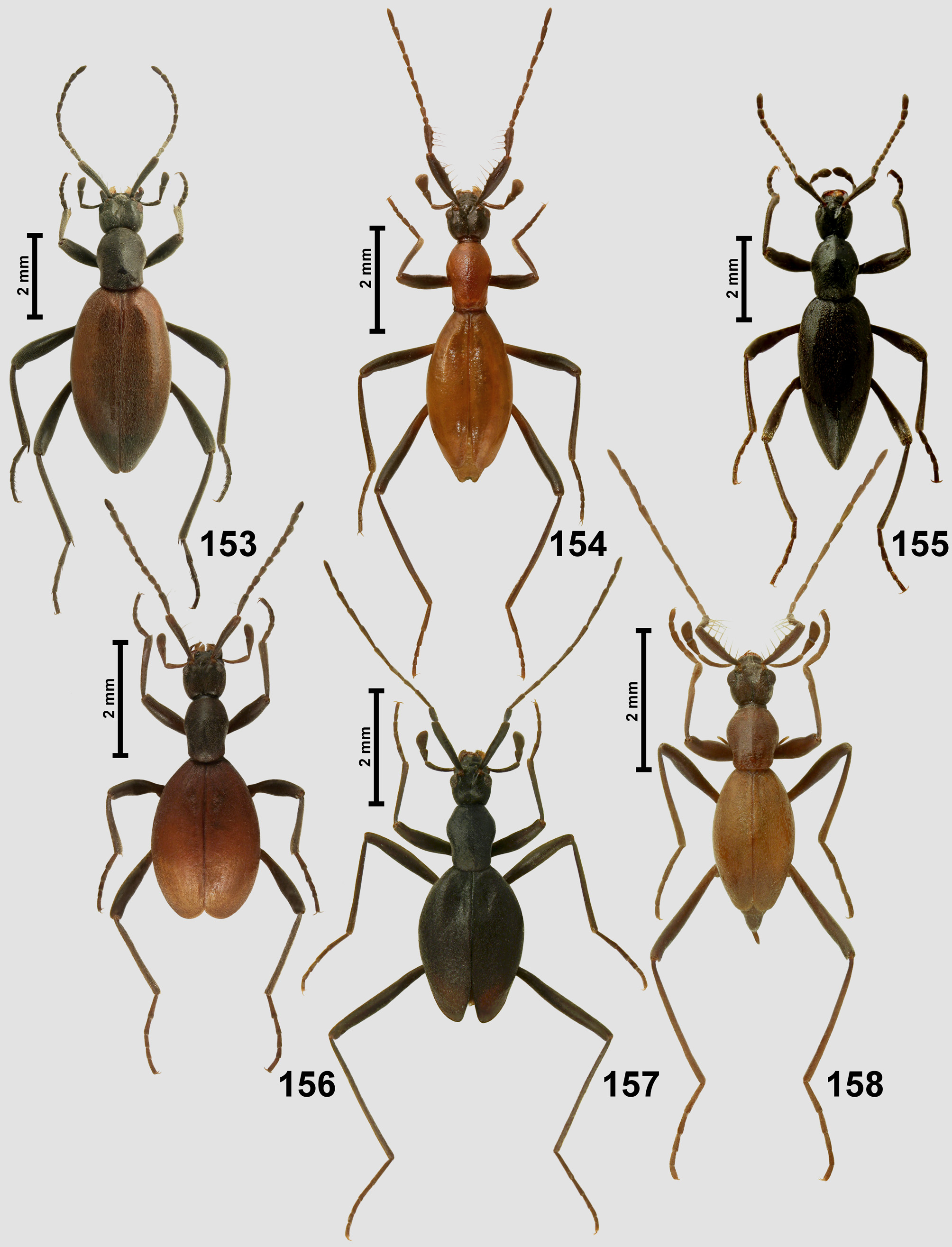
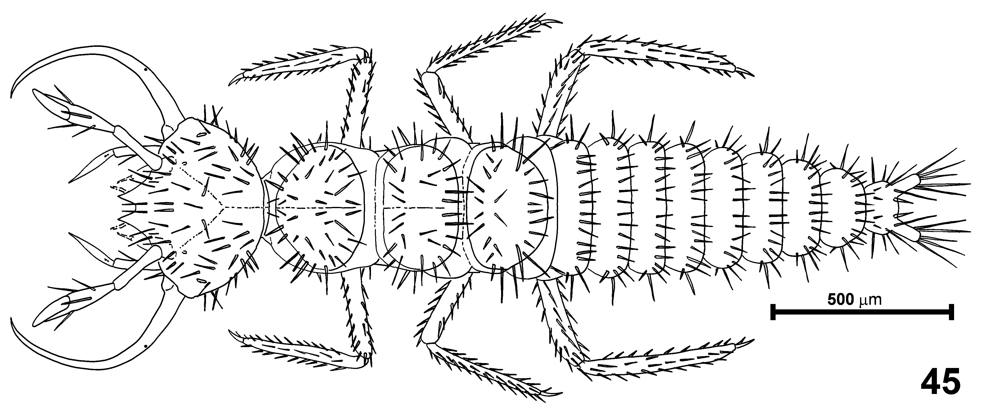
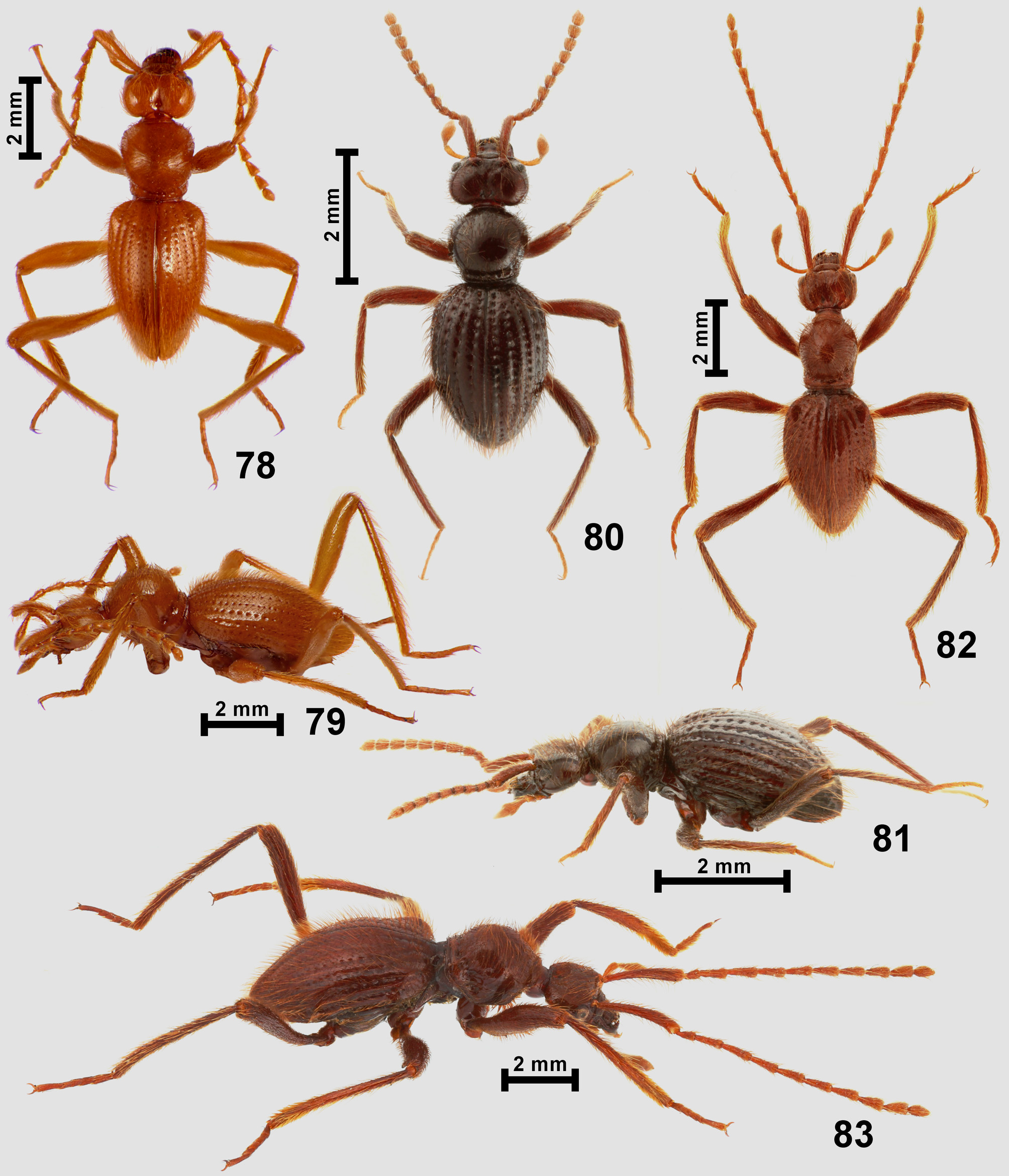
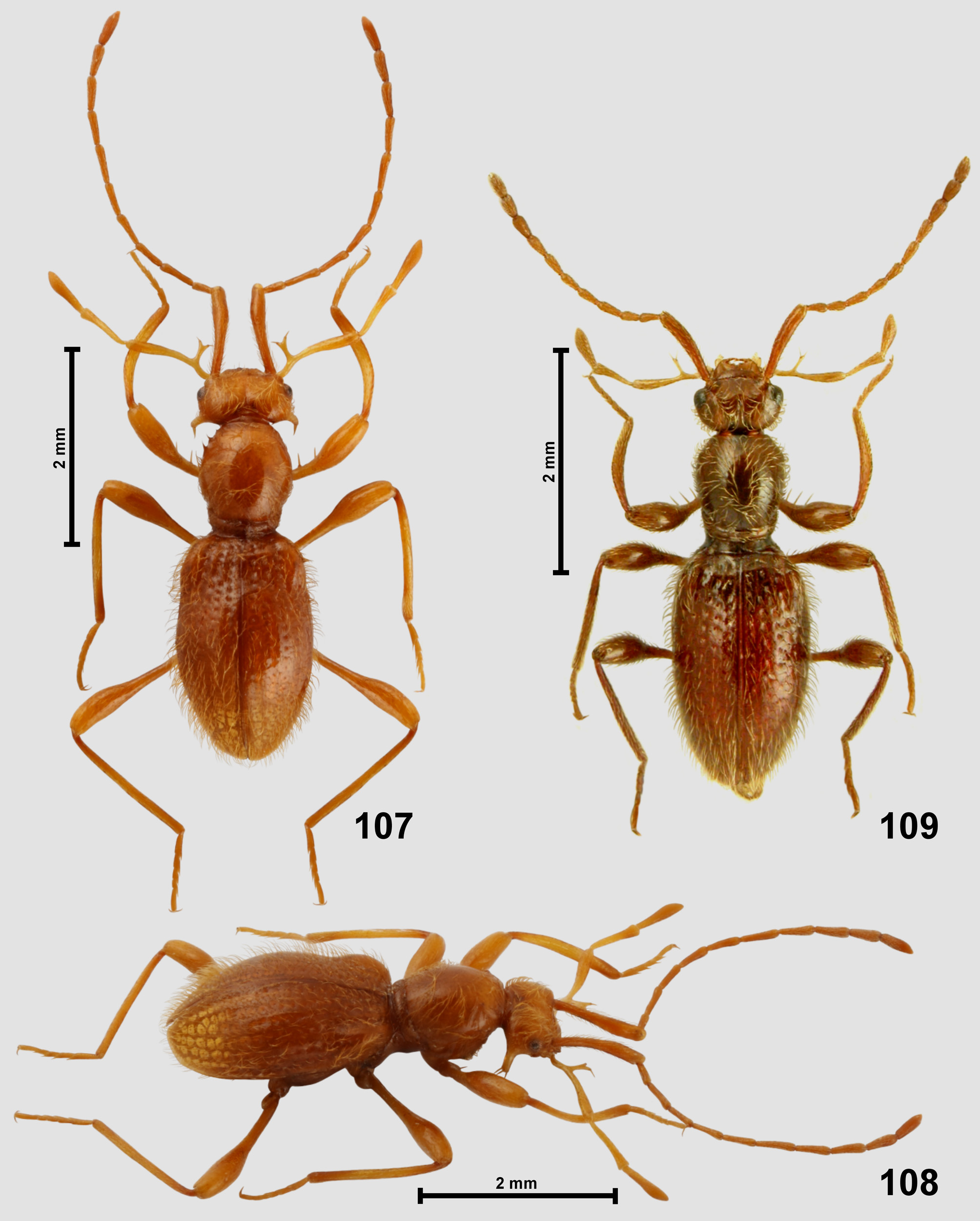
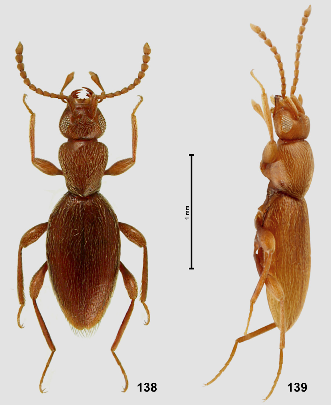
Characteristics. Adults. Body ( Figs 153–163 View FIGURES 153–158 View FIGURES 159–162 View FIGURE 163 ) large, 3.10̄ 7.50 mm in length, yellowish-brown to black, in some species head or head and pronotum dark brown to nearly black and elytra testaceous, brown or reddish, strongly convex, dorsally densely but finely setose, setae short (often extremely so) and recumbent, unmodified except for long and thick bristles on scape and pedicel.
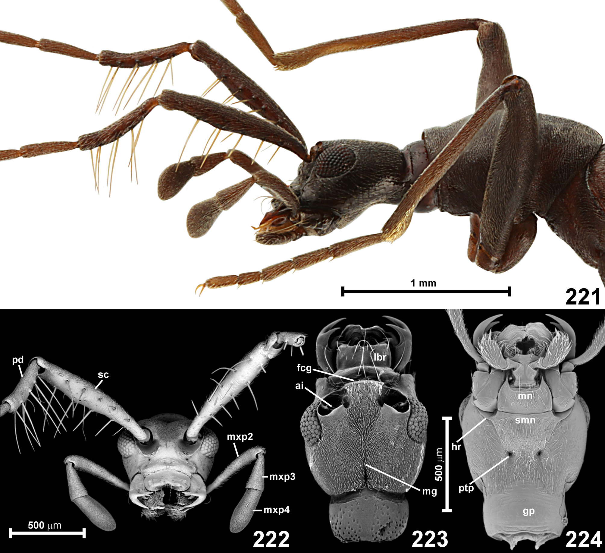
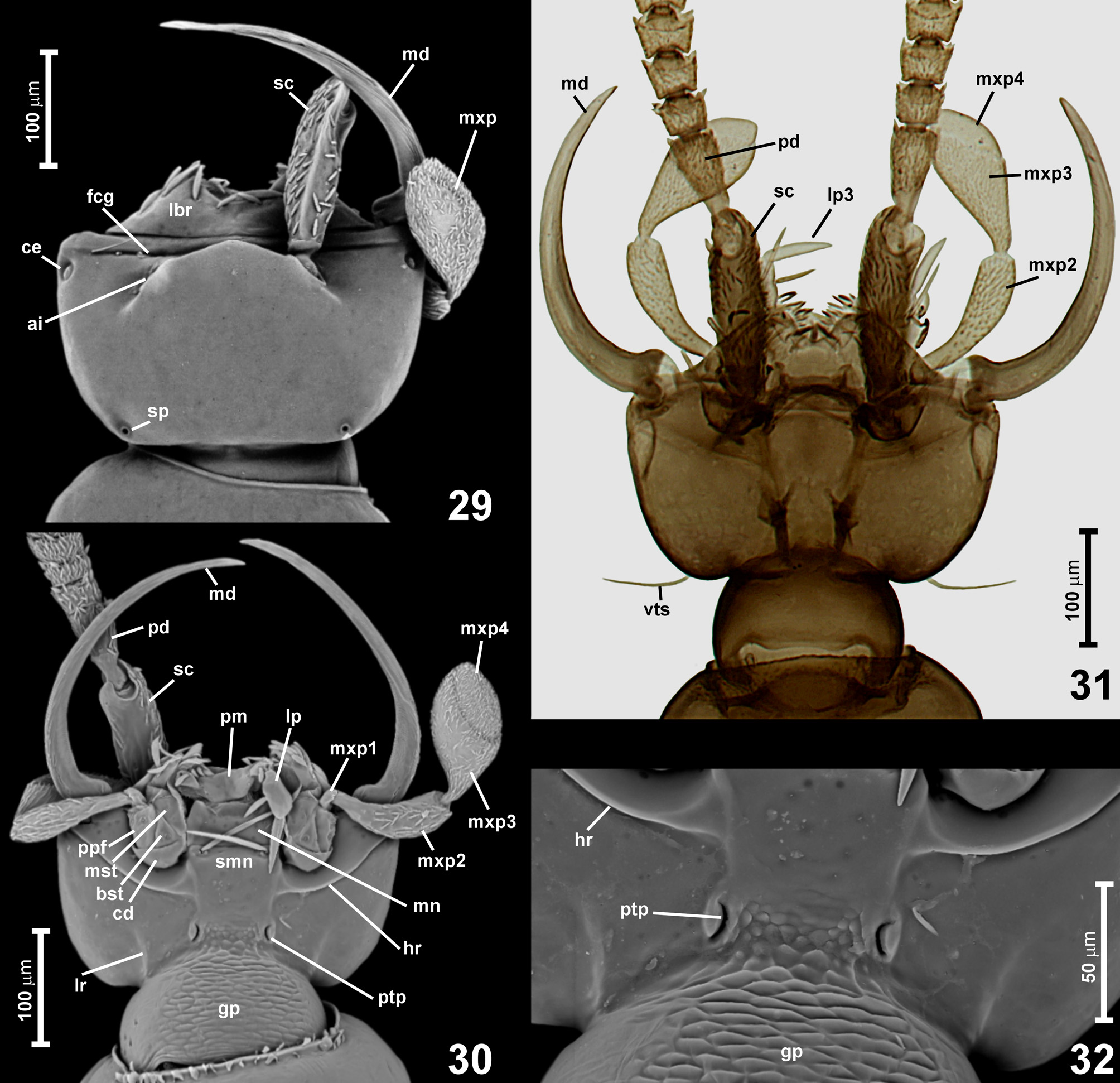
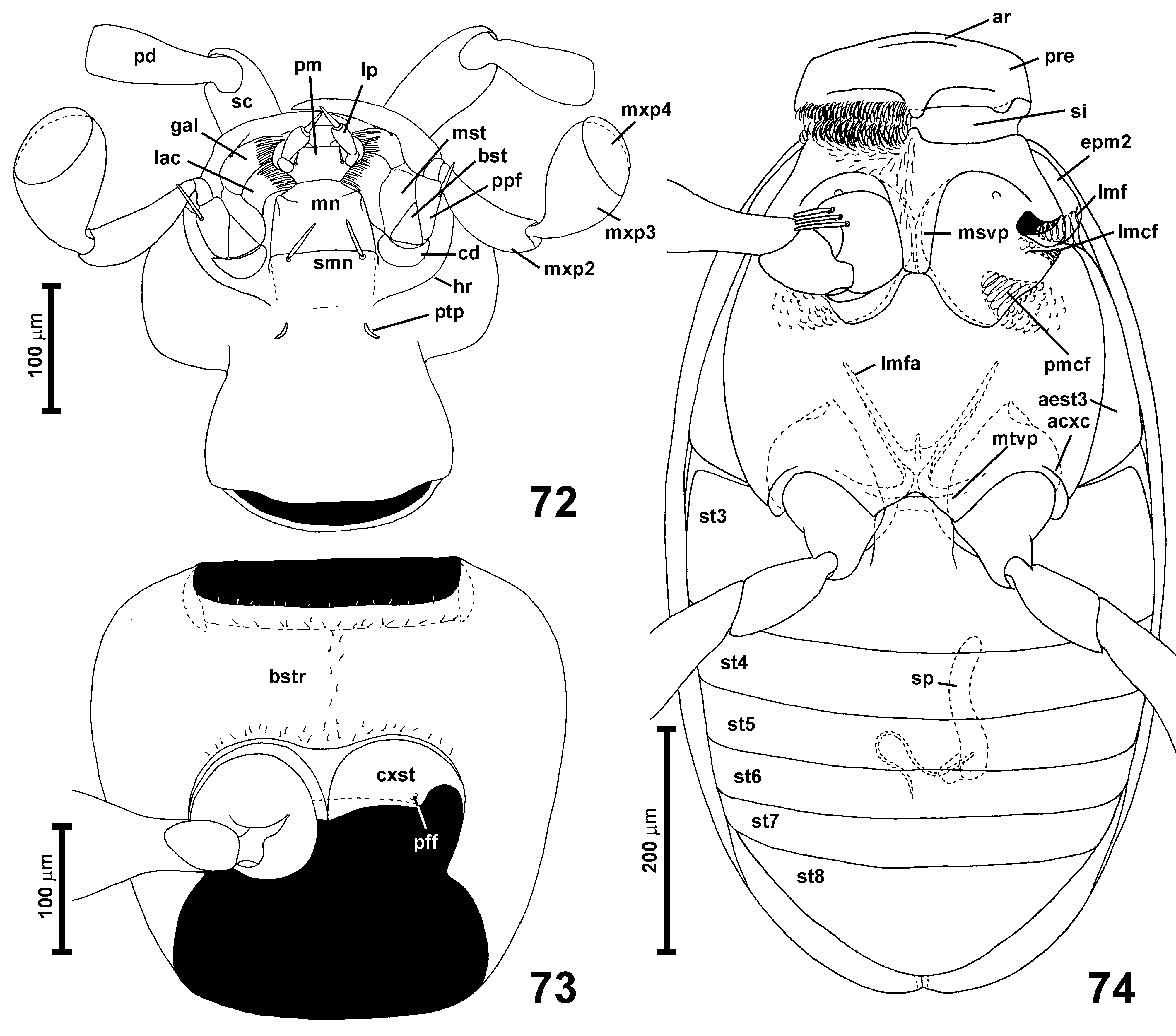
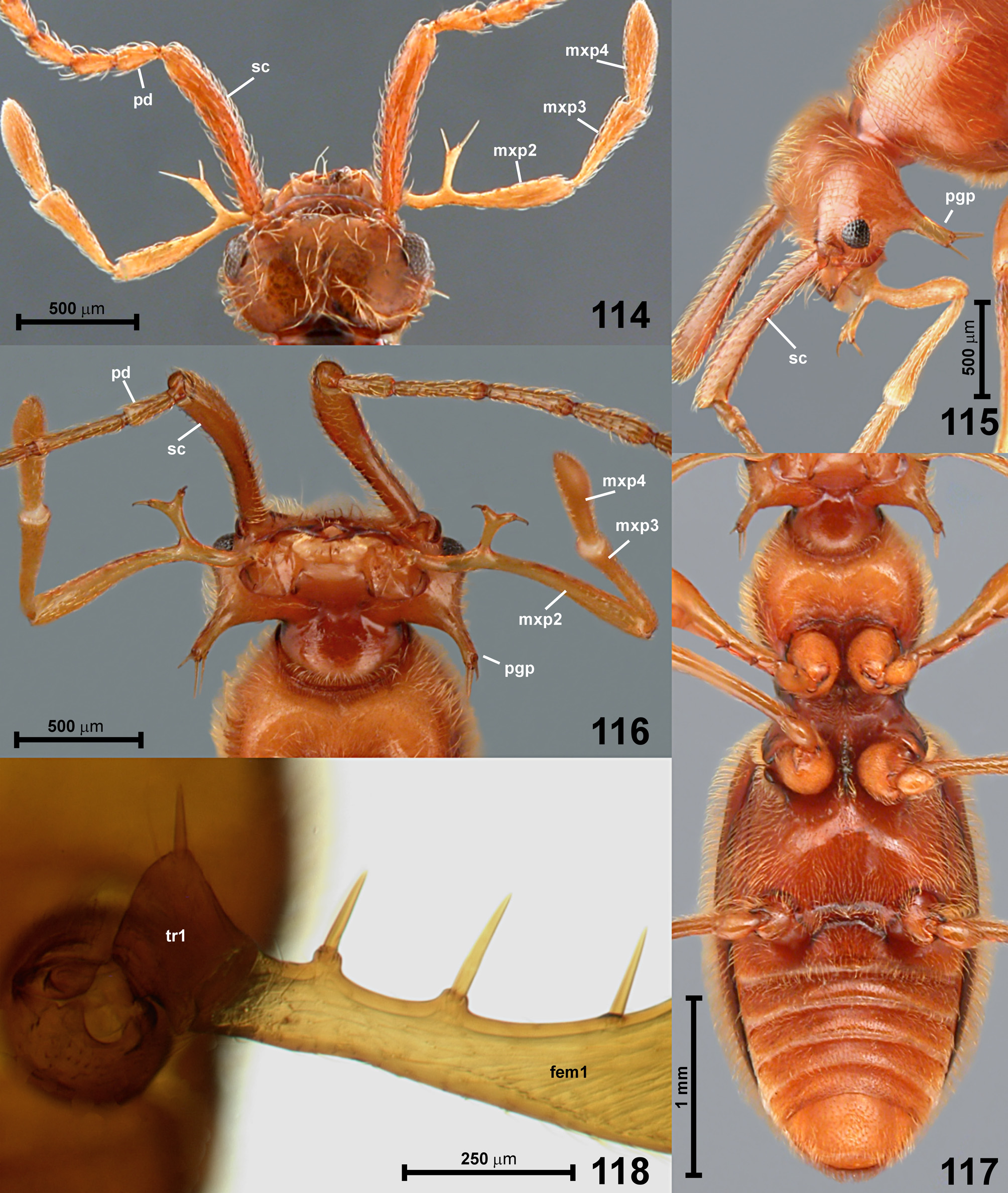
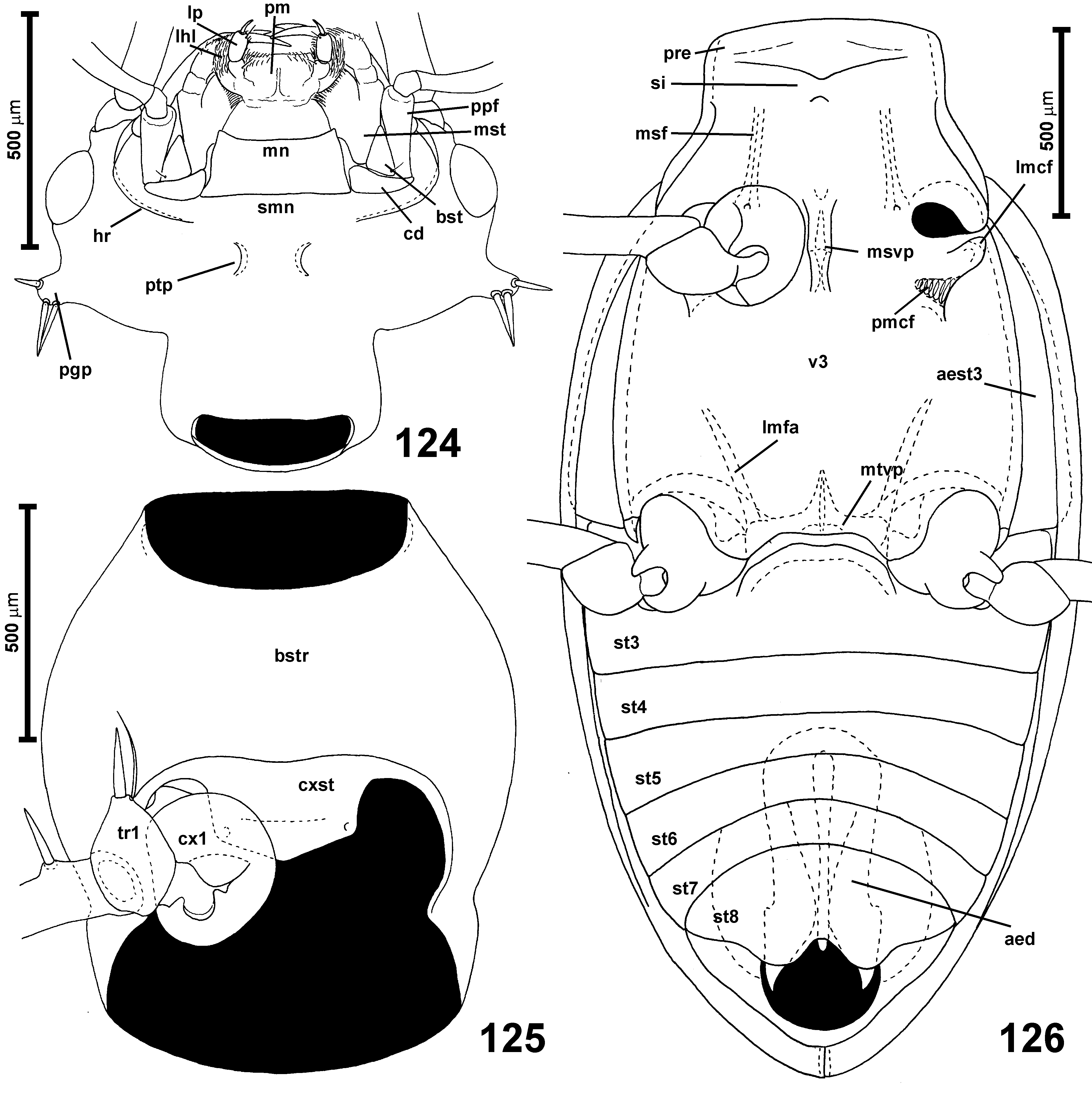
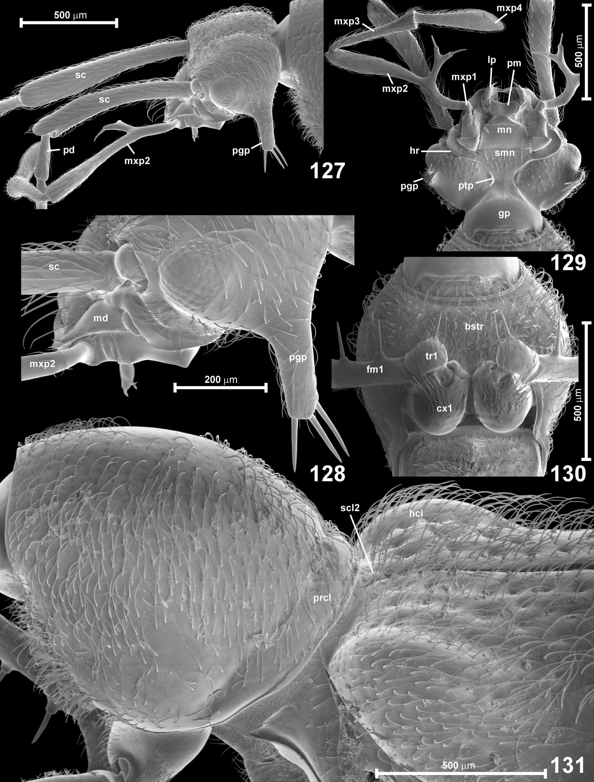
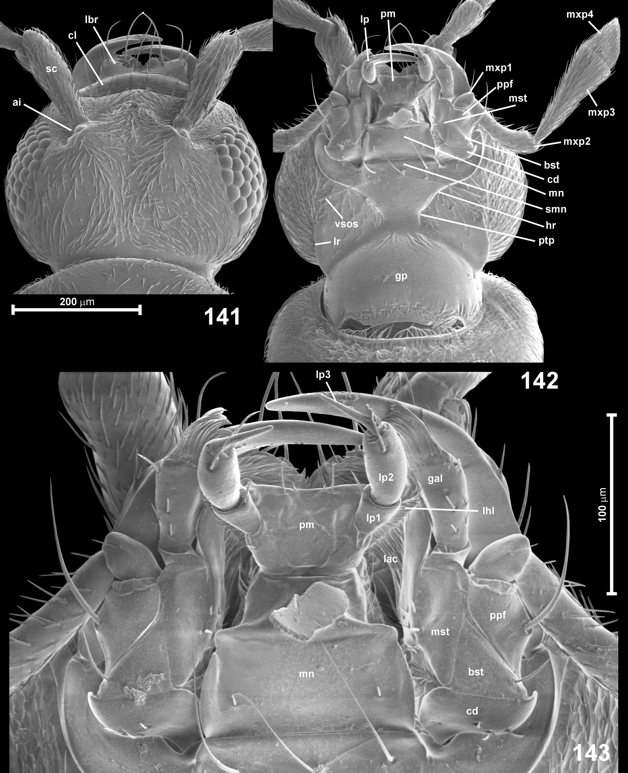
Head capsule ( Figs 167–168 View FIGURES 167–171 , 172 View FIGURES 172–175 , 176–177 View FIGURES 176–179 , 221–224 View FIGURES 221–224 ) divided into large and exposed anterior part and much smaller, subcylindrical 'neck' region retracted into prothorax and demarcated by distinct occipital constriction; 'neck' region much broader than half width of head. Anterior part of head flattened, subequal in width with prothorax, distinctly elongate, broadest near middle. Composite eyes dorsolateral, moderately large, composed of numerous small ommatidia, not projecting or weakly projecting from silhouette of the head, broadly separated from mandibular bases. Vertex and frons divided by a distinct median longitudinal groove ( Figs 167 View FIGURES 167–171 , 176 View FIGURES 176–179 , 223 View FIGURES 221–224 ; mg); vertex strongly transverse, convex at sides, with posterior margin nearly straight or slightly concave. Tempora much longer than eyes, weakly rounded. Frons between antennal insertions not forming a demarcated 'platform', weakly convex or flattened, anteriorly demarcated by a deep and very short frontoclypeal groove largely obliterated at sides. Clypeus very short and broad, with nearly straight sides slightly convergent anterad or parallel. Antennal insertions ( Figs 176 View FIGURES 176–179 , 223 View FIGURES 221–224 ; ia) located anterodorsally, relatively narrowly separated. Gular plate ( Figs 172 View FIGURES 172–175 , 177 View FIGURES 176–179 , 224 View FIGURES 221–224 ; gp) lacking sutures, indistinctly transversely reticulate; posterior tentorial pits ( Figs 172 View FIGURES 172–175 , 177 View FIGURES 176–179 , 224 View FIGURES 221–224 ; ptp) circular or oval, in front of broad and diffuse transverse impression demarcating 'neck' region ventrally; hypostomal ridges ( Figs 177 View FIGURES 176–179 , 224 View FIGURES 221–224 ; hr) arcuate, posteriorly reaching middle between anterior submental margin and posterior tentorial pits. Head finely to strongly punctate, densely setose (e.g. Fig. 176 View FIGURES 176–179 ).
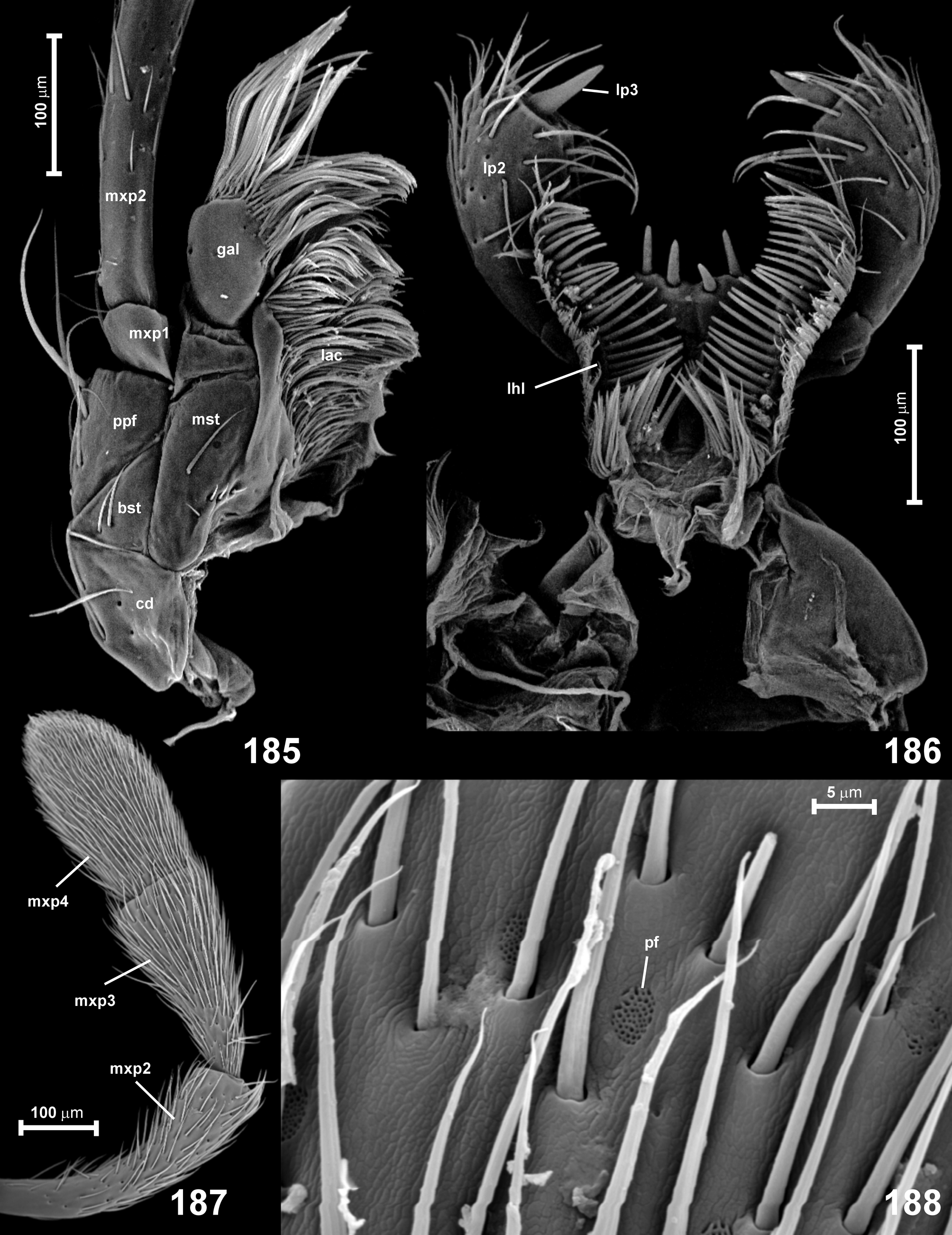
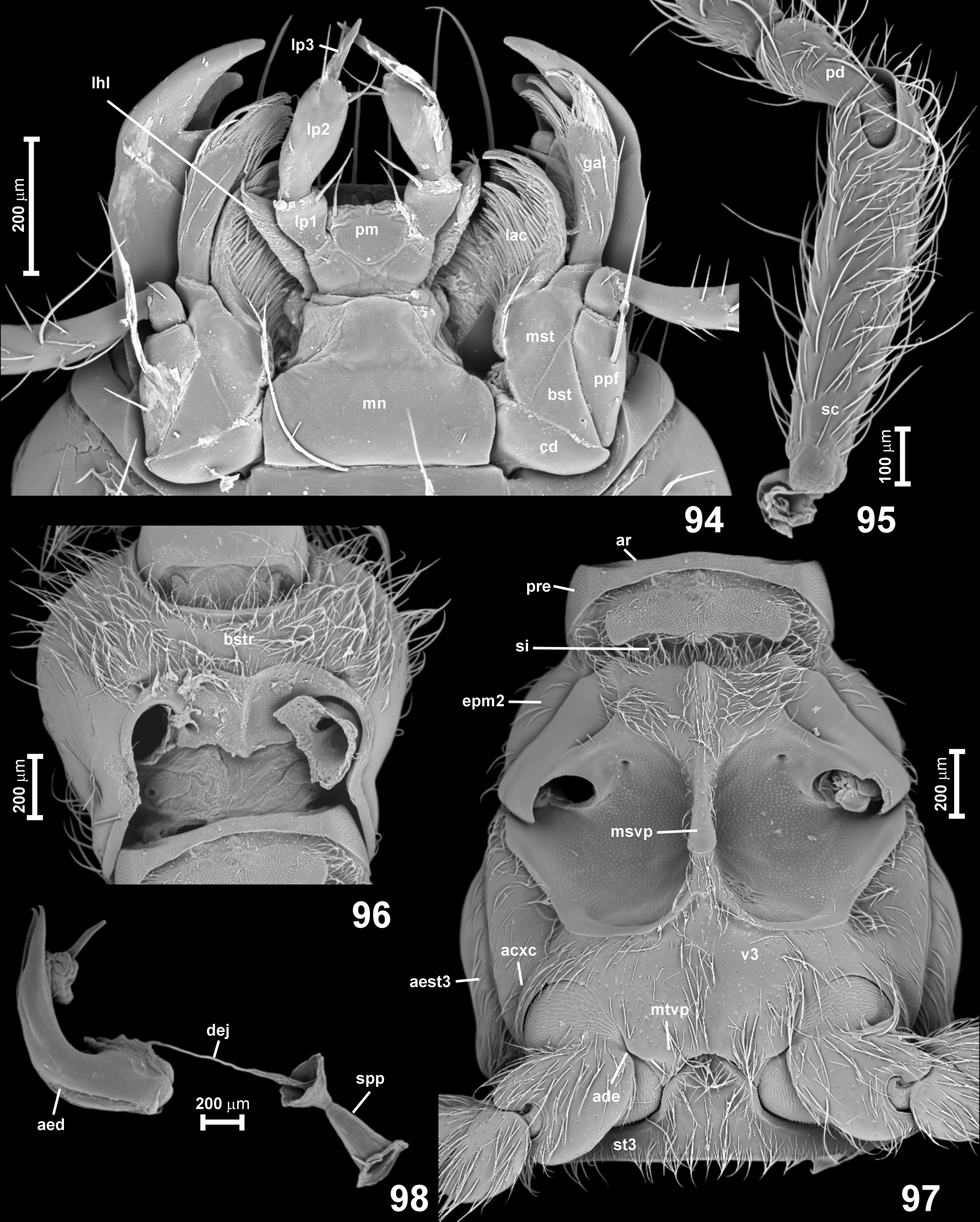
Antennae ( Figs 153–163 View FIGURES 153–158 View FIGURES 159–162 View FIGURE 163 , 168–170 View FIGURES 167–171 , 178–179 View FIGURES 176–179 , 221–222 View FIGURES 221–224 ) long and slender, shorter than body or subequal in length; scape ( Figs 168–169 View FIGURES 167–171 , 178 View FIGURES 176–179 , 222 View FIGURES 221–224 ; sc) 5–10 or even more times as long as broad, thickened, much longer than head, with lateroventral (more ventral than lateral) emargination; pedicel ( Figs 168–169 View FIGURES 167–171 , 178 View FIGURES 176–179 , 222 View FIGURES 221–224 ; pd) also conspicuously enlarged, slightly to much shorter and narrower than scape, typically 5–8 times as long as broad, broadening from narrow base to subapical region. Both scape and pedicel with two ventral longitudinal rows of several long and thick bristles with papillate insertions, area between bristles with variously densely distributed porous fields ( Figs 170 View FIGURES 167–171 , 179 View FIGURES 176–179 ; pf; unknown in fossils); antennomeres III–XI distinctly narrower than pedicel, elongate (often strongly so), each slightly thickened distad, basal stalks not exposed in intact beetles, basal rings absent or indistinct; antennomere XI elongate and indistinctly asymmetrical. Antennomeres covered with variously dense, long setae; surface of antennomeres smooth.
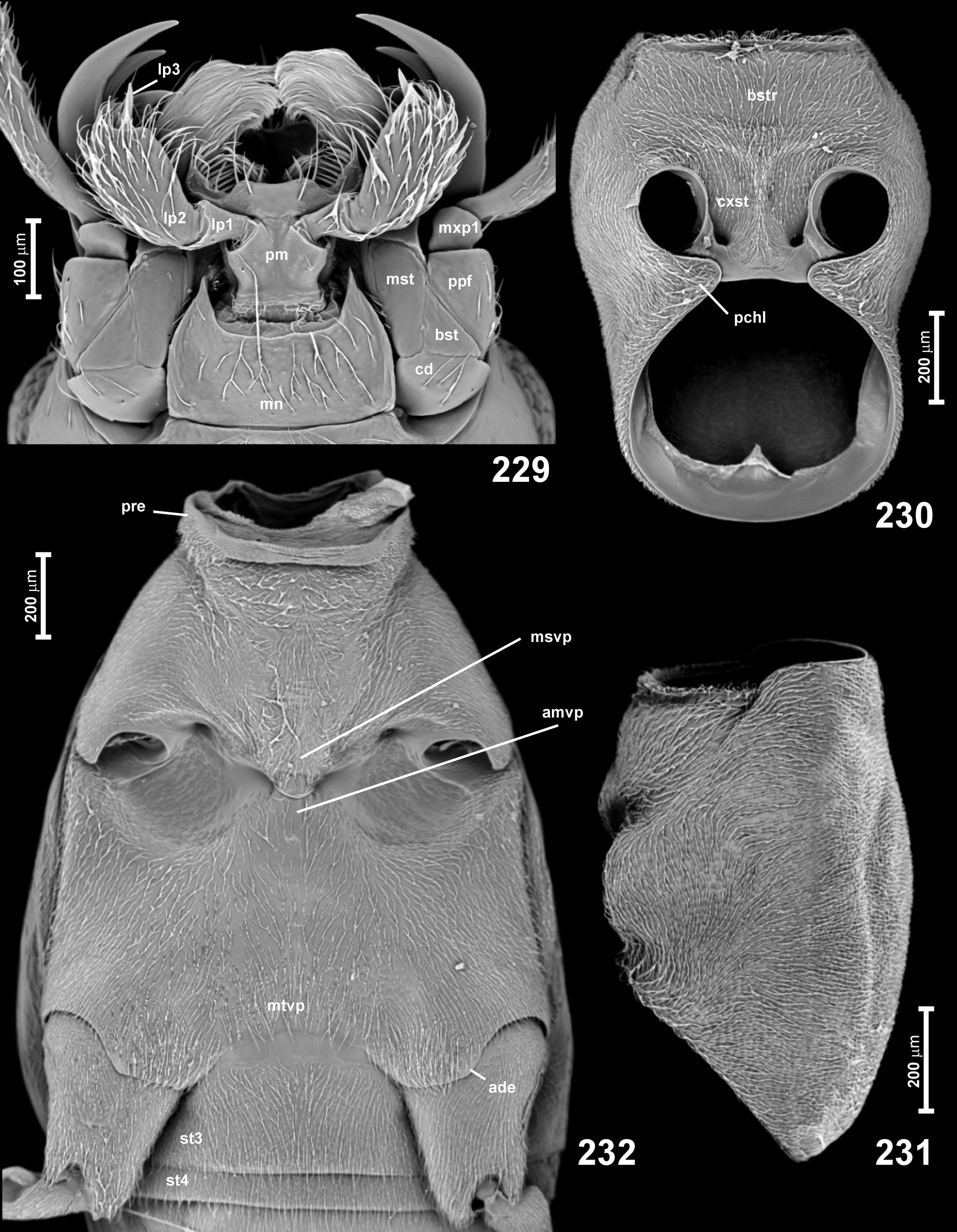
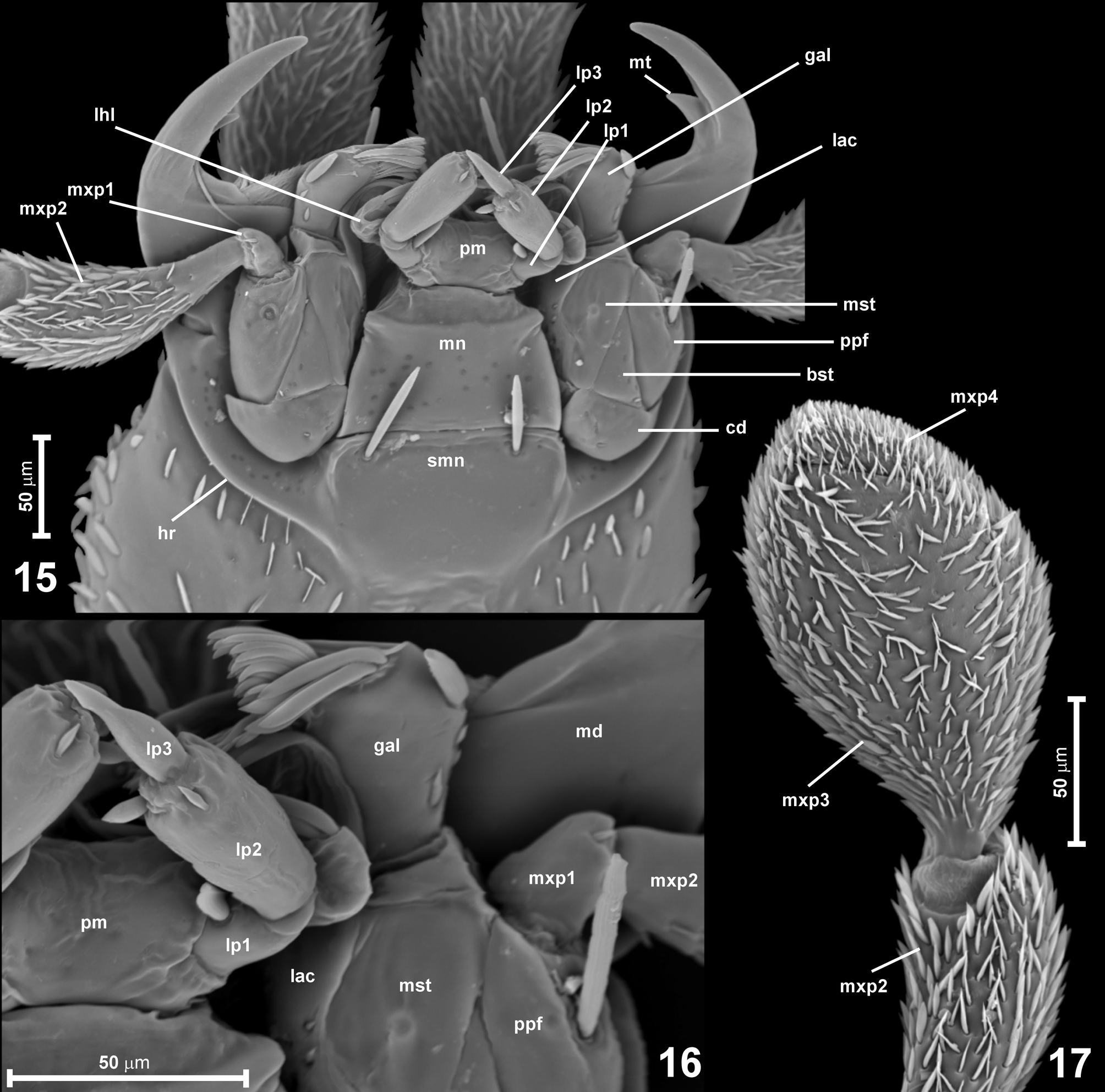
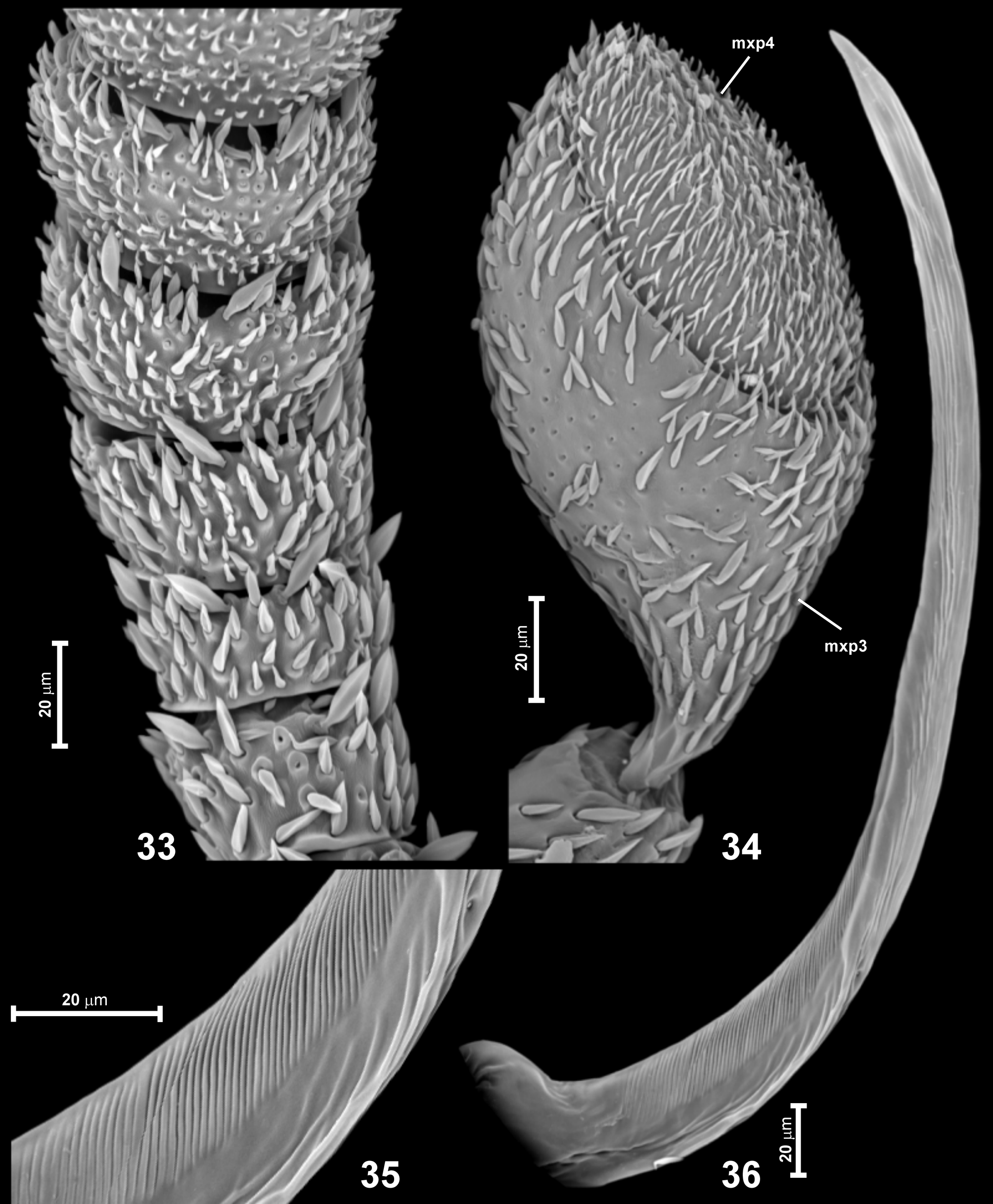
Mouthparts. Labrum ( Figs 176 View FIGURES 176–179 , 180–181 View FIGURES 180–183 , 225–226 View FIGURES 225–228 ; lbr) strongly transverse, with lateral margins slightly convergent anterad or parallel and weakly rounded, and with anterior margin weakly concave, with a pair of broad and short sublateral teeth broadly separated at middle by a very shallow emargination, and with two partly irregular transverse rows of long and short setae; epipharynx ( Fig. 181 View FIGURES 180–183 ; eph) smooth, with dense lateral trichia. Mandibles ( Figs 173 View FIGURES 172–175 , 176 View FIGURES 176–179 , 180, 182–183 View FIGURES 180–183 , 222–226 View FIGURES 221–224 View FIGURES 225–228 ) symmetrical, subtriangular and robust, each with one dorsal and a group of 2–3 ventral mesal teeth, setose prostheca ( Figs 182–183 View FIGURES 180–183 ; pst) present and long, its setae extending to dorsal and ventral surface of basal half of mandible. Maxilla ( Figs 172–173 View FIGURES 172–175 , 177 View FIGURES 176–179 , 184–185, 224, 229) with large, long cardo ( Fig. 185 View FIGURES 185–188 ; cd); basistipes ( Fig. 185 View FIGURES 185–188 ; bst) subtriangular and elongate; mediostipes ( Fig. 185 View FIGURES 185–188 ; mst) large and sharply demarcated from lacinia ( Fig. 185 View FIGURES 185–188 ; lac) and galea ( Fig. 185 View FIGURES 185–188 ; gal), which are both elongate and each with conspicuously dense group of thin distal setae; palpifer ( Fig. 185 View FIGURES 185–188 ; ppf) broad and elongate; maxillary palp slightly to much longer than head capsule, composed of minute palpomere I ( Figs 172–173 View FIGURES 172–175 , 177 View FIGURES 176–179 , 185 View FIGURES 185–188 , 229 View FIGURES 229–232 ; mxp1), slender, curved, distinctly but only slightly broadening distad palpomere II ( Figs 167–168 View FIGURES 167–171 , 172 View FIGURES 172–175 , 177 View FIGURES 176–179 , 187 View FIGURES 185–188 , 222 View FIGURES 221–224 ; mxp2), palpomere III ( Figs 16 7–168 View FIGURES 15–17 View FIGURES 7–9 View FIGURES 10–14 View FIGURES 18–21 View FIGURES 22–23 View FIGURES 24–28 View FIGURES 29–32 View FIGURES 33–36 View FIGURES 37–39 View FIGURES 40–41 View FIGURES 42–44 View FIGURE 45 View FIGURES 46–51 View FIGURES 52–56 View FIGURES 57–63 View FIGURES 64–68 View FIGURES 69–71 View FIGURES 72–74 View FIGURES 75–77 View FIGURES 78–83 View FIGURES 84–85 View FIGURES 86–88 View FIGURE 89 View FIGURES 90–93 View FIGURES 94–98 View FIGURES 99–100 View FIGURES 101–106 View FIGURES 107–108 View FIGURES 110–112 View FIGURE 113 View FIGURES 114–118 View FIGURES 119–123 View FIGURES 124–126 View FIGURES 127–131 View FIGURES 132–134 View FIGURES 135–137 View FIGURES 138–139 View FIGURE 140 View FIGURES 141–143 View FIGURES 144–147 View FIGURES 148–152 View FIGURES 153–158 View FIGURES 159–162 View FIGURE 163 View FIGURES 164–165 View FIGURE 166 View FIGURES 167–171 , 172 View FIGURES 172–175 , 177 View FIGURES 176–179 , 187 View FIGURES 185–188 , 222 View FIGURES 221–224 ; mxp3) strongly elongate, strongly and gradually broadened distad, with transverse or (rarely) slightly obtuse distal margin, palpomere IV ( Figs 167–168 View FIGURES 167–171 , 172 View FIGURES 172–175 , 177 View FIGURES 176–179 , 187 View FIGURES 185–188 , 222 View FIGURES 221–224 ; mxp4) slightly asymmetrical, shorter, about as long as, or longer than III, suboval or indistinctly subtriangular, broadening from base to about distal third and with subtriangular or rounded apex, or broadly boomerang-shaped (with one margin convex and the other one concave), elongate. Surface of palpomere IV, and sometimes also III, with sparsely distributed porous fields ( Fig. 188 View FIGURES 185–188 ; pf). Palpomeres III and IV slightly (sometimes indistinctly) flattened, II–IV covered with relatively long and dense setae. Labium ( Figs 172–173 View FIGURES 172–175 , 177 View FIGURES 176–179 , 184, 186, 224, 229) with broad and short submentum ( Figs 172 View FIGURES 172–175 , 177 View FIGURES 176–179 , 224 View FIGURES 221–224 ; smn) posteriorly not demarcated from gular region, densely setose and lacking an outstanding pair of anterior or subanterior lateral setae; mentum ( Figs 173 View FIGURES 172–175 , 184, 229; mn) subtrapezoidal and strongly transverse, with anterior margin very deeply emarginate, so that anterolateral corners form triangular and usually pointed lobes projecting anterad; prementum ( Figs 173 View FIGURES 172–175 , 184, 229; pm) long, subtrapezoidal, broadest distally, lacking demarcated ligula, with several pairs of submedian anterior setae, with broadly separated bases of labial palps; lateral hypopharyngeal lobes ( Fig. 186 View FIGURES 185–188 ; lhl) moderately large, with conspicuously sparse, thick mesal setae; hypopharynx ( Fig. 186 View FIGURES 185–188 ) with two lateral groups of long trichia; labial palp composed of three palpomeres: palpomere I ( Figs 173 View FIGURES 172–175 , 184, 229; lp1) small, elongate, strongly broadening distad, palpomere II ( Figs 173 View FIGURES 172–175 , 184, 229; lp2) largest, conspicuously enlarged, long and broad, approximately barrelshaped, palpomere III ( Figs 173 View FIGURES 172–175 , 184, 229; lp3) very small and narrow in relation to III, as long as about half length of III and very narrow, pointed.
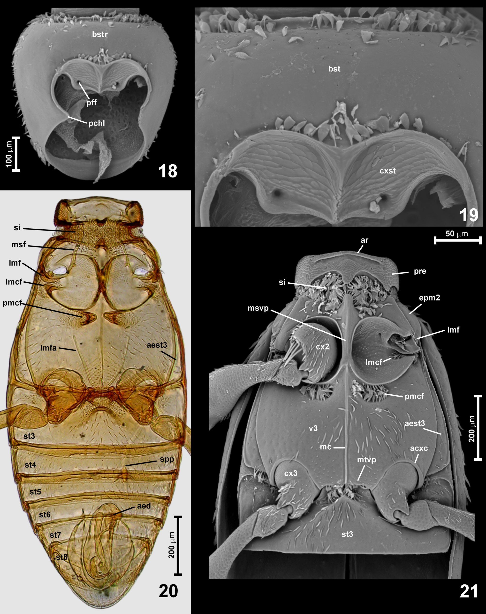
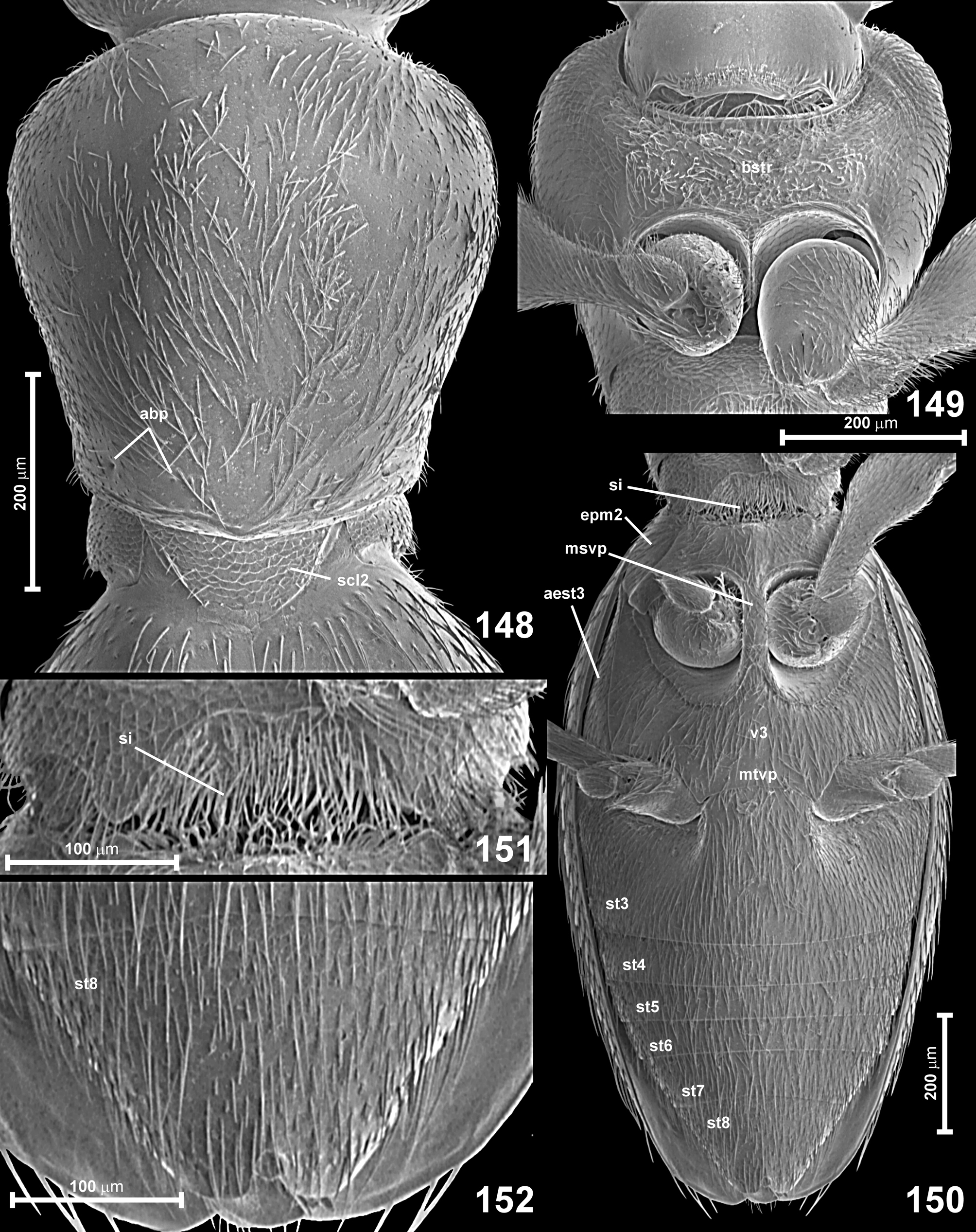
Prothorax ( Figs 153–162 View FIGURES 153–158 View FIGURES 159–162 , 174 View FIGURES 172–175 , 189 View FIGURES 189–193 , 230–231 View FIGURES 229–232 ) elongate (usually strongly so), strongly convex but usually with flattened dorsum, broadest near anterior third. Pronotum with anterior and posterior margins arcuate (anterior margin sometimes nearly straight), sides rounded in anterior half and sinuate or (rarely) nearly straight in posterior half; anterior and posterior corners obtuse-angled or broadly rounded; pronotal base lacking pits and groove. Prosternum ( Figs 174 View FIGURES 172–175 , 189 View FIGURES 189–193 , 230 View FIGURES 229–232 ) with basisternal part ( Figs 174 View FIGURES 172–175 , 189 View FIGURES 189–193 , 230 View FIGURES 229–232 ; bstr) usually slightly shorter than or equal in length to coxal part ( Fig. 174 View FIGURES 172–175 , 189 View FIGURES 189–193 , 230 View FIGURES 229–232 ; cxst). Prosternum laterally completely fused with hypomera. Coxal region anteriorly and laterally without marginal carina; postcoxal hypomeral lobes ( Fig. 174 View FIGURES 172–175 , 189 View FIGURES 189–193 , 230 View FIGURES 229–232 ; pchl) conspicuously large, rounded and strongly projecting mesad or anteromesad and overlapping with (but not fused to) posterolateral lobes of prosternum, so that procoxal cavities are not open, but entirely delimited posterioly by hypomeral lobes. Prosternal intercoxal process developed as a narrow and weakly elevated carina in intact beetles hidden between procoxae (character not studied in extinct taxa). Ventral surface of prothorax densely setose.
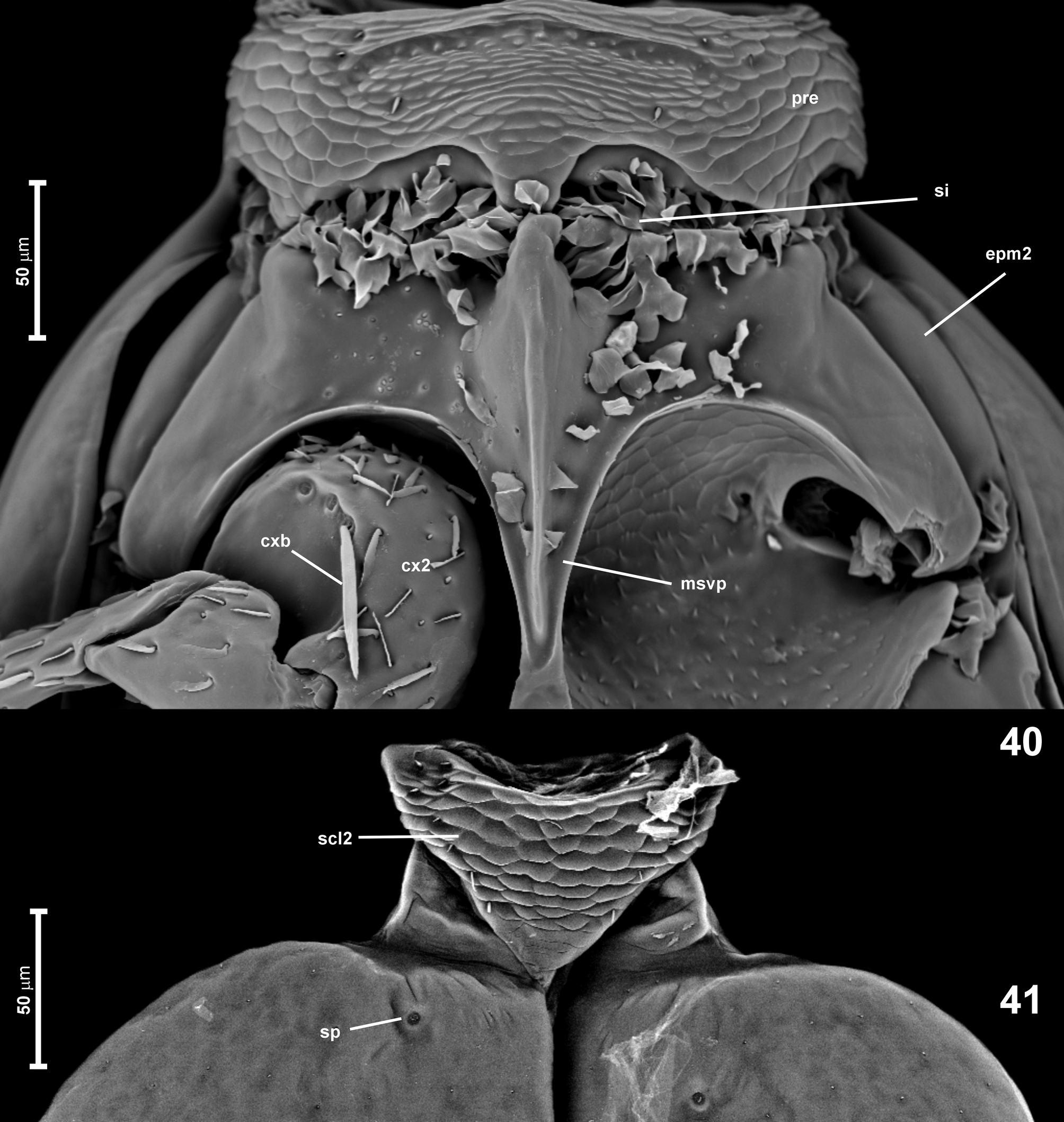
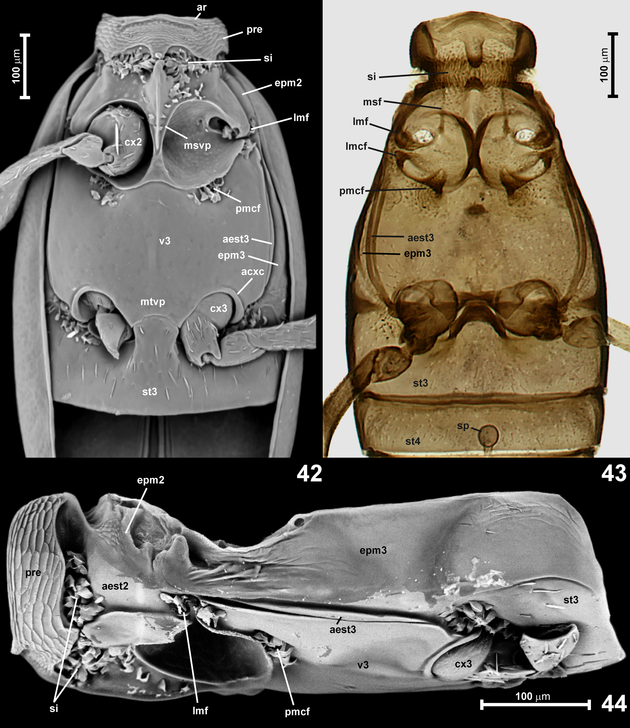
Mesoventrite ( Figs 171 View FIGURES 167–171 , 175 View FIGURES 172–175 , 191–192 View FIGURES 189–193 , 232 View FIGURES 229–232 , 233 View FIGURES 233–235 ) subtrapezoidal, broadening posteriorly. Prepecti ( Fig. 191 View FIGURES 189–193 ; pre) moderately long and together with anteromedian mesoventral area forming a relatively short 'collar', which is weakly impressed just behind its anterior ridge, anterior margin of impression typically with a short subtriangular posteromedian projection. Impressed area can be shallow, short and diffuse, posteriorly gradually becoming shallower to indistinguishably fuse with median precoxal portion of mesoventrite ( Figs 175 View FIGURES 172–175 , 232 View FIGURES 229–232 ), or forming a deep and broad, well-defined setose impression densely filled with long setae, with its posterior margin abruptly elevated and sharply demarcated from median precoxal portion of mesoventrite ( Fig. 191 View FIGURES 189–193 ; si). Mesoventral intercoxal process ( Figs 171 View FIGURES 167–171 , 175 View FIGURES 172–175 , 191–192 View FIGURES 189–193 , 232 View FIGURES 229–232 , 233 View FIGURES 233–235 ; msvp) reaching middle of mesocoxal cavities; in extant taxa short and very broad, subtriangular or subtrapezoidal, weakly convex, broadly separating mesocoxae, posteriorly fused with metaventrite or separated from the latter by transverse groove, and then with its posterior margin subtriangular, rounded or nearly straight; in one extinct genus mesoventral process relatively narrow, longer than broad, broadest between anterior margins of mesocoxae, slightly narrowing caudad and with truncate apex. Mesanepisterna ( Fig. 233 View FIGURES 233–235 ; aest2) relatively narrow and strongly elongate, demarcated from median part of mesoventrite by a distinct ridge and from mesepimera by complete suture; mesepimera ( Fig. 233 View FIGURES 233–235 ; epm2) elongate, indistinctly demarcated from metepimera, not exposed in ventral view.
Mesonotum with cordiform, broad mesoscutellum ( Fig. 190 View FIGURES 189–193 ; scl2) with pointed apex, in intact specimens not visible between elytral bases, or only its very tip discernible; scutoscutellar suture absent (mesonotum not studied in fossils).
Metanotum ( Fig. 193 View FIGURES 189–193 ) partly reduced, with lightly sclerotized mesoscutum, but only slightly shortened alacristae ( Fig. 193 View FIGURES 189–193 ; ala); hind wings absent (not studied in fossils).
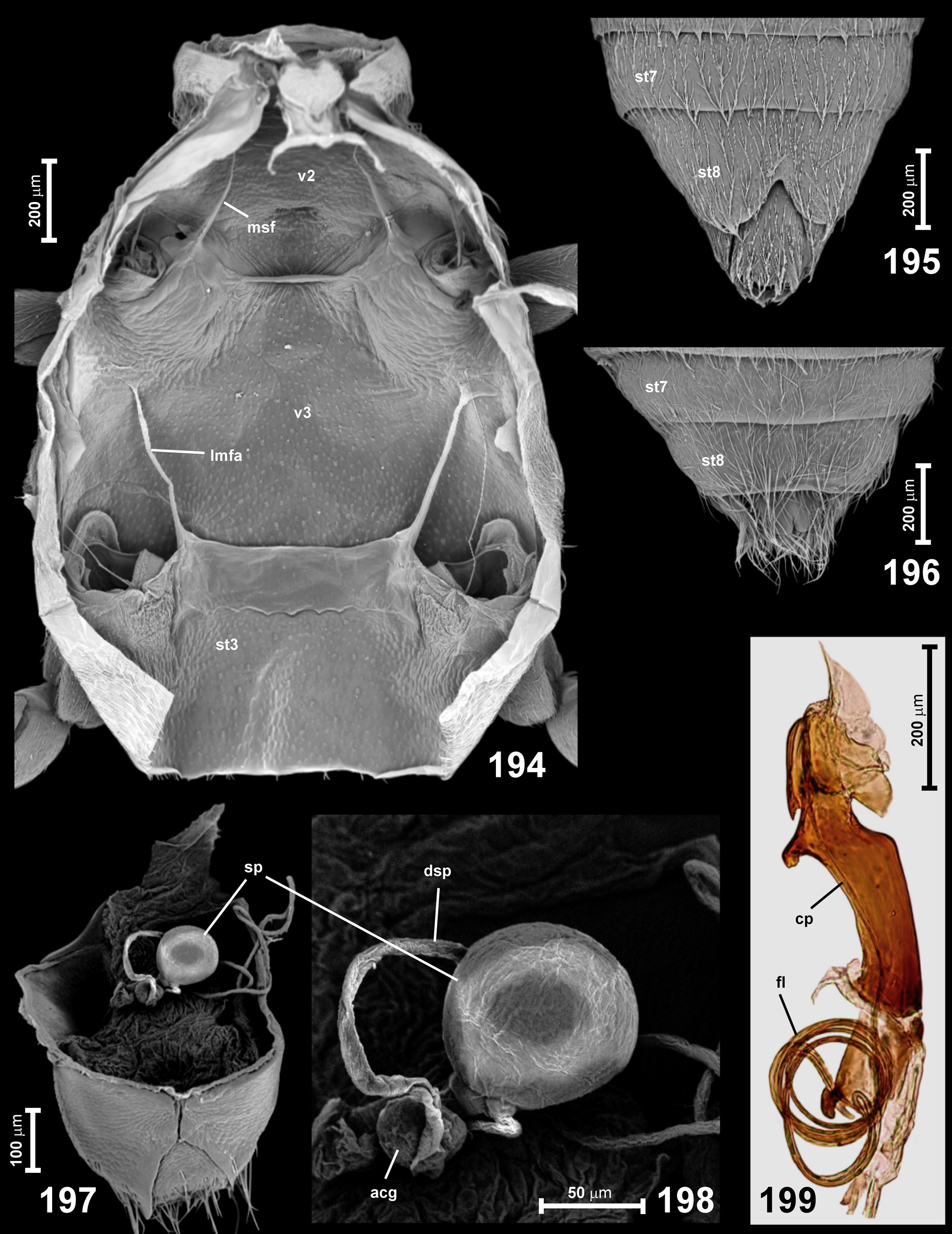
Metaventrite ( Figs 171 View FIGURES 167–171 , 175 View FIGURES 172–175 , 191 View FIGURES 189–193 , 194 View FIGURES 194–199 , 232 View FIGURES 229–232 , 233 View FIGURES 233–235 ) short, subrectangular and usually strongly transverse, with lateral margins rounded; mesocoxal cavities with all margins non-carinate; posterior margin of metaventrite deeply bisinuate laterally (in front of each metacoxa) and with a broad metaventral intercoxal process ( Figs 191 View FIGURES 189–193 , 232 View FIGURES 229–232 ; mtvp) with shallowly concave or straight posterior margin; anteriorly metaventrite forming a short (in extant forms) or elongate (in extinct species) anterior metaventral process ( Figs 175 View FIGURES 172–175 , 191 View FIGURES 189–193 , 232 View FIGURES 229–232 ; amvp) similar in shape to mesoventral intercoxal process and meeting the latter at middle of mesocoxal cavities; metaventral foveae absent. External admetacoxal part of posterior metaventral margin lacking adcoxal carinae, but with adcoxal expansions ( Figs 191 View FIGURES 189–193 , 232 View FIGURES 229–232 ; ade). Metanepisterna ( Fig. 233 View FIGURES 233–235 ; aest3) relatively narrow, partly visible in ventral view, narrowing posterad; metepimera ( Fig. 233 View FIGURES 233–235 ; epm3) 2–3 times as broad as metanepisterna, with inner and outer components not demarcated, posteriorly extending far behind metacoxae.
Metendosternite (metafurca) with stem much broader than long and with broadly separated, divergent lateral furcal arms ( Fig. 194 View FIGURES 194–199 ; lmfa), lacking median longitudinal projection (metafurca not studied in fossils).
Legs ( Figs 153–163 View FIGURES 153–158 View FIGURES 159–162 View FIGURE 163 , 171 View FIGURES 167–171 , 174 View FIGURES 172–175 , 191 View FIGURES 189–193 , 221 View FIGURES 221–224 , 232 View FIGURES 229–232 ) very long and slender (often extremely so). Pro- and mesocoxa short subconical, metacoxa with nearly hemispherical basal part and subconical distal part. Mesocoxa lacking coxal bristles. All trochanters short and subtriangular (sometimes with various male secondary sexual characters); femora weakly clavate; tibiae slender; tarsi long and slender, nearly subcylindrical, tarsomeres I–V reducing in length, tarsomere V strongly elongate, with curved and slender claws lacking elongate costae; empodial region was not studied.
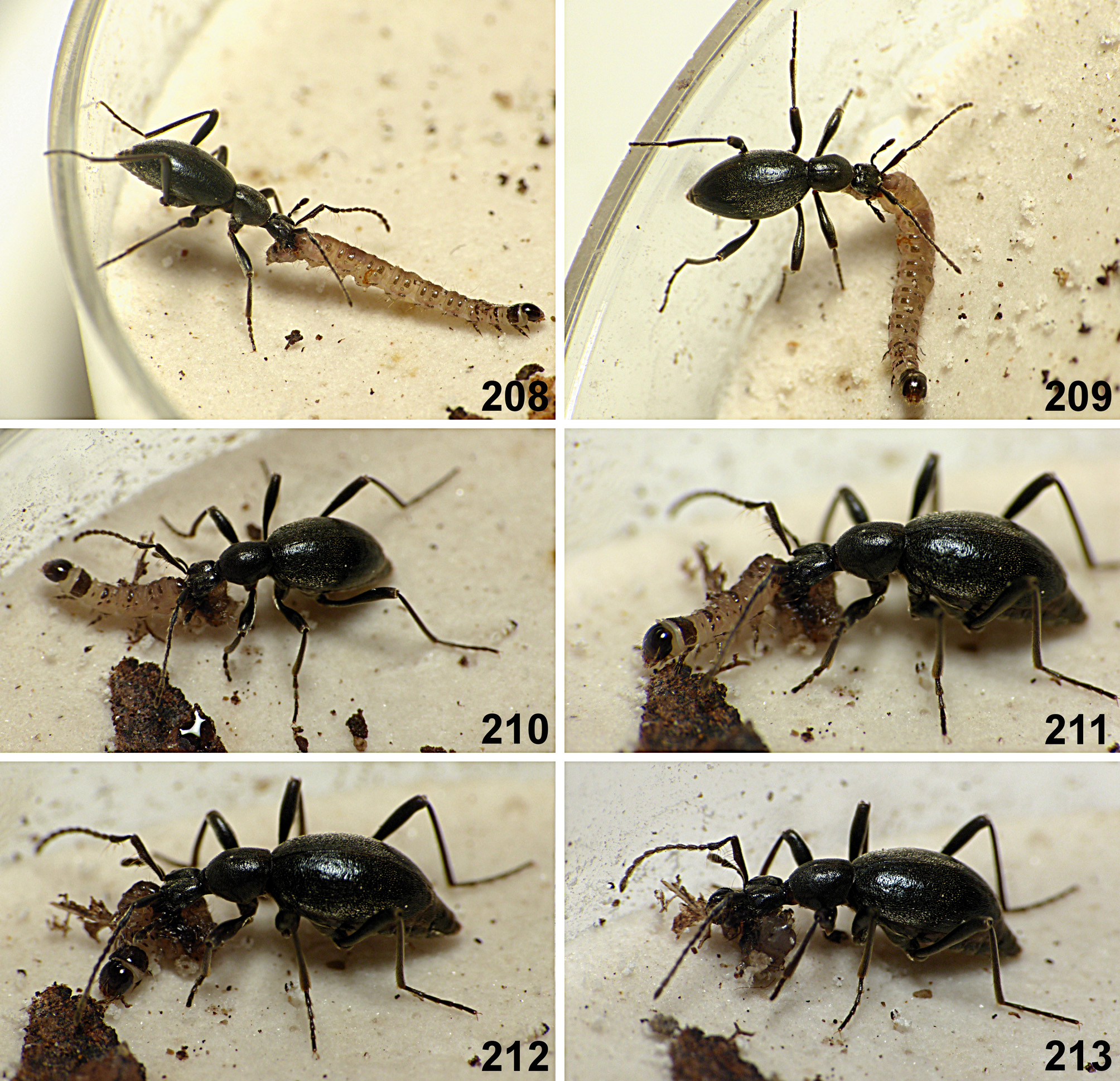
Elytra ( Figs 153–163, 2–235 View FIGURES 153–158 View FIGURES 159–162 View FIGURE 163 View FIGURES 1–6 View FIGURES 7–9 View FIGURES 10–14 View FIGURES 15–17 View FIGURES 18–21 View FIGURES 22–23 View FIGURES 24–28 View FIGURES 29–32 View FIGURES 33–36 View FIGURES 37–39 View FIGURES 40–41 View FIGURES 42–44 View FIGURE 45 View FIGURES 46–51 View FIGURES 52–56 View FIGURES 57–63 View FIGURES 64–68 View FIGURES 69–71 View FIGURES 72–74 View FIGURES 75–77 View FIGURES 78–83 View FIGURES 84–85 View FIGURES 86–88 View FIGURE 89 View FIGURES 90–93 View FIGURES 94–98 View FIGURES 99–100 View FIGURES 101–106 View FIGURES 107–108 View FIGURES 110–112 View FIGURE 113 View FIGURES 114–118 View FIGURES 119–123 View FIGURES 124–126 View FIGURES 127–131 View FIGURES 132–134 View FIGURES 135–137 View FIGURES 138–139 View FIGURE 140 View FIGURES 141–143 View FIGURES 144–147 View FIGURES 148–152 View FIGURES 164–165 View FIGURE 166 View FIGURES 167–171 View FIGURES 172–175 View FIGURES 176–179 View FIGURES 180–183 View FIGURES 185–188 View FIGURES 189–193 View FIGURES 194–199 View FIGURES 200–201 View FIGURES 202–207 View FIGURES 208–213 View FIGURES 214–216 View FIGURES 217–220 View FIGURES 221–224 View FIGURES 225–228 View FIGURES 229–232 View FIGURES 233–235 ) oval, strongly convex, lacking humeral calli and basal impressions, with rounded or pointed apices; elytral disc in extant forms with superficial, indistinct and incomplete longitudinal rows of fine punctures, often barely discernible, obscured by chaotic secondary punctures or coarse microscultpure; in one extinct genus elytra with complete and deeply impressed longitudinal striae. Elytra densely setose, setae short and nearly recumbent.
Abdomen ( Figs 171 View FIGURES 167–171 , 195–196 View FIGURES 194–199 ) with sternite III firmly fused with metaventrite (so that during disarticulation it is almost impossible to separate intact abdomen), much longer than sternite IV, but shorter than IV–VI together; sternite VIII in male distinctly, often deeply emarginate ( Fig. 195 View FIGURES 194–199 ).
Aedeagus (illustrated e.g. in Bordoni & Castellini (1973), Jałoszyński ( 2012c; d; e), Jałoszyński et al. (2015), Leleup (1968), Lhoste (1936, 1937)) elongate, sometimes extremely so, with asymmetrical median lobe and asymmetrical parameres, one shorter than the other, sometimes vestigial or completely obliterated; flagellum very long and forming several coils ( Fig. 199 View FIGURES 194–199 ; fl). Ejaculatory duct with elongate and narrow sperm pump lacking funnel-like structures. Aedeagus with elongate copulatory piece ( Fig. 199 View FIGURES 194–199 ; cp) with membranous endophallus permanently everted, and only inflated during copulation, with distal end of flagellum permanently fixed, so that flagellum is not extricable. Aedeagus in repose positioned asymmetrically inside abdomen, with basal orifice lateral or dorsolateral.
Spermatheca ( Figs 197, 198 View FIGURES 194–199 ; sp) subglobose, with relatively large accessory gland.
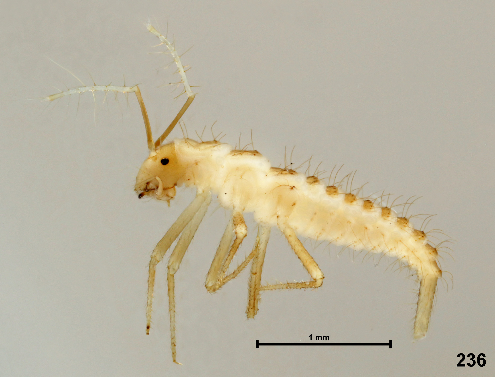
Characteristics. Larvae. Larvae ( Figs 200–201 View FIGURES 200–201 , 236 View FIGURE 236 ) are known for two extant genera. Campodeiform, subparallel or with strongly narrowing abdomen, slightly flattened; head, tergal and sternal plates brown to nearly black, remaining areas whitish or yellowish, or nearly entire larva orange. Body sparsely covered with long unmodified setae and dense asperities forming patterns among smooth areas of tergal plates, additionally with sparse short leaf-like setae with elongate ribs; frontal impression with setae covered with irregular convexities. Head prognathous and slightly tilted ventrad, lacking 'neck', with one stemma at each side; epicranial stem and frontal sutures distinct but short, together with antennal insertions shifted to posterior half of head capsule; nasale with a row of several short setae with papillate insertions. Head with large glandular impression at the junction of epicranial stem and frontal sutures, impression surrounded by or filled with short modified setae (first instar larvae without impression). Antenna 2–4 times longer than head, very slender, antennomeres subcylindrical and not broadened distad, antennomeres I and II very long and similarly broad, antennomere II subdivided into three sections, antennomere III vestigial, developed as a barely discernible papilla adjacent to base of strongly elongate, slightly asymmetrical, subconical and pointed accessory appendage. Mandibles falciform, moderately slender, pointed, each with one submedian mesal tooth; stipital projection of maxilla divided into two very short and broad, densely setose lobes; maxillary palp longer than head, with palpomere I short and II and III strongly elongate; labial palp with palpomere I longer than II, both subconical. Thoracic tergites with ecdysial line visible as a smooth elongate median stripe among lateral fields of dense asperities. Abdomen with ten segments, all except X (or IX and X) transverse; segment X elongate; urogomphi absent. Sternal plates on thorax and abdomen reduced to small, paired (2–4) and setose sclerites. Legs conspicuously long and slender, with particularly densely setose tibiotarsi. Spiracles annular, lateral, nine pairs: one on mesothorax and eight pairs on abdominal segments I–VIII.
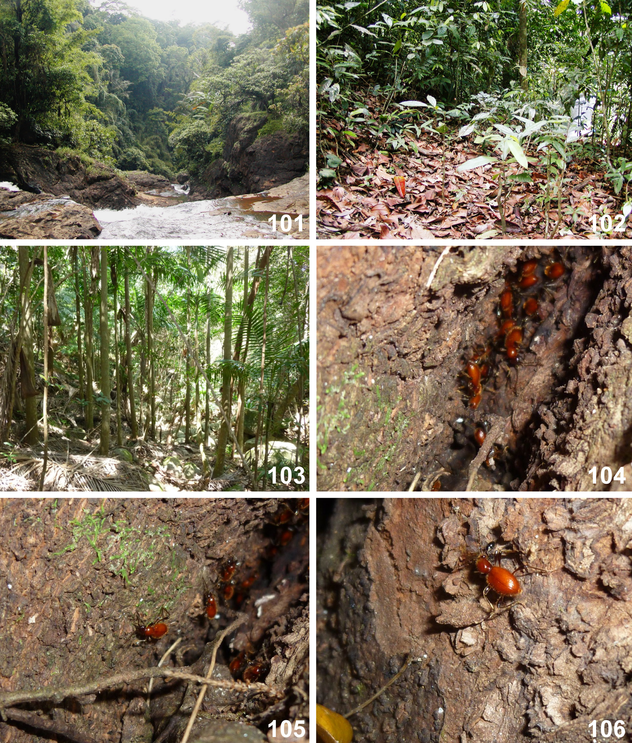
Composition and distribution. Mastigini include three extant and one extinct genera, comprising 56 extant species and subspecies distributed in southern Europe and south Africa ( Figs 164–166 View FIGURES 164–165 View FIGURE 166 ), and three extinct species known from Cenomanian Myanmar amber.
Remarks. Mastigini and †Baltostigini form a monophyletic and distinct group characterized, among some less conspicuous structures, by strongly enlarged and spinose scape and pedicel. All known Mastigini , including the extant genus Clidicostigus Jałoszyński et al., 2017 , have asymmetrical aedeagi twisted inside abdomen in repose and lack wings and humeral calli. †Baltostigini differ in symmetrical aedeagus, which in repose is symmetrically positioned inside the abdomen, and are winged, with prominent humeral calli. Moreover, Mastigini comprise large and strikingly elongate beetles, whereas adults of †Baltostigini are small and stout. The shape of maxillary palpomere IV also differentiates these tribes; it is elongate in Mastigini , and broader than long, axe-shaped in †Baltostigini.


No known copyright restrictions apply. See Agosti, D., Egloff, W., 2009. Taxonomic information exchange and copyright: the Plazi approach. BMC Research Notes 2009, 2:53 for further explanation.
|
Kingdom |
|
|
Phylum |
|
|
Class |
|
|
Order |
|
|
Family |
|
|
SubFamily |
Scydmaeninae |
|
SuperTribe |
Mastigitae |