
Trichogorgia muzikae, Cordeiro, 2019
|
publication ID |
https://doi.org/10.11646/zootaxa.4706.4.2 |
|
publication LSID |
lsid:zoobank.org:pub:728CBA79-31A4-45F9-94B8-13AF43B5CCAA |
|
DOI |
https://doi.org/10.5281/zenodo.5207498 |
|
persistent identifier |
https://treatment.plazi.org/id/2614B657-5B2E-FFEE-82E0-FAABFC5782E4 |
|
treatment provided by |
Plazi |
|
scientific name |
Trichogorgia muzikae |
| status |
sp. nov. |
Trichogorgia muzikae View in CoL sp. nov.
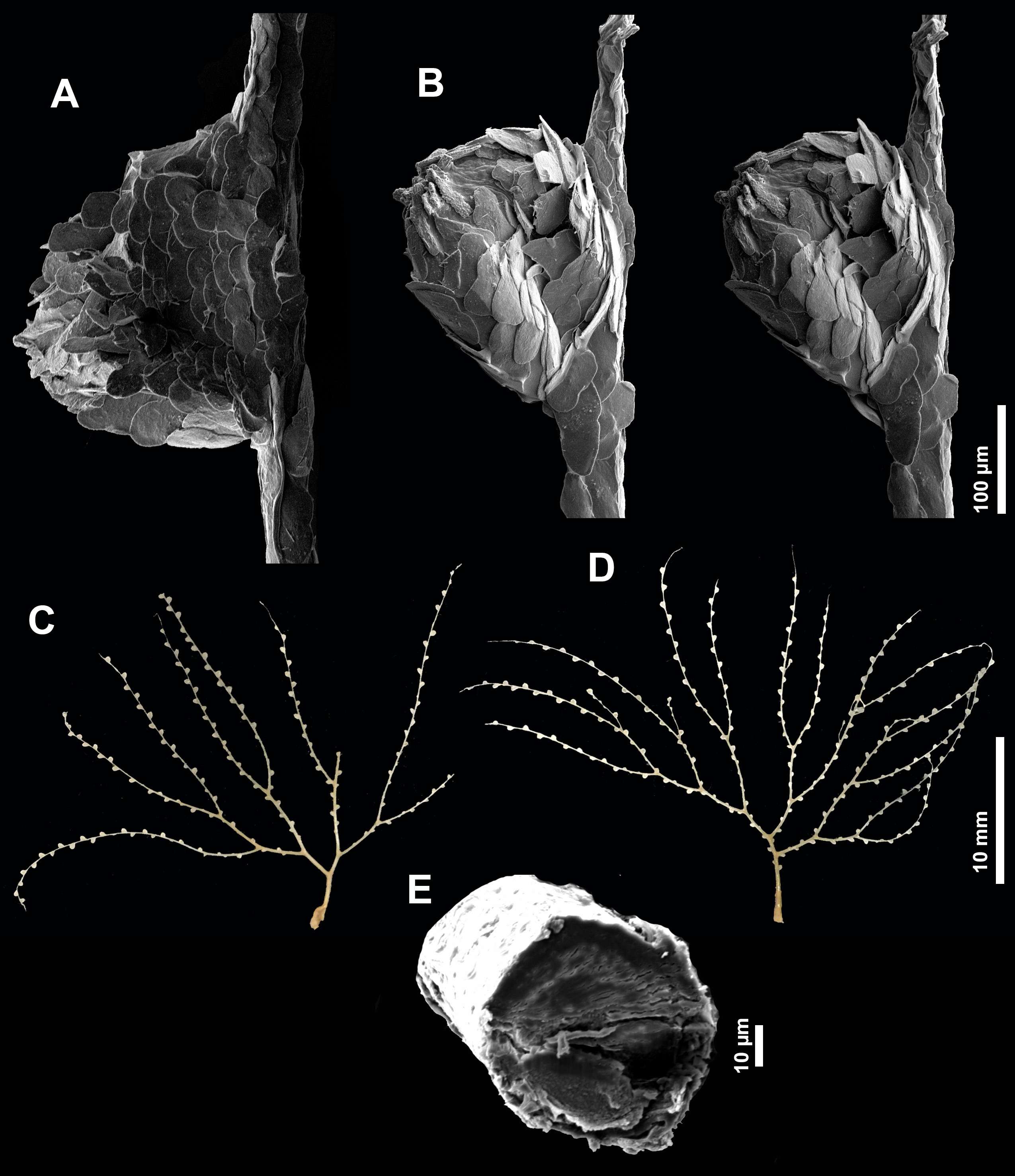
Fig. 4–5 View FIGURE 4 View FIGURE 5 , 6D View FIGURE 6
Holotype: 24°25’12”N, 123°49’48”E, 25 m, Iriomote , Indabishi, Japan, coll. by Katherine Muzik on July 18 th 1982 ( USNM 77155 About USNM , one colony) ( Fig. 4D View FIGURE 4 ). GoogleMaps
Paratype: same collection data of the holotype ( USNM 77155 About USNM , three complete colonies) ( Fig. 4C View FIGURE 4 ) .
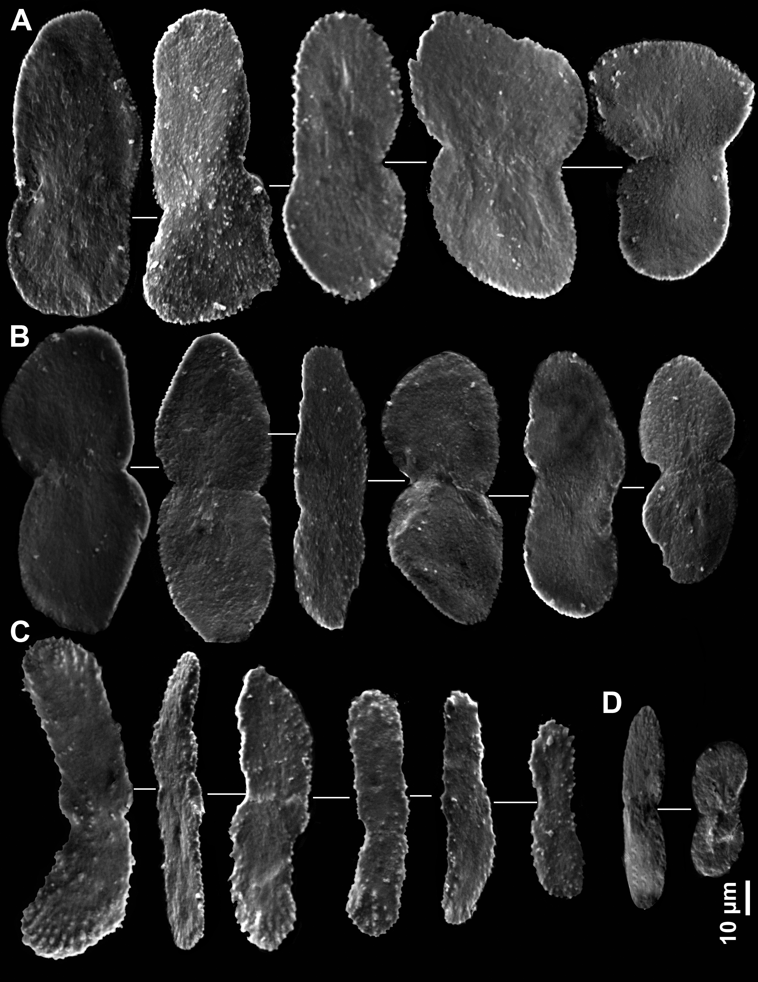
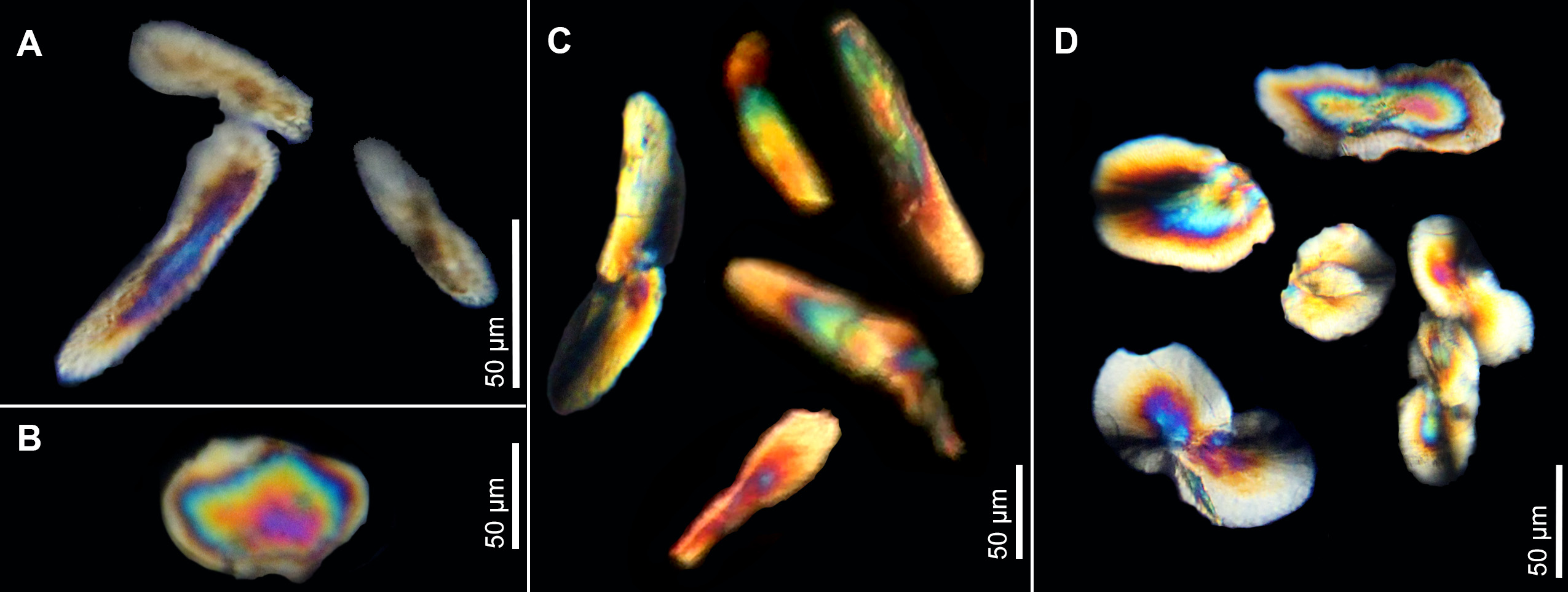
Description: Small, delicate, uniplanar, flexible flabellate colonies, dichotomously branched, not lyrate, up to 2.8 cm tall and 4 cm wide. Colonies with attachment scars, but lacking calcareous holdfasts. Calcified axis, golden to pale yellow, circular in cross-section ( Fig. 4E View FIGURE 4 ), layers concentric (not undulated) up to 0.5 mm in diameter. First order branchlets 0.8–3.3 mm long and internodes 3.0– 7.4 mm long. Polyps verruciform 0.18–0.23 mm tall and 0.2–0.3 mm wide when contracted, occurring in an alternate manner in two rows along branchlets and rarely on the main stem, slightly turned to one face of the colony, occurring in a density of 10–14 polyps/cm. Interpolypar distances from 0.17 proximally, decreasing to 0.04 mm towards branchlet tips. Sclerites present in both polyps and coenenchyme as very flattened smooth eight-shaped scales with a median waist. Main stem usually devoid of polyps and up to 4.6 mm long. Branchlets up to the 6 th order and terminal branches up to 20 mm long. Sclerites show a concentric extinction pattern under polarized light. Polyp body wall with up to eight rows of transversely oriented scales 55–100 µm long (81.87± 12.98 µm) and 25–41 µm wide (32.75± 4.86 µm), becoming more irregularly shaped and longitudinally oriented toward the oral portion ( Fig. 5A View FIGURE 5 ). Tentacular scales longitudinally arranged, slightly more sculptured, elongate, usually lacking a median waist 50–92 µm long (70.57± 12.59 µm) and 15–27 µm wide (19.43± 4.35 µm) ( Fig. 5C View FIGURE 5 ). Pinnular scales similar to small tentacular scales, but slightly less sclulptured ( Fig. 5D View FIGURE 5 ). Coenenchymal sclerites broader than those from body wall, occurring on the entire axis and between zooids, 40–100 µm long (74.4± 16.06 µm) and 40–60 µm wide (48.6±5.27) ( Fig. 5B View FIGURE 5 ).
Comparisons: Trichogorgia muzikae belongs to a group of species with conspicuous sclerites, which excludes T. capensis , T. lyra and T. utinomii sp. nov. The Southwestern Atlantic T. brasiliensis Castro, Medeiros & Loiola, 2010 and the Indian T. insulaeuropensis Weinberg, 2013 are similar in overall colony shape, differing from each other in the absence of coenenchymal sclerites in the latter. Both species differ from T. muzikae by having terminal branchlets usually reaching two-thirds of colony length; by showing dozens of sclerite rows around polyp body walls, more elongate irregular scales and some lacking median waist. In addition, scales are longitudinally placed in T. isulaeuropaensis , in contrast with the transversally arranged scales in T. muzikae . Trichogorgia flexilis Hickson, 1904 is morphologically closer, but yet different from the new species in having longitudinally arranged sclerites and dozens of sclerite rows in the body wall. Trichogorgia viola Deichmann, 1936 was described based on poorly preserved specimens and its description prevents comparisons either with the new species or with T. flexilis . The remaining proximal parts of T. viola (MCZ 4834— holotype) reach 6–9 cm in height, whereas complete colonies of T. muzikae are usually less than 3 cm tall. Further differences may arise pending a re-description of Deichmann’ species.
Remarks: Specimens are so small and delicate that they can easily be mistaken for hydroids, with which they share space.
Etymology: Named in honor to Dr. Katherine Muzik, who dedicated several years studying chrysogorgiids and also collected the specimens described herein.
Distribution: Known only from the type locality, Indabishi, Japan, 25 m.
No known copyright restrictions apply. See Agosti, D., Egloff, W., 2009. Taxonomic information exchange and copyright: the Plazi approach. BMC Research Notes 2009, 2:53 for further explanation.
|
Kingdom |
|
|
Phylum |
|
|
Class |
|
|
SubClass |
Octocorallia |
|
Order |
|
|
SubOrder |
Calcaxonia |
|
Family |
|
|
Genus |