
Myzophyllobothrium rubrum Shipley and Hornell, 1906
|
publication ID |
https://doi.org/10.11646/zootaxa.4999.3.1 |
|
publication LSID |
lsid:zoobank.org:pub:051E68AE-6A5B-44AC-8F90-AEECB8997883 |
|
DOI |
https://doi.org/10.5281/zenodo.5118882 |
|
persistent identifier |
https://treatment.plazi.org/id/2A0987A5-FFCF-FFF1-FF7A-3C9C2075FC86 |
|
treatment provided by |
Plazi |
|
scientific name |
Myzophyllobothrium rubrum Shipley and Hornell, 1906 |
| status |
|
Myzophyllobothrium rubrum Shipley and Hornell, 1906
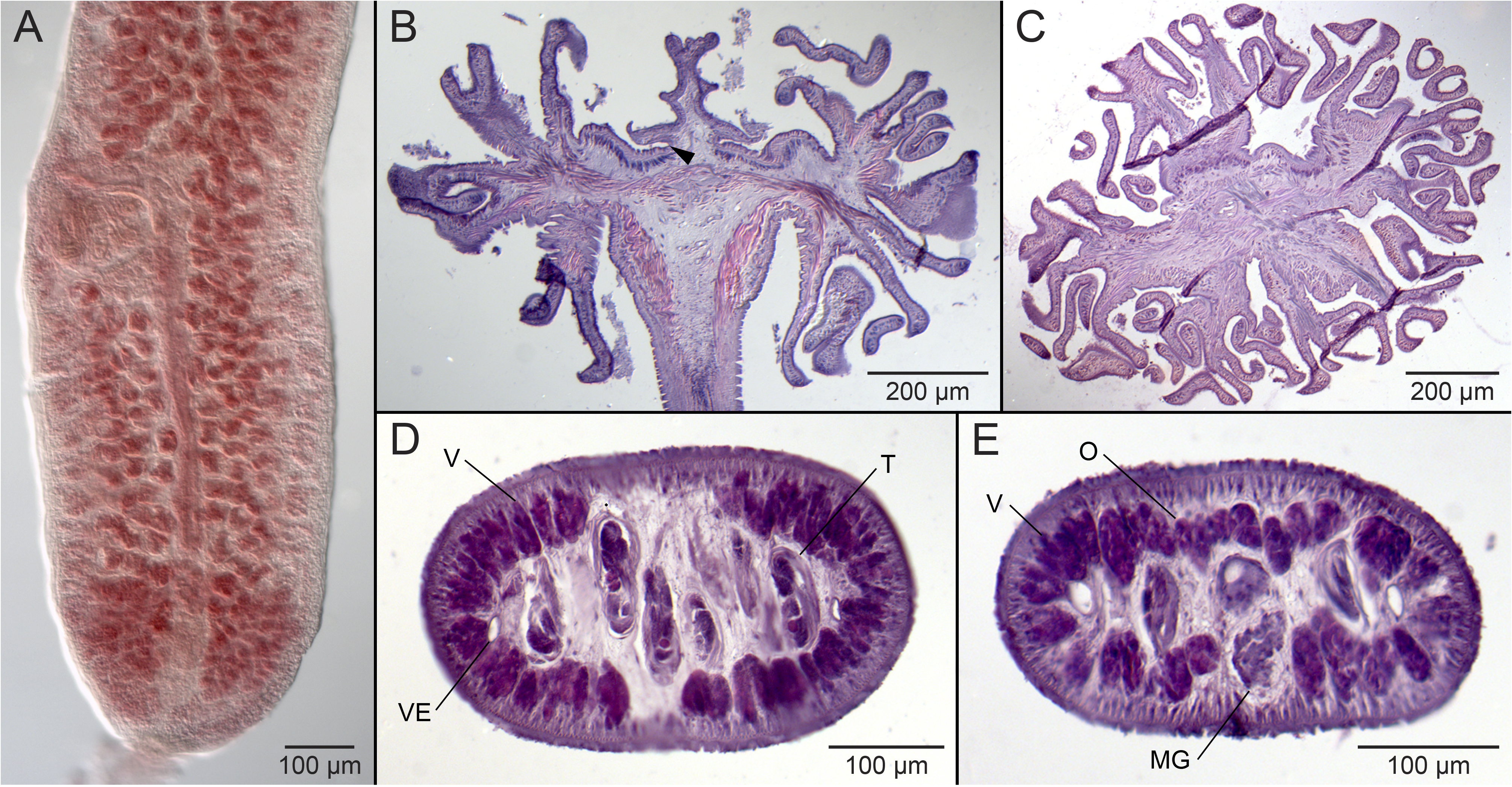
( Fig. 4A View FIGURE 4 )
Type and only known host: Whitespotted eagle ray, Aetobatus ocellatus (as Aetobatis [sic] narinari ) ( Myliobatiformes : Aetobatidae ).
Type locality: Puttalam Lake , Sri Lanka (as Ceylon) .
Additional localities: None.
Site of infection: Spiral intestine.
Specimens examined: BMNH nos. 2006.7.14.1–3, three slides identified by Southwell (1925) as Myzophyllobothrium rubrum (see Jensen and Caira 2006).
Sequence data: None.
Remarks. Joyeux and Baer (1961) resolved the issue surrounding the two different interpretations of the scolex of this species presented by Shipley and Hornell (1906). In their figure of the scolex of one of the co-types of the species, Joyeux and Baer (1961; fig. 290) illustrated the anterior region of the scolex to clearly consist of four biloculate bothridia rather than four simple suckers. Unfortunately, we have been unable to locate the co-types of this species. The specimens of M. rubrum on the three slides examined here were reported by Southwell (1925) as having been collected from the type host and locality. Unfortunately, all of these specimens are in relatively poor condition, and include only one scolex. We did not consider this material to be of sufficient quality to allow re-description of this species. Nonetheless, given the paucity of information available on the proglottid anatomy of M. rubrum , a photograph of the posterior region of a mature proglottid of one of these specimens is presented in Fig. 4A View FIGURE 4 .
No known copyright restrictions apply. See Agosti, D., Egloff, W., 2009. Taxonomic information exchange and copyright: the Plazi approach. BMC Research Notes 2009, 2:53 for further explanation.
|
Kingdom |
|
|
Phylum |
|
|
Class |
|
|
Order |
|
|
Family |
|
|
Genus |