
Simulium nunesdemelloi Hamada, Pepinelli & Hernández, 2006
|
publication ID |
https://doi.org/10.5281/zenodo.273647 |
|
DOI |
https://doi.org/10.5281/zenodo.6255339 |
|
persistent identifier |
https://treatment.plazi.org/id/2C68E82A-FFA9-8D14-0F3E-2273D1AFFC0B |
|
treatment provided by |
Plazi |
|
scientific name |
Simulium nunesdemelloi Hamada, Pepinelli & Hernández |
| status |
sp. nov. |
Simulium nunesdemelloi Hamada, Pepinelli & Hernández View in CoL , new species
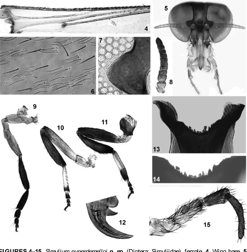
Female ( Figs. 1–22 View FIGURES 1 – 3 View FIGURES 4 – 15 View FIGURES 16 – 22 ). General color brownish; lateral body length (from anterior region of head to abdominal apex) 2.09 mm ( n = 1); lateral thorax length (from neck to wing base) 0.56–0.62 mm ( n = 2). Wing length 2.16–2.21 mm ( n = 2), width 0.96 mm ( n = 2).
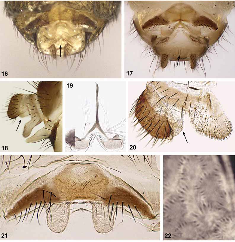
Frons, clypeus, and occiput with silvery blue pruinosity; frons longer than wide ( Fig. 5 View FIGURES 4 – 15 ); fronto-ocular suture absent, fronto-ocular triangle small, as in Fig. 7 View FIGURES 4 – 15 . Antenna ( Fig. 8 View FIGURES 4 – 15 ) length 0.91–0.99 mm, with silver pubescence; pedicel, scape, and first flagellomere brownish yellow, following flagellomeres increasingly dark brown. Palpus dark brown; sensory vesicle length approximately 1/3 length of palpomere III, with wide mouth ( Fig. 15 View FIGURES 4 – 15 ), palpomere V 1.4 times as long as palpomere III and 1.6 times as long as palpomere IV. Mandible with 2 or 3 weak, external serrations and 25–28 internal teeth ( n = 2). Lacinia with 25–30 retrorse teeth ( n = 2). Cibarium with sclerotized cornuae, medial area with strong and sharp teeth ( Figs. 13, 14 View FIGURES 4 – 15 ). Scutum dark brownish orange ( Figs. 1–3 View FIGURES 1 – 3 ); independent of light incidence, scutum with 1 median and 1+1 submedian dark longitudinal thin bands, extending from anterior to posterior region ( Figs. 1, 2 View FIGURES 1 – 3 ); covered with silver hairs, clumped in small groups ( Fig. 6 View FIGURES 4 – 15 ). Anepisternum and katepisternum light brown. Scutellum brownish orange, with golden and dark brown hairs; postnotum brown. Costa with spines and setae, Sc and base of R bare, with exception of 1 female with 1 seta on distal region of Sc ( Fig. 4 View FIGURES 4 – 15 ). Foreleg ( Fig. 9 View FIGURES 4 – 15 ) with coxa, trochanter, and femur light brown; tibia, basitarsus, and tarsomeres I–IV brown. Middle leg ( Fig. 10 View FIGURES 4 – 15 ) with coxa, trochanter, and femur light brown; tibia brown; basitarsus mostly whitish, except small portion distally, brown; tarsomere I whitish basally, remainder brown, tarsomeres II–IV brown. Hind leg ( Fig. 11 View FIGURES 4 – 15 ) with coxa, femur, and tibia brown; trochanter and basal 2/3 of basitarsus light brown, distal 1/3 brown; tarsomeres I–IV dark brown, except base of tarsomere I light brown; calcipala as broad as long, reaching pedisulcus. Tarsal claws with subbasal tooth ( Fig. 12 View FIGURES 4 – 15 ). Femora and tibiae of middle and hind legs with narrow scale-like setae. Basal fringe of abdomen with short, brownish golden hairs. Tergite II with silver pruinosity (best seen in lateral view); tergites VI–VIII shiny brown. In lateral view, cercus with apex wider than its base ( Figs. 18, 20 View FIGURES 16 – 22 ), anal lobe subtriangular ( Fig. 20 View FIGURES 16 – 22 ); in ventral view, in situ, as in Fig. 16 View FIGURES 16 – 22 , after clearing as in Fig. 17 View FIGURES 16 – 22 . Cercus with internal, membranous folded region in ventral view ( Figs. 16, 17 View FIGURES 16 – 22 ) and lateral view ( Figs. 18, 20 View FIGURES 16 – 22 ). Hypogynial valves ( Fig. 21 View FIGURES 16 – 22 ) not sclerotized, subrectangular, with microtrichia, valves widely separated. Genital fork ( Fig. 19 View FIGURES 16 – 22 ) with stem long, lateral arms forming V-shape at junction with main stem, apodemes strong. Spermatheca subspherical, with cuticular microspines ( Fig. 22 View FIGURES 16 – 22 ); spermathecal duct and area of attachment unpigmented.
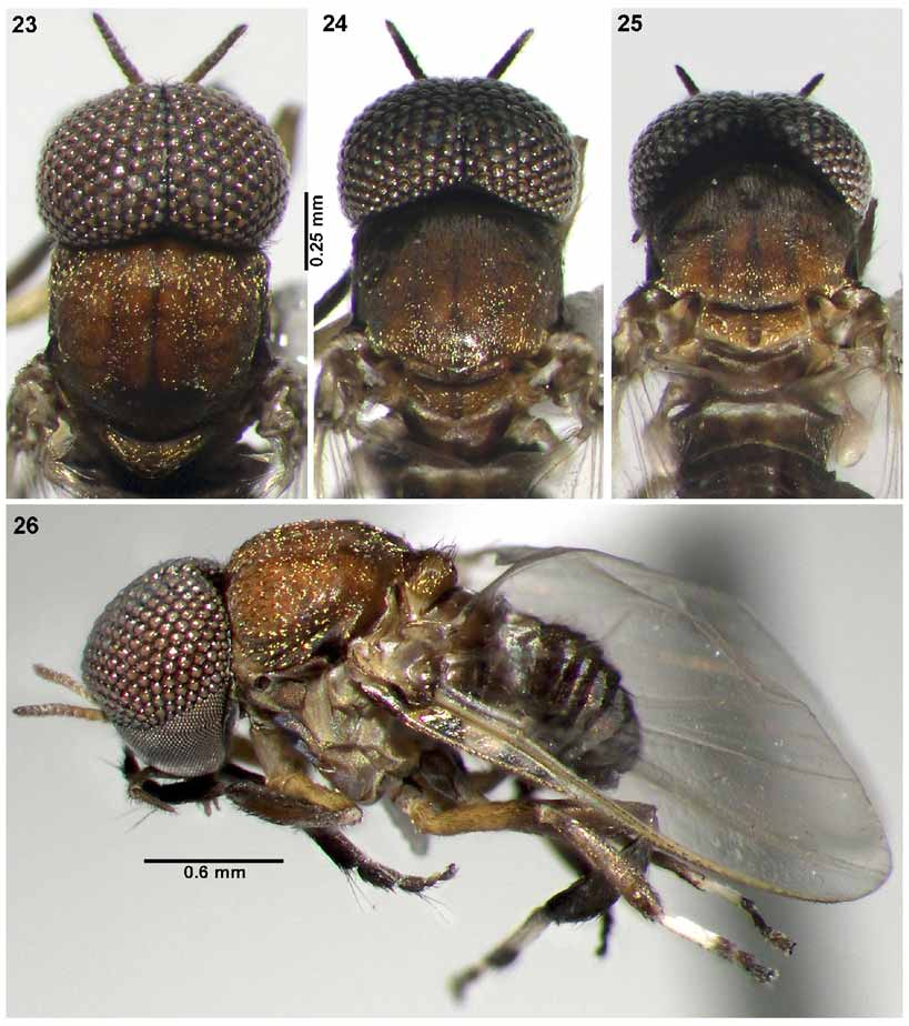
Male ( Figs. 23–34 View FIGURES 23 – 26 View FIGURES 27 – 34 ). General body color brownish, body length 2.0– 2.4 mm ( n = 2); lateral thorax length (from neck to anterior region of wing insertion) 0.41–0.54 mm (mean = 0.47, SD = 0.06, n = 3). Wing length 2.3 mm ( n = 1), width 0.9 mm ( n = 1).
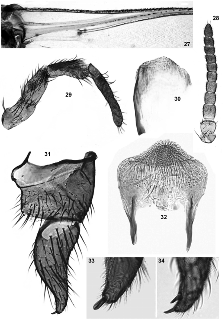
Antenna ( Fig. 28 View FIGURES 27 – 34 ) length 0.43–0.47 mm; pedicel and scape brownish yellow, flagellomeres increasingly dark. Palpus ( Fig. 29 View FIGURES 27 – 34 ) brown, palpomere V about 1.94 times as long as palpomere III and 1.85 times as long as palpomere IV; sensory vesicle small, subspherical, approximately 1/4 length of palpomere III. Scutum brownish-orange ( Figs. 23–26 View FIGURES 23 – 26 ), with 3 dark longitudinal thin bands running from anterior to posterior region ( Figs. 23–25 View FIGURES 23 – 26 ); covered with golden hairs, clumped in small groups ( Figs. 23–26 View FIGURES 23 – 26 ).
Anepisternum and katepisternum light brown. Scutellum brownish-orange ( Figs. 23–25 View FIGURES 23 – 26 ) with thin golden hairs; with posterior light ( Figs. 24, 25 View FIGURES 23 – 26 ), with thin, dark band in central region. Postnotum black with silver pruinosity. Wing with Sc and base of R bare ( Fig. 27 View FIGURES 27 – 34 ). Legs with same color pattern as in female. Abdominal tergites black; basal fringe with thin, long, black hairs and golden highlights; tergites II–VII with silver pruinosity. Gonocoxite and gonostylus ( Fig. 31 View FIGURES 27 – 34 ) dark brown, gonocoxite wider than long; gonostylus almost double gonocoxite length, longer than wide, bearing 1 or 2 spinules ( Figs. 33, 34 View FIGURES 27 – 34 ). Ventral plate ( Fig. 32 View FIGURES 27 – 34 ), in ventral view, longer than wide, subrectangular, with domeshaped keel. Median sclerite not seen. Paramere and aedeagal membrane as in Fig. 30 View FIGURES 27 – 34 .
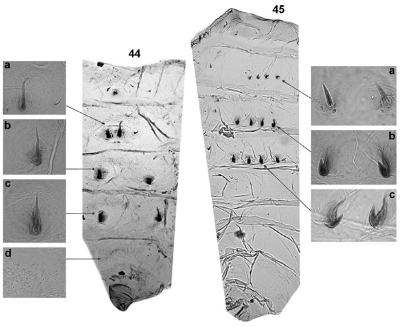
Pupa ( Figs. 35–45 View FIGURES 35 – 38 View FIGURES 39 – 43 View FIGURES 44 – 45 ). Mean length 2.4 mm (SD = 0.2, n = 5). Cocoon ( Figs. 35–38 View FIGURES 35 – 38 ) boot-shaped. Mean length along dorsal surface 2.5 mm (SD = 0.1, n = 5). Head projecting downward, with 3 pairs of trichomes, 1 frontal pair, 3–5 branched, longer than 2 dorsal simple or bifid pairs ( Fig. 41 View FIGURES 39 – 43 ); tubercles absent or scarce on dorsal region, mostly present on facial region. Gill with 19 or 20 thick filaments, varying in length and thickness ( Figs. 39, 40 View FIGURES 39 – 43 ), distributed in 3 dimensions ( Fig. 36 View FIGURES 35 – 38 ), and with sclerotized distal end ( Fig. 43 View FIGURES 39 – 43 ).
Thorax almost without tubercles, some rounded and pointed tubercles present on posterior region of thorax and on area below trunk of gill filaments. Five pairs of 2–4 branched trichomes ( Fig. 42 View FIGURES 39 – 43 ) and 1 pair of bifid, thick, long lateral trichomes. Abdomen as in Figs. 44, 45 View FIGURES 44 – 45 . Tergite I with 1 + 1 setae sublaterally. Tergite II with 4 + 4 stout, short setae. Tergites III and IV with 4 + 4 anteriorly directed hooks on posterior margin. No comb-like spines present on tergites. Sternites III–VIII with anterior group of microspines; sternites V–VII with 2 + 2 long hooks.
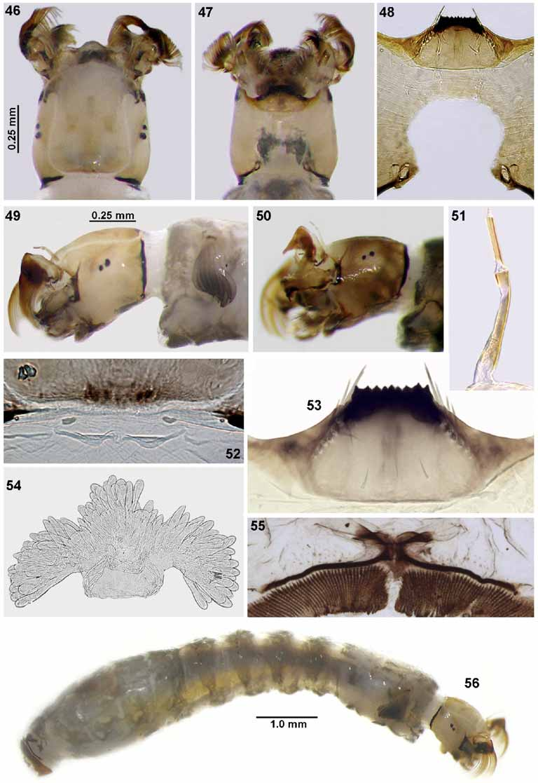
Larva (final instar) ( Figs. 46–56 View FIGURES 46 – 56 ). Mean body length 6.2 mm (SD = 0.71, n = 5); head capsule, mean lateral length 0.50 mm (SD = 0.02, n = 5). General coloration variable, from light ( Fig. 49 View FIGURES 46 – 56 ) to dark grayish green ( Fig. 50 View FIGURES 46 – 56 ) (in Carnoy's solution). Head capsule (in dorsal view) with dark region on midline and along basal margin ( Fig. 46 View FIGURES 46 – 56 ), with small, simple setae. Cervical sclerites small, elliptical, free in membrane ( Fig. 52 View FIGURES 46 – 56 ). Postgenal cleft ( Fig. 48 View FIGURES 46 – 56 ) wide and round, postgenal bridge 0.57 times as long as hypostoma. Subesophageal ganglion slightly pigmented ( Fig. 47 View FIGURES 46 – 56 ). Antenna ( Fig. 51 View FIGURES 46 – 56 ) as long as labralfan stalk; medial article longer than distal and proximal articles, distal article longer than proximal article, proportions of articles (proximal to distal, excluding apical sensillum) 1: 1.4–1.75: 0.8–0.9. Labral fan with 41–43 primary rays. Hypostoma ( Fig. 53 View FIGURES 46 – 56 ) with 9 teeth, median tooth and sublateral teeth not well differentiated; hypostoma lateral margin with 2 paralateral teeth and 3–5 lateral serrations per side; with 4–6 sublateral setae per side. Lateral mandibular process not seen; mandibular teeth: 1 apical, 2 small external; 3 subapical (first subequal to third and both smaller than second, or all subequal), 6 or 7 internal teeth; 1 mandibular serration and 1 small mandibular sensillum ( sensu Craig & Craig 1986). Body with simple setae on dorsal region of abdomen; ventral tubercles absent. Gill histoblast in situ ( Fig. 49 View FIGURES 46 – 56 ) large, with sclerotized distal end of filaments pointing toward ventral region of body; gill histoblast dissected with 19 or 20 filaments. Arms of anal sclerite as in Fig. 55 View FIGURES 46 – 56 , anterior arms 0.3 times as long as posterior arms, associated with some thin and few enlarged setae. Posterior circlet bearing 143–150 rows ( n = 2) with 21–24 hooks ( n = 4). Rectal papilla with 3 branches ( Fig. 54 View FIGURES 46 – 56 ), each with approximately 29 or 30 finger-shaped lobes.
Holotype. Male (M), collected in the Rio Negro, São Gabriel da Cachoeira County, 0 0o 10’S, 67o01’W, collectors N. Hamada, R.L.M Ferreira & L. Aquino, 08/X/1998 ( INPA).
Paratypes. Same locality and collectors as holotype, 10 pupae (P), 2 larvae (L), 08/X/ 1998 ( INPA); 8 P, 3/X/1998 ( INPA); same locality as holotype, collectors N. Hamada & R. Ale-Rocha 10 L, 11/XI/1999 ( INPA); 3 P, 4 L ( INPA), 3 P ( BMNH), 1 F (pinned), 1 F (on slide), 2 M (pinned), 1 M (on slide), collectors N. Hamada & R. Ale-Rocha, 16-17/XI/ 1999, Rio Tiquié, Pari Cachoeira Community, 0 0o 13’N, 69o40’W, Coll. N. Hamada & R.
Ale-Rocha, 10 L, 2 P, 12/XI/1999, ( INPA).
Etymology. This species is named in honor of Professor José Alberto Sampaio Nunes de Mello, a dear friend and former Master’s dissertation advisor of N. Hamada and one of the pioneers in black fly studies at INPA, in the Amazon region.
Diagnosis and taxonomic discussion. We do not include S. nunesdemelloi in any subgenus within the Simuliidae because it shares characters with Trichodagmia and Thyrsopelma . The male and female of the new species are similar to those of S. scutistriatum Lutz and S. nigrimanum Macquart. The males of all of these species have a ventral plate without a developed central region ( Fig. 32 View FIGURES 27 – 34 ), and the females have a subtriangular anal lobe with microtrichia ( Figs. 16, 20 View FIGURES 16 – 22 ). However, these species can be distinguished in the pupal stage by the number of pupal gill filaments: 19 or 20 in S. nunesdemelloi , 12 in S. scutistriatum , and 18 in S. nigrimanum . In this respect, S. nunesdemelloi is most similar to S. orbitale but the gill filaments are much thicker and more crenulated ( Figs. 39–40 View FIGURES 39 – 43 ) than those of S. orbitale . Also, the cephalic and thoracic trichomes of the new species are not spiniform ( Figs. 41–42 View FIGURES 39 – 43 ) as in S. orbitale . Based on Coscarón (1991) and Miranda Esquivel & Coscarón (2001), the following combination of characters can distinguish larvae of S. nunesdemelloi n. sp. from those of other species with known larvae in the subgenera Thyrsopelma and Trichodagmia : absence of racketshaped scales on the dorsal region of the abdomen, rounded postgenal cleft ( Fig. 48 View FIGURES 46 – 56 ), dissected histoblast with 19 or 20 gill filaments with heavily sclerotized tips, and in situ histoblast with gill filament tips pointing toward the ventral region of the body ( Fig. 49 View FIGURES 46 – 56 ).
Bionomics. This species was not collected biting humans during the fieldwork. Larvae and pupae were collected in forested areas, in large rivers (Rio Negro and Rio Tiquié, with widths of 1.65 km and 70 m, respectively), on Podostemaceae leaves growing on rocky substrate in highly turbulent water and, usually, at depths below 0.5 m. River-water temperature was approximately 26o C, pH 4.5–5.2 and electrical conductivity less than 10 ΜS/cm. Immatures of S. orbitale also use Podostemaceae leaves as substrate. But usually the new species is collected in low density, whereas S. orbitale is collected in high density and nearer the surface. Most of the collected pupae of S. nunesdemelloi n. sp. had a silk mesh covering the opening of the cocoon ( Figs. 37, 38 View FIGURES 35 – 38 ); the mesh holds the larval exuviae inside the cocoon.


| INPA |
Instituto Nacional de Pesquisas da Amazonia |
No known copyright restrictions apply. See Agosti, D., Egloff, W., 2009. Taxonomic information exchange and copyright: the Plazi approach. BMC Research Notes 2009, 2:53 for further explanation.
|
Kingdom |
|
|
Phylum |
|
|
Class |
|
|
Order |
|
|
Family |
|
|
Genus |