
Stenostygnus huberi, Mamani & Porto & Iglesias & González, 2021
|
publication ID |
https://doi.org/10.11646/zootaxa.4984.1.15 |
|
DOI |
https://doi.org/10.5281/zenodo.5213031 |
|
persistent identifier |
https://treatment.plazi.org/id/2D0A576D-FF94-312F-A895-B4E4FCD8FCCA |
|
treatment provided by |
Plazi |
|
scientific name |
Stenostygnus huberi |
| status |
sp. nov. |
Stenostygnus huberi View in CoL spec. nov.
Figs 7–12 View FIGURES 7 View FIGURES 8 View FIGURES 9 View FIGURES 11 View FIGURES 12
urn:lsid:zoobank.org:act:
Type material. VENEZUELA, Lara: H olotype male (α) ( MIZA 0103461 About MIZA ), Parque Nacional Yacambu , 15–16 Dec 2002, A. Pérez-González, A. Giupponi & Bernhard A. Huber, O. Villarreal M. & B. Striffler . Paratypes: 2 males (ss) ( one male SEM voucher) , 6 females, 1 juvenile ( MNRJ 2429 View Materials ), with same data as for holotype . 1 male (ss) ( SEM voucher) (MACN-Ar 41020), with same data as for holotype . 1 male (ss), 1 female, 1 juvenile (MACN-Ar 41019), with same data as for holotype .
Etymology. Patronymic in honor of the Austrian arachnologist and friend Bernhard A. Huber in recognition of his remarkable contribution to arachnology, and also for being one of the collectors of the type series; name in the genitive case.
Diagnosis. Differs from all other species of Stenostygnus by the presence of granules in the interocular region ( Fig. 8 A View FIGURES 8 ); patella IV of males with strong spiniform apophysis on the distal-retrolateral surface (Figs 10 A–B); ventral plate of the penis with a deep cleft basally fused and apically “V” shaped ( Fig. 11 View FIGURES 11 ). It can be distinguished from Stenostygnus pusio by the lateral borders of the scutum almost parallel and mesotergal areas almost equally wide ( Fig. 8 A View FIGURES 8 ) versus scutum lateral borders slightly arched (giving an oval appearance to the habitus) ( Pinto-da-Rocha 1995, fig. 1); presence on the pedipalp of a meso-medial spine on the femur and a meso-distal spine on the patella ( Fig. 9 D View FIGURES 9 ) versus without spines in S. pusio ; pars distalis ventrally convex versus concave in S. pusio (Pinto-da- Rocha 1995, fig. 7). This species can be easily separated from S. martensi spec. nov. by the presence of scattered granules on the interocular area and mesotergal areas I to IV ( Fig. 8 A View FIGURES 8 ) versus smooth areas in S. martensi spec. nov. ( Fig. 2 A View FIGURES 2 ); presence of granules on the coxae III and IV ( Fig. 8 B View FIGURES 8 ) versus smooth in S. martensi spec. nov. ( Fig. 2 B View FIGURES 2 ); femur without ectal spines ( Fig. 9 E View FIGURES 9 ) versus two small ectal spines in S. martensi spec. nov. ( Fig. 3 F View FIGURES 3 ); lamina apicalis medially divided by a cleft that creates a “V” shape ( Fig. 11 A View FIGURES 11 ) versus divided into two halves medially in contact in S. martensi spec. nov. ( Fig. 5 C View FIGURES 5 ).
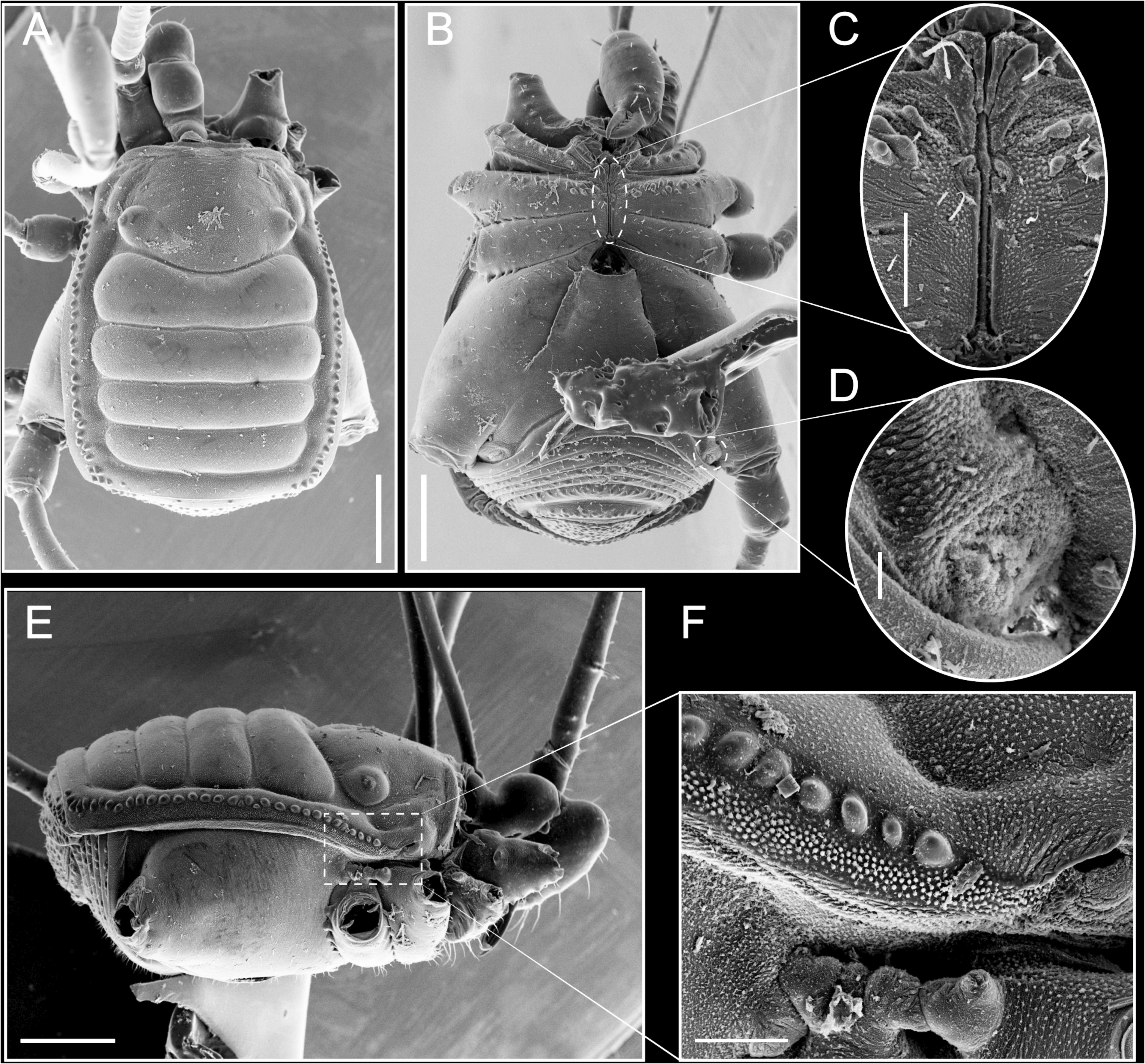
Description. Male ( Holotype): measurements in Table 2 View TABLE 2 . Dorsum: Habitus slightly oval (almost rectangular) in shape ( Figs 7 A View FIGURES 7 ; 8 A View FIGURES 8 ). Carapace wider than long with the anterior border straight and unarmed. Cheliceral sockets not marked. Eyes widely separated; eye mounds very low, near sulcus I; interocular area with scattered granules, with greater concentration on the eye mounds ( Figs 7 C View FIGURES 7 ; 8 A View FIGURES 8 ). Carapace in lateral view slightly convex ( Figs 7 C View FIGURES 7 ; 8 E View FIGURES 8 ). Mesotergum in lateral view slightly convex ( Fig. 8 E View FIGURES 8 ), divided into five well defined areas ( i.e. with sulci well marked). Mesotergal areas I to IV with scattered granules. Lateral margins and posterior border of scutum with a row of small granules ( Fig. 8 A View FIGURES 8 ). Ozopore with an oval and narrow orifice with a descending channel toward the posterior region ( Fig. 8 F View FIGURES 8 ). Free tergites granulated ( Fig. 8 A, E View FIGURES 8 ).
Venter: Without any conspicuous armature ( Fig. 8 B, C View FIGURES 8 ). Spiracles visible ( Fig. 8 B, D View FIGURES 8 ). Coxa I to III with minutes granules. Coxa II with tubercles in the medial region. Coxa IV smooth. ( Figs 7 B View FIGURES 7 ; 8 B View FIGURES 8 ).
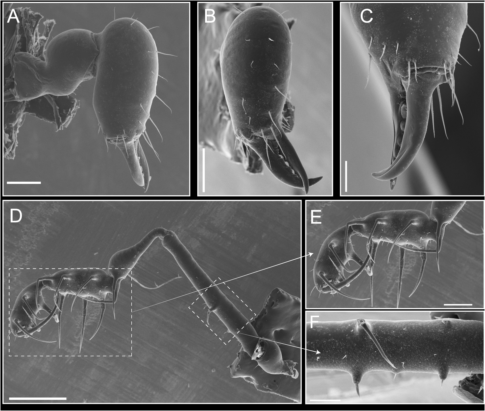
Chelicerae: Without remarkable armature. Basichelicerite with a short bulla. Cheliceral hand with sparse setae ( Fig. 9 A View FIGURES 9 ). Fingers thin, with uniform small teeth ( Fig. 9 B, C View FIGURES 9 ).
Pedipalps: Raptorial and long, with spines concentrated on tibia and tarsus ( Fig. 9 D View FIGURES 9 ). Coxa elongated, surpassing the anterior border of the carapace ( i.e. can be seen in dorsal view) ( Fig. 7 A View FIGURES 7 ) proximally with one small dorsoectal protuberance; trochanter small and smooth; femur thin and cylindrical, with a meso-medial spine ( Fig. 9 E View FIGURES 9 ); patella clavate, swollen distally, with a meso-distal spine ( Fig. 9 D View FIGURES 9 ); tibia with one ventro-basal spine and armed ventrally with three ectal and three mesal spines; tarsus mesal and ectally with three spines on each side ( Fig. 9 E View FIGURES 9 ).
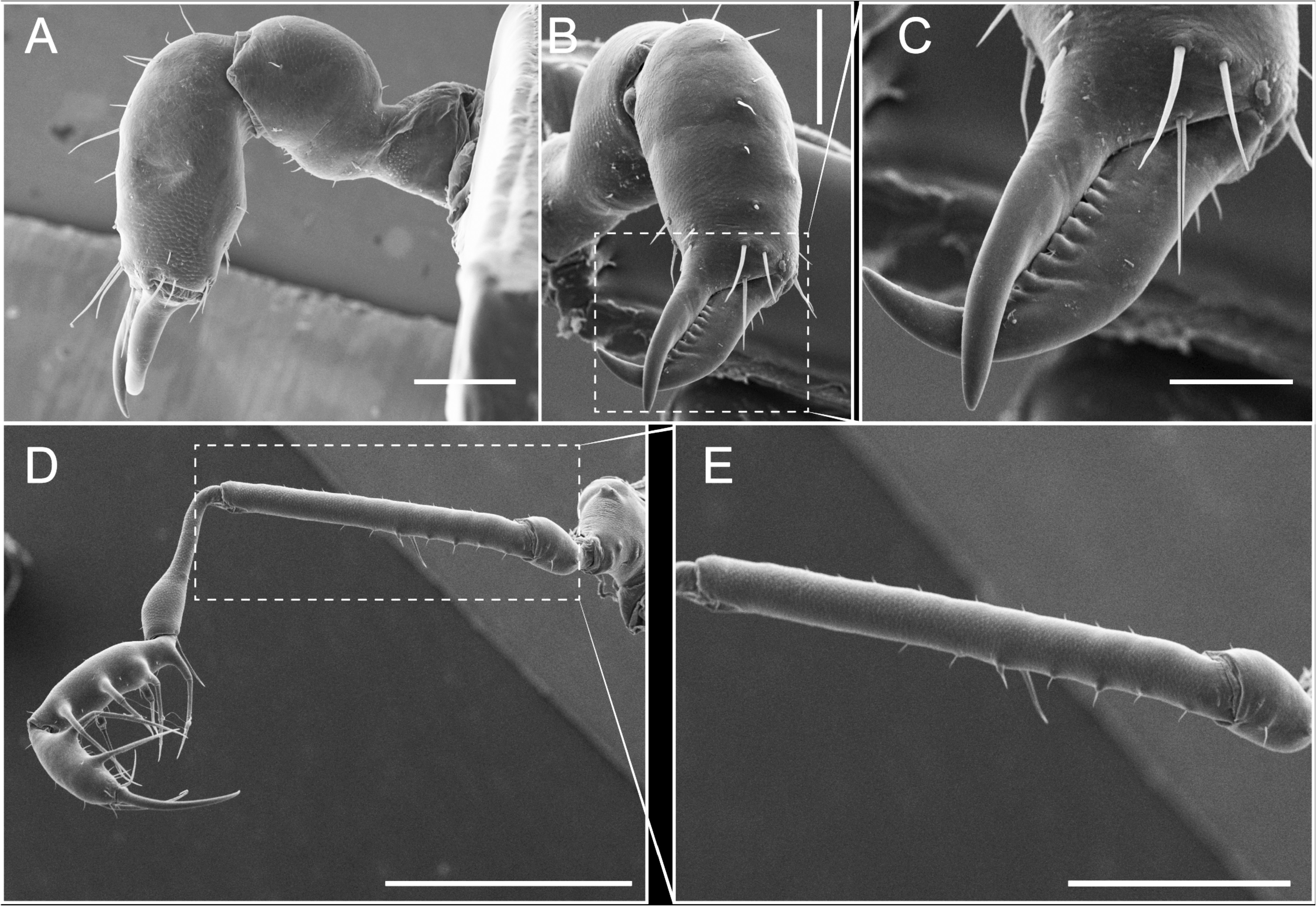
Legs: Thin, cylindrical, and elongated (Fig. 10 A). Coxa IV without remarkable armature; femur IV slightly curved; patella I–III short and swollen; patella IV with a prolateral-distal spiniform apophyses and dorsally roughly granulated (some of the granules looks like small pointed tip tubercles) (Fig. 10 B); tibia IV slightly swollen distally (Fig. 10 B); tarsi III and IV with dense scopula (Fig. 10 C, D). Tarsal formula: 6(3):11(3):6:6.
Color (specimen preserved in 80% ethanol): Body yellowish. Mesotergal areas, with slight brown reticulations ( Fig. 7 View FIGURES 7 ).
Figure 10. Stenostygnus huberi spec. nov., male (ss) ( paratype, MNRJ 2429), left leg IV (A retrolateral, B detail of patella and tibia, C detail of tarsus, D detail of scopula). Scale bars: A= 2 mm; B= 500 µm; C= 200 µm; D= 50 µm.
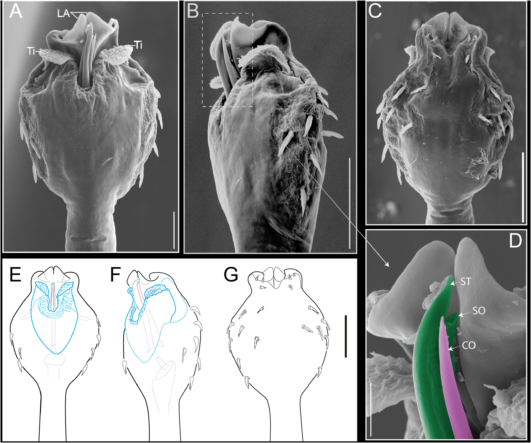
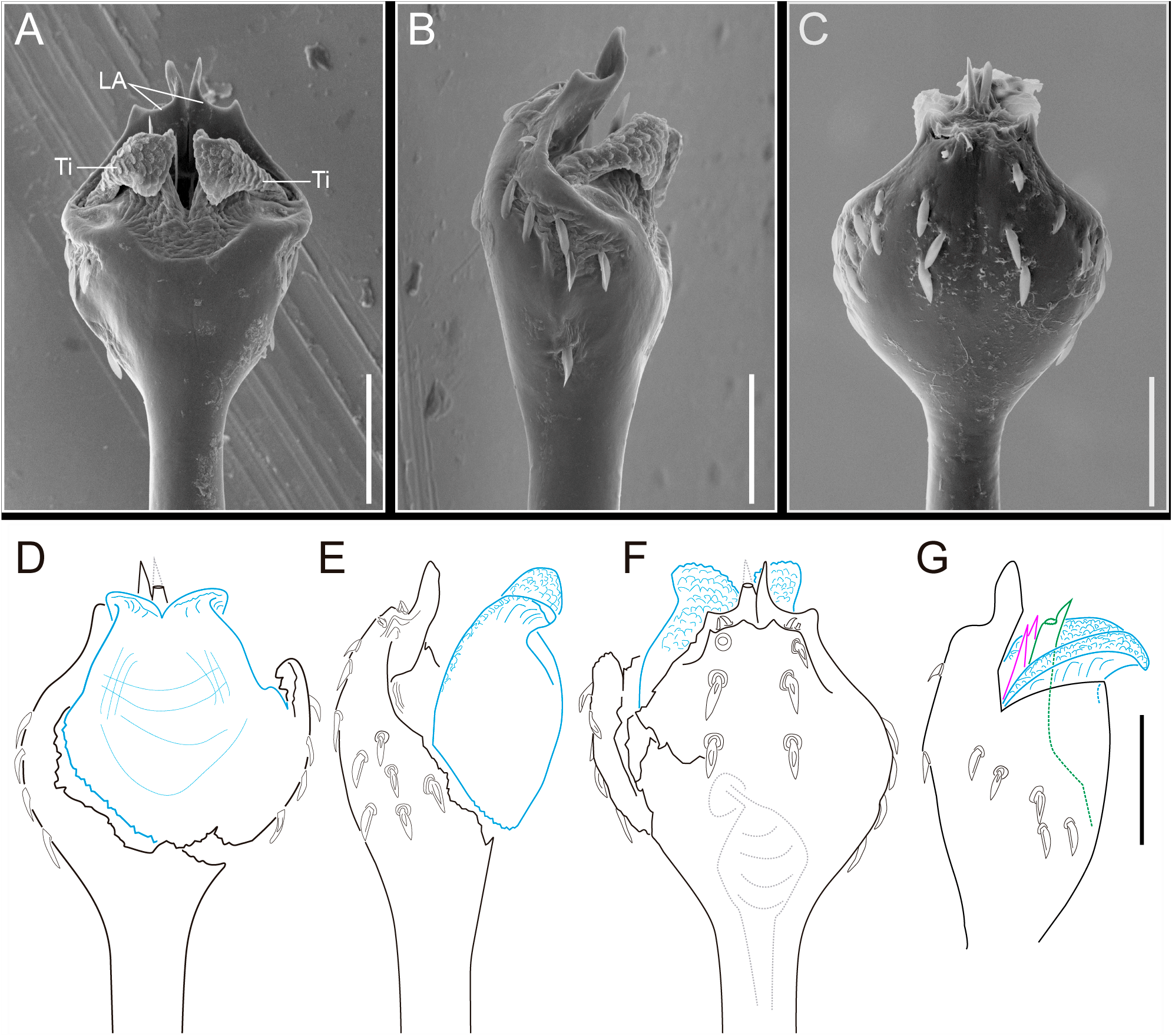
Genitalia: Penis with pars basalis and distalis well demarcated, pars distalis is markedly rounded contrasting with the thin and tubular pars basalis ( Fig. 11 A, D View FIGURES 11 ). Pars distalis ending in a lamina apicalis medially divided by a cleft with a “V” shaped ( Fig. 11 C, F View FIGURES 11 ). Lateral border of pars distalis armed with 6 pairs of leaf-like macrosetae and 2 subapical pairs of very short (truncated, not leaf-like) setae ( Fig. 11 B, F View FIGURES 11 ). Dorsally the capsula externa has two wide and apically rounded titillators. The apical borders of the titillators are dorsally projected. The internal surface of titillators is covered by small digitiform projections ( Fig. 11 A, B View FIGURES 11 ). Capsula interna with two laminar and sharply pointed conductors bearing an also laminar and pointed stylus with a ventral subapical opening of ductus ejaculatorius ( Fig. 11 G View FIGURES 11 ). The cuticular area of the lateral macrosetae has a rugose (folding) appearance ( Fig. 11 B View FIGURES 11 ), this could be an effect of thinner or less chitinous cuticular layer in this region, that could be (hypothetically) related to the region of pars distalis that became swollen during the turgescent stage in the hydraulic penial functioning, this inflation could be responsible for the macrosetae erection necessary for functional interaction with the ovipositor during copulation. These putative expansible areas could functionally correspond to the spongy body (‘Schwellkörper’ in Martens 1978) commonly observed in Asian biantids.
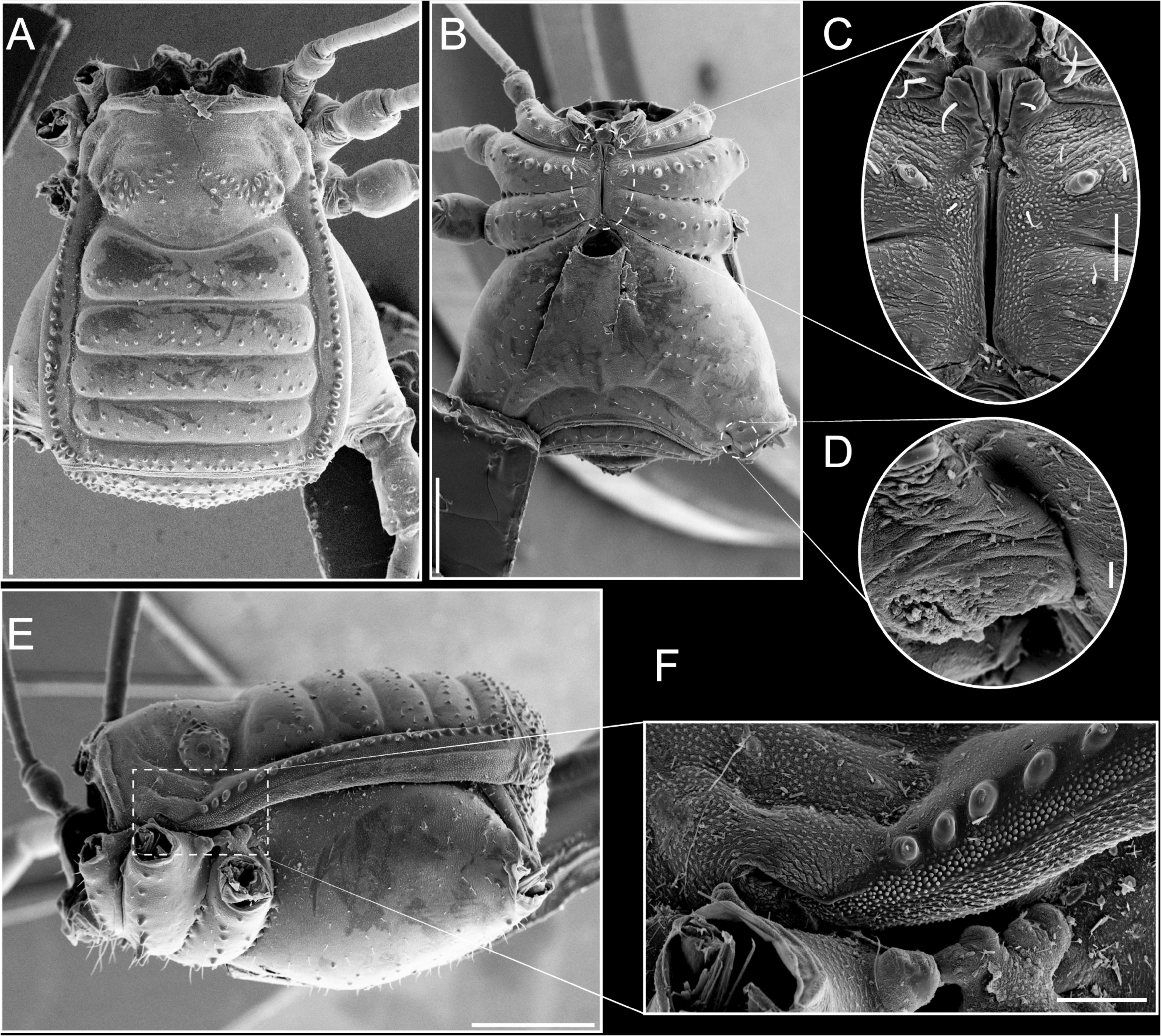
Male (ss) paratype (MNRJ 2429): measurements as in Table 2 View TABLE 2 . Similar to the male (α) in relation to the armature of the dorsal scutum ( Fig. 12 B, C View FIGURES 12 ). Sexual dimorphism evident in patella IV as in the holotype but male ( ss) with prolateral-distal spiniform apophyses and dorsal tubercles smaller in size ( Fig. 12 D View FIGURES 12 ).
Female ( paratype, MNRJ 2429): measurements as in Table 2 View TABLE 2 . Similar to male in relation to the armature of the dorsal scutum ( Fig. 12 A, C, E View FIGURES 12 ), but without prolateral-distal spiniform apophyses and dorsal tubercles in patella IV ( Fig. 12 B View FIGURES 12 ).
Variation. Measurements ( Table 2 View TABLE 2 ): Males (n=5): total body length (1.92–2.35), carapace length (0.71–0.75), mesotergal length (1.70–1.93), carapace maximum width (1.08–1.18), mesotergal scutum maximum width (1.42– 1.61), leg I (5.63–6.30), leg II (14.07–14.68), leg III (7.99–9.39), leg IV (11.61–13.77); Females (n=7): total body length (1.86–2.40), carapace length (0.64–0.73), mesotergal length (1.59–1.86), carapace maximum width (1.07– 1.10), mesotergal scutum maximum width (1.48–1.62), leg I (4.97–5.39), leg II (11.82–13.14), leg III (6.88–7.55), leg IV (9.37–10.44). Tarsal formula (n=12): 6–7(3):9–11(3):6:6.
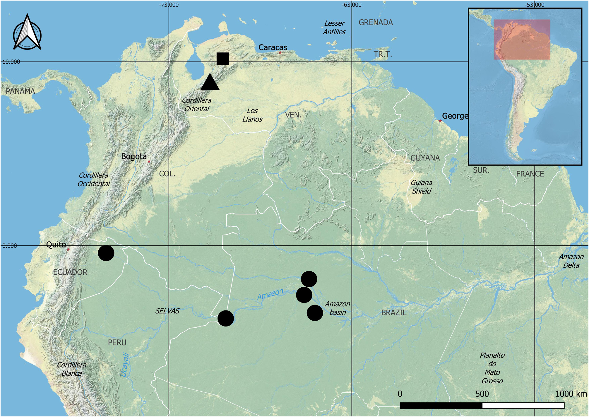
Distribution. Known only from the type locality ( Fig.13 View FIGURE 13 ).


No known copyright restrictions apply. See Agosti, D., Egloff, W., 2009. Taxonomic information exchange and copyright: the Plazi approach. BMC Research Notes 2009, 2:53 for further explanation.
|
Kingdom |
|
|
Phylum |
|
|
Class |
|
|
Order |
|
|
Family |
|
|
SubFamily |
Stenostygninae |
|
Genus |