
Agalenocosa Mello-Leitão
|
publication ID |
https://doi.org/10.11646/zootaxa.3790.1.1 |
|
publication LSID |
lsid:zoobank.org:pub:F5C77E2B-0FE0-4740-BF00-2421E7061EC6 |
|
DOI |
https://doi.org/10.5281/zenodo.6133785 |
|
persistent identifier |
https://treatment.plazi.org/id/367587E8-FF90-FFF7-FF1B-5461A477DB51 |
|
treatment provided by |
Plazi |
|
scientific name |
Agalenocosa Mello-Leitão |
| status |
|
Agalenocosa Mello-Leitão View in CoL View at ENA
Agalenocosa Mello-Leitão 1944: 335 View in CoL ( type species by original designation Agalenocosa singularis Mello-Leitão View in CoL ). Roewer, 1955: 200. Capocasale, 2001: 270.
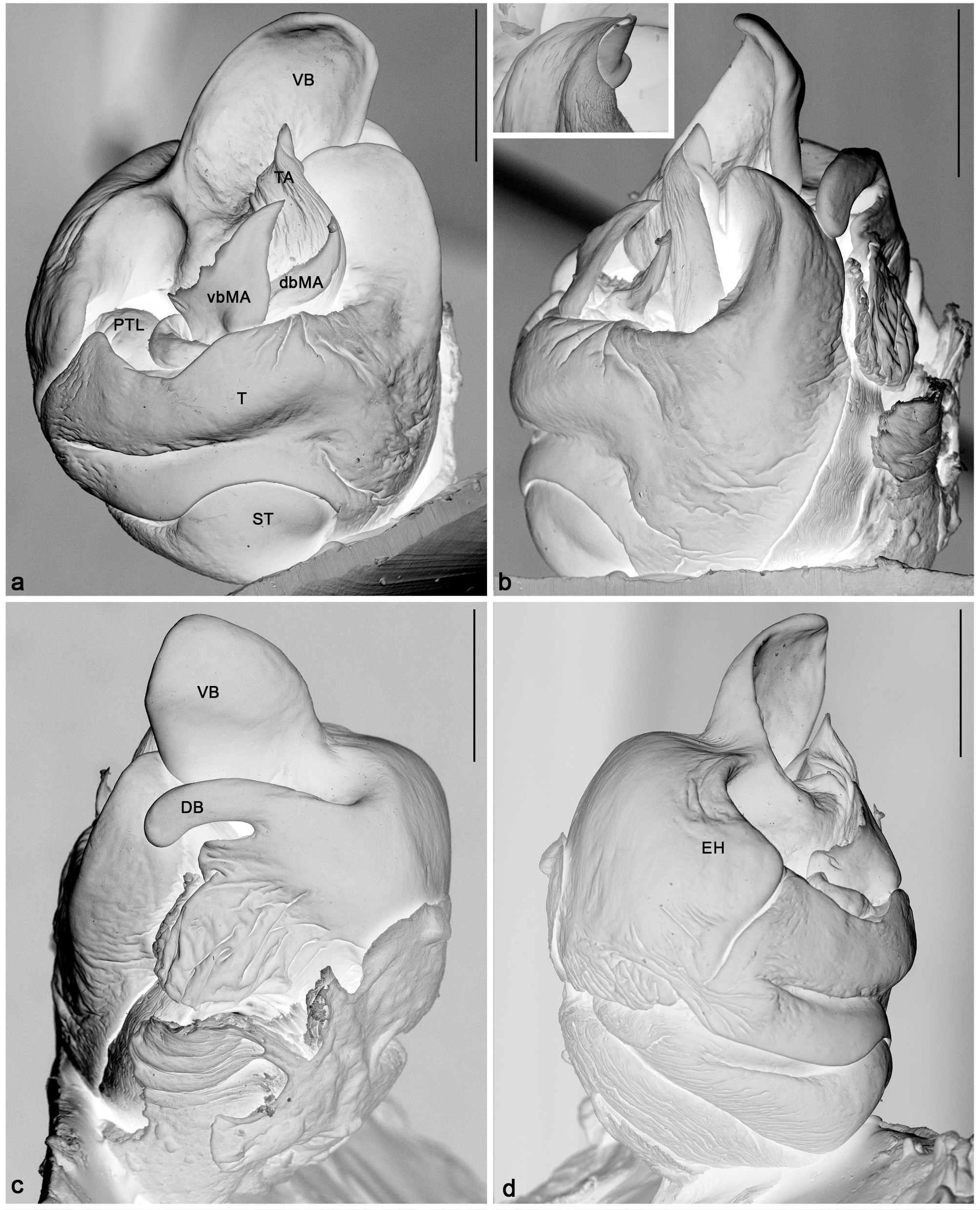
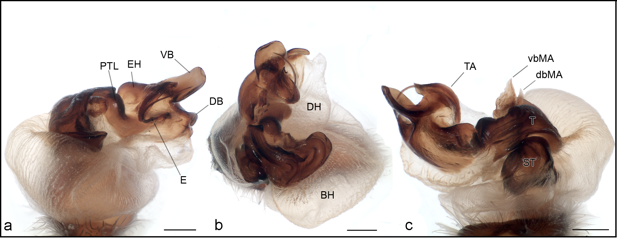
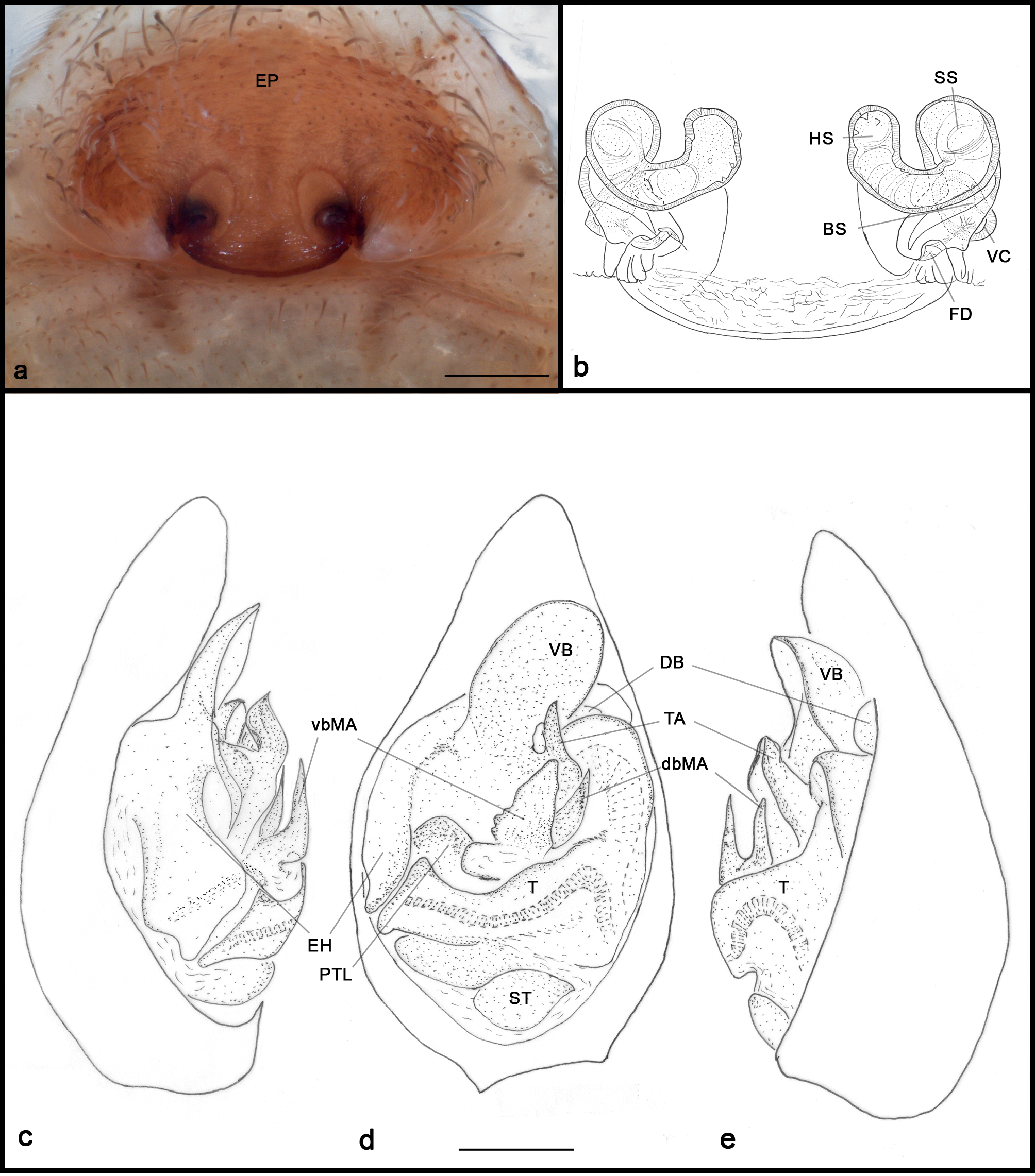
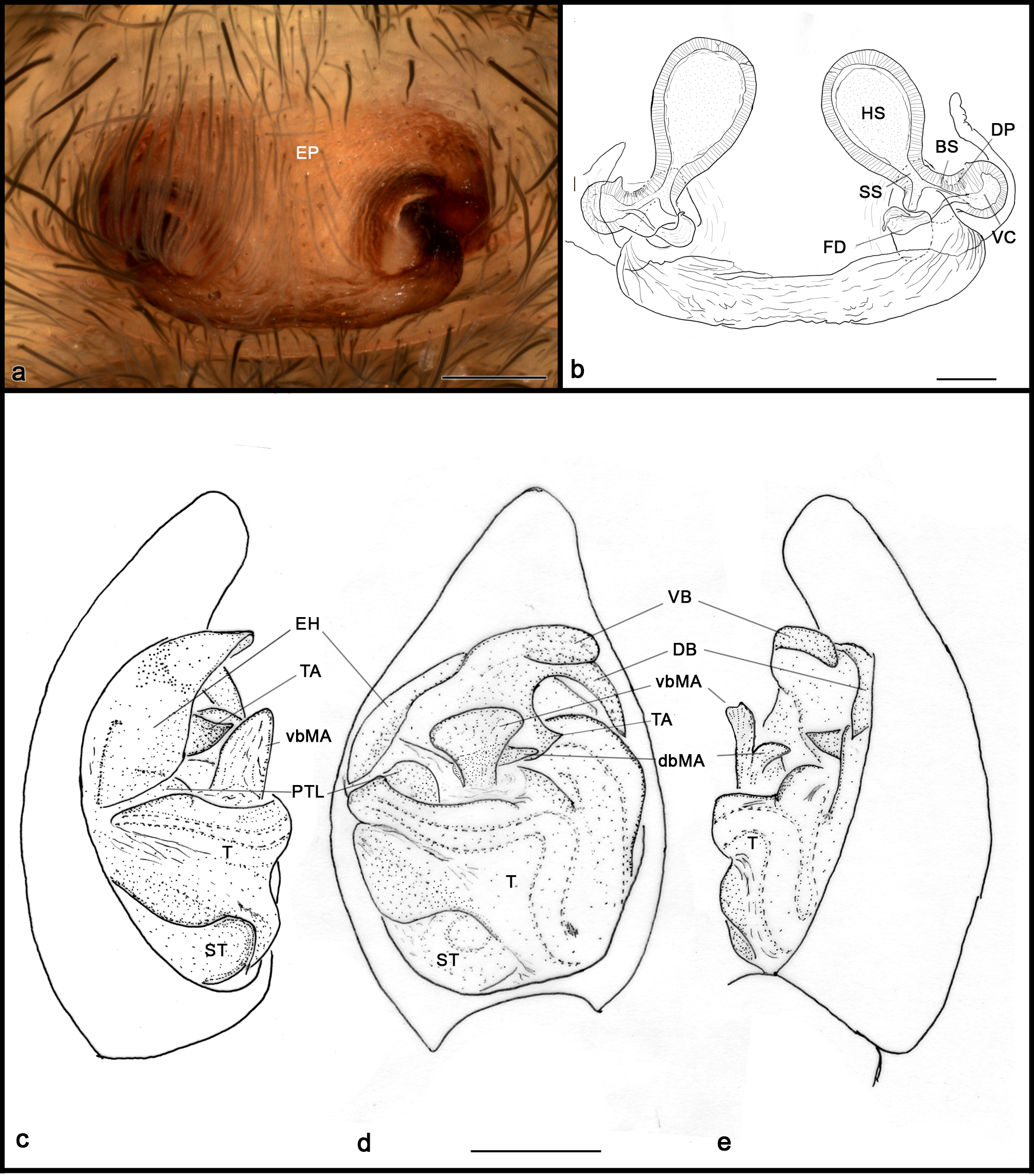
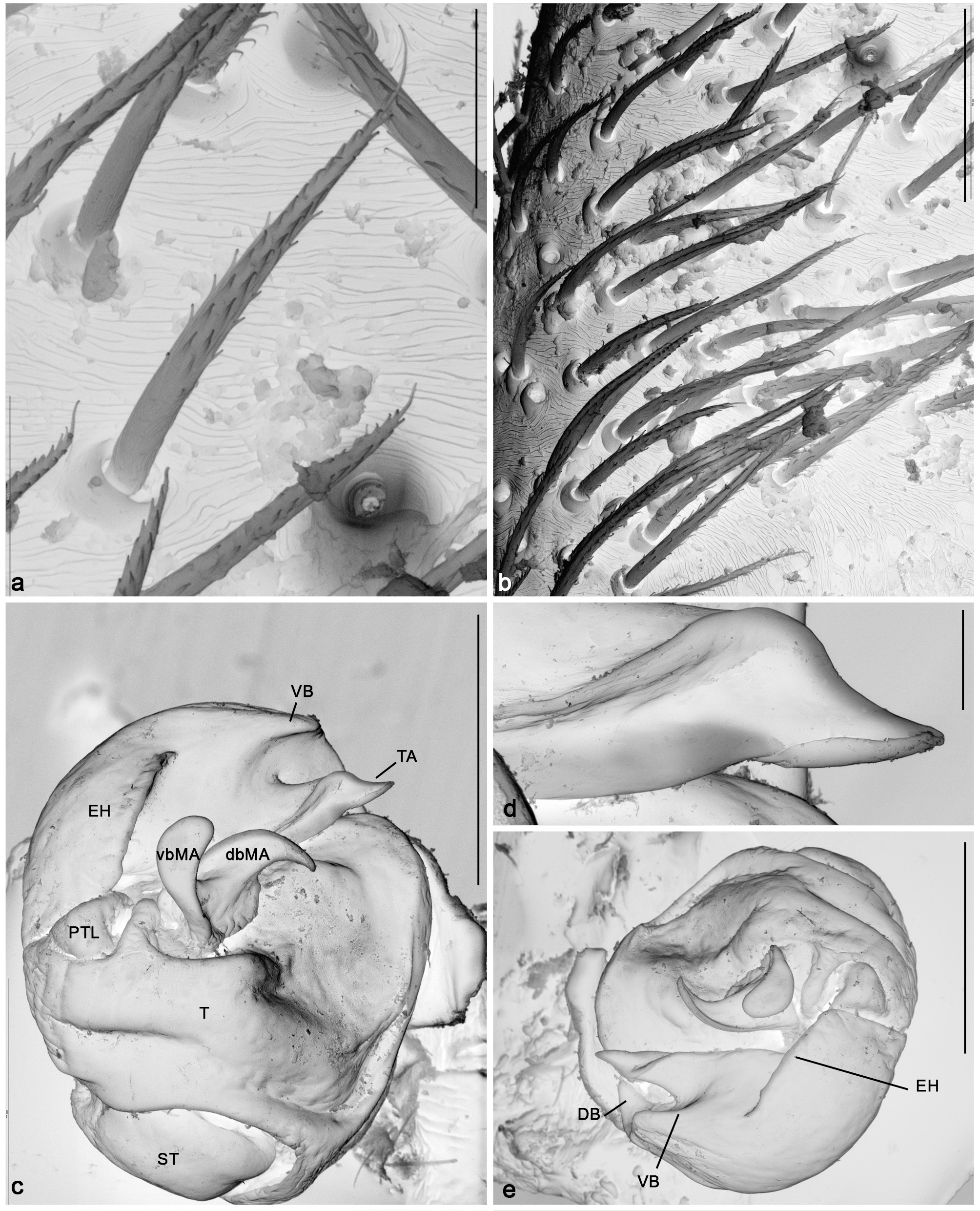
Diagnosis. Males of this genus can be recognised by the presence of a basal retrolateral apophysis on the tibia of the pedipalp ( Fig. 8 View FIGURE 8 ), armed with short, stout setae ( Fig 3 View FIGURE 3 a) except in the case of A. grismadoi sp. nov., which has stout setae but no apophyses ( Figs 20 View FIGURE 20 a,b). The embolic division has an sclerotised area divided in two branches, one large ventral (VB in Fig. 2 View FIGURE 2 a–d; sclerotised ridge in A. grismadoi sp. nov., Figs 19 View FIGURE 19 d, 20c, e) and the other smaller and dorsal (DB in Fig. 2 View FIGURE 2 a–d), which is sometimes not visible in the unexpanded bulb (i.e. in the case of A. velox , Fig. 10 View FIGURE 10 d). Females resemble those of Pirata Sundevall, 1833 and Piratula Roewer, 1960 , but can be differentiated by the lack of the posterior lobes of the epigyne (Figs 42–43 and fig. 70 in Omelko et al. 2011).
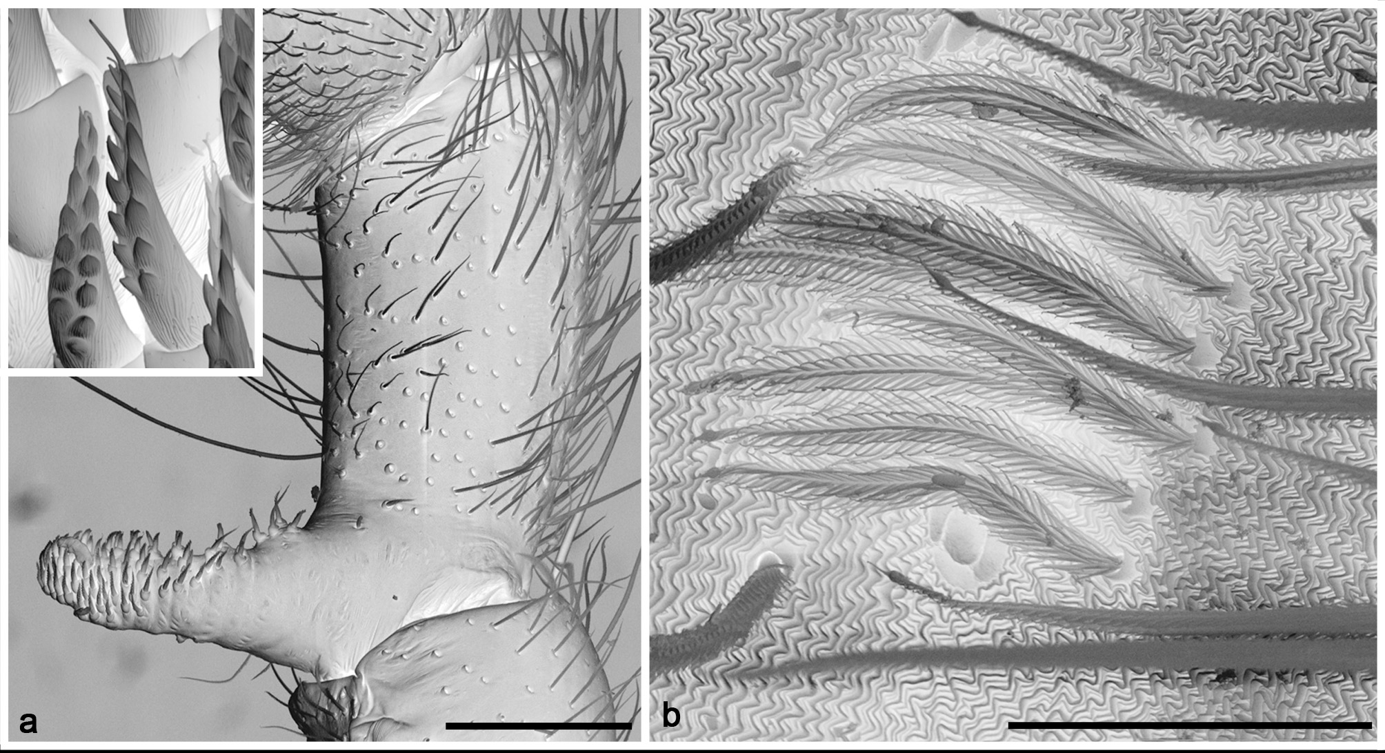
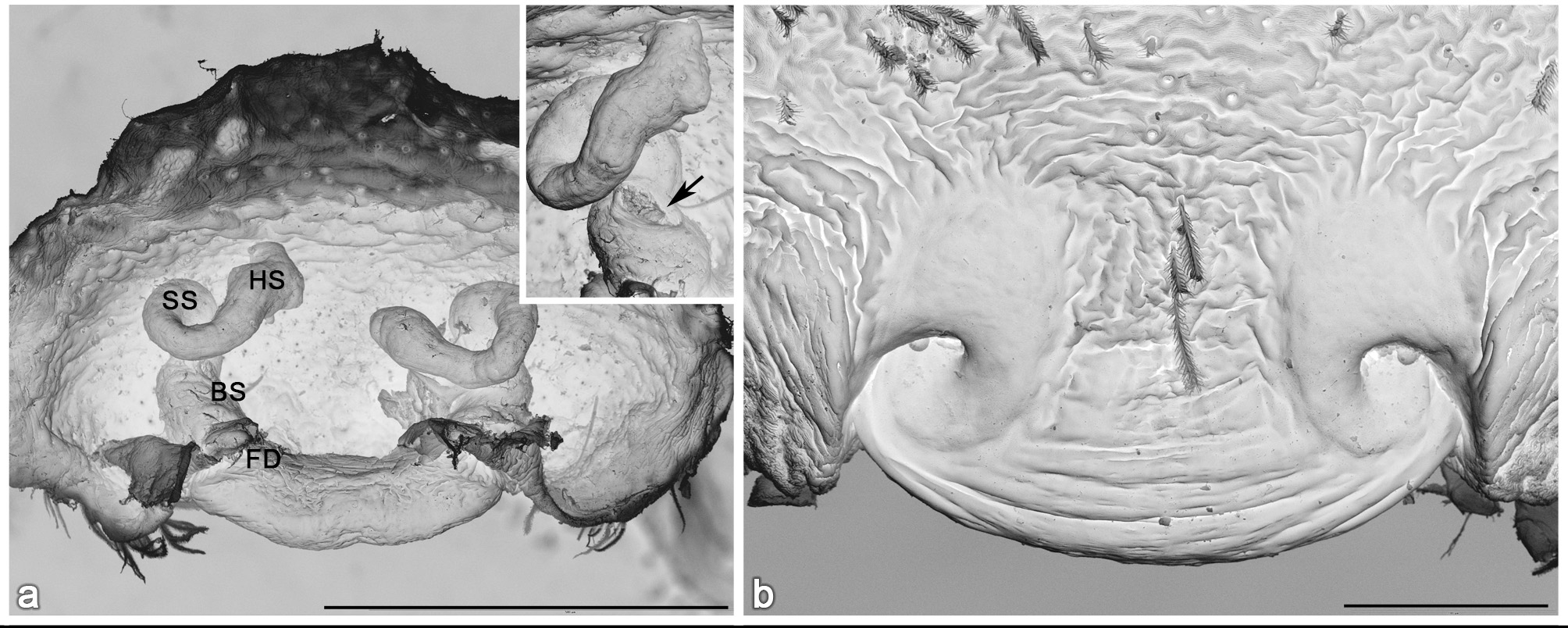
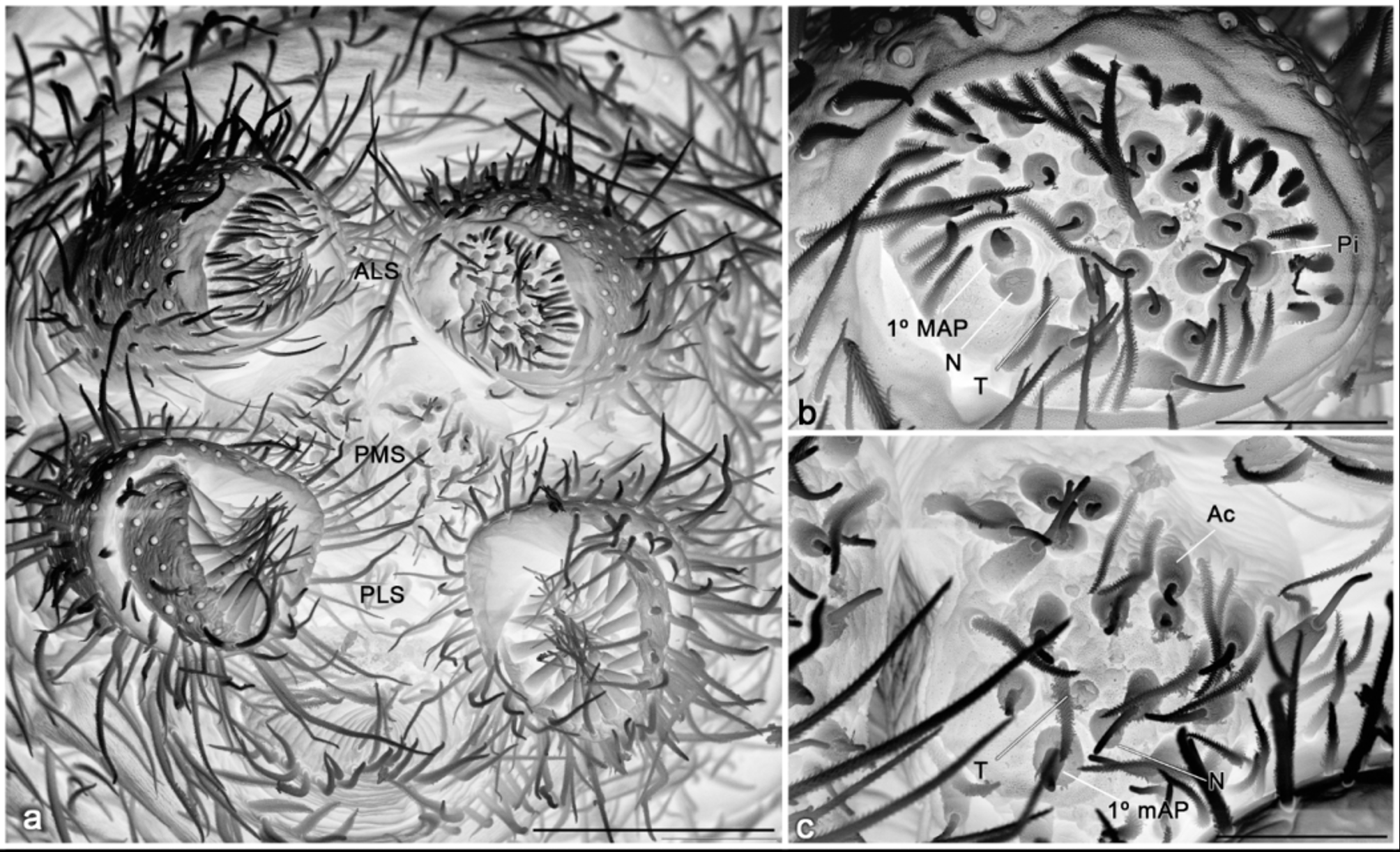
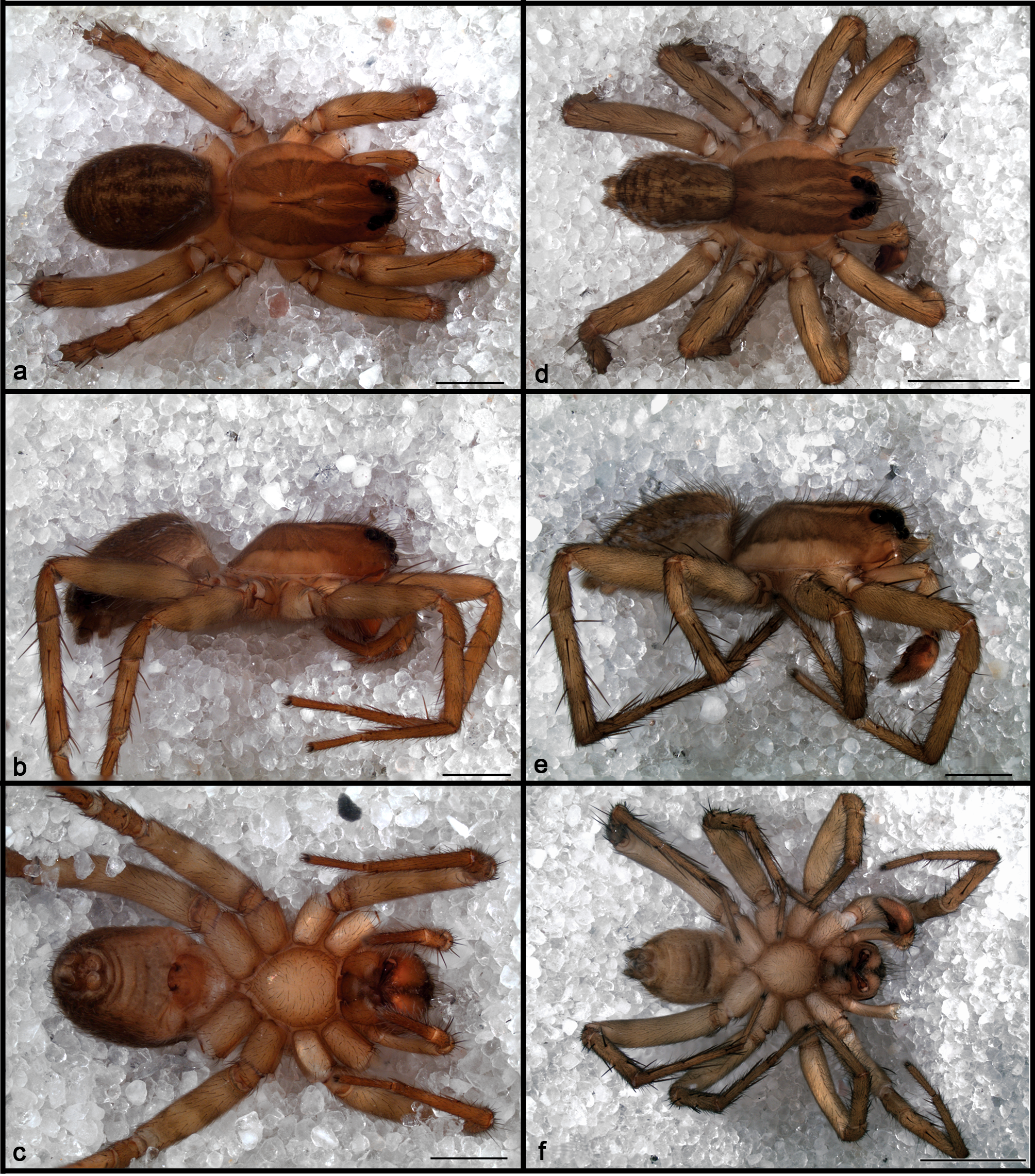
Description. Medium-sized wolf spiders (TL, 4.20–9.98 mm). Females larger than males. Carapace brown with a light brown median band and a Y-shaped mark that extends from fovea to the PLE (tenuous in A. grismadoi sp. nov. and A. punctata , absent in A. luteonigra ). Eyes surrounded by black pigment. Dorsum of abdomen brown to dark olive-grey, with light yellow lanceolate mark in the cardiac area and a pattern of white spots laterally and posterior to the cardiac mark, composed by glistening setae ( Fig. 3 View FIGURE 3 b) (except in A. luteonigra , which have a black abdomen). Carapace longer than wide, dorsal line straight in lateral view ( Figs 9 View FIGURE 9 b, e; domed in A.grismadoi sp. nov. – Figs 18 View FIGURE 18 b, e). Caput flanks in frontal view steep in males, gently sloping in females. AME larger than ALE, AER slightly procurved in anterior view ( Fig. 24 View FIGURE 24 ). Chelicerae with three promarginal and three retromarginal teeth. Abdomen oval. Colulus a fleshy triangular lobe, with several setae (fig. 5a). The spinnerets have a similar conformation to what Townley and Tillinghast (2003) reported for Lycosidae , with six spinnerets, ALS and PLS two segmented, PMS with a single segment ( Figs 5 View FIGURE 5 a, 6a). ALS with two major ampullate gland spigots on the mesal margin (the posterior one reduced to a nubbin in the male ( Fig. 6 View FIGURE 6 b)) and piriform gland spigots ( Fig. 5 View FIGURE 5 b). PMS with about 30 aciniform and two cylindrical gland spigots in the female ( Fig. 5 View FIGURE 5 c) and about 20 aciniform in the male and without cylindrical gland spigots ( Fig. 6 View FIGURE 6 c), with a few setae between them. Female with two minor ampullate gland spigot with a nubbin and a tartipore close to them on the PMS ( Fig. 5 View FIGURE 5 c) and only one minor ampullate gland spigot in the male ( Fig. 6 View FIGURE 6 c). PLS long, tubular, distal segment short, conical, with aciniform and cylindrical gland spigots, with setae among them ( Fig. 5 View FIGURE 5 d).
Leg formula IV> I> II> III or IV> I> III> II, except in A. luteonigra where the leg formula is IV>III> I> II. Tibia I with two pairs of ventral spines (2-2-0), except in A. pirity sp. nov. (0-2-0) and A. grismadoi sp. nov. (2- 0-0).
Femur of male palp without modifications. Tibia with a basal retrolateral apophysis, covered with strong setae ( Fig. 3 View FIGURE 3 a), reduced in A. grismadoi sp. nov. ( Fig. 8 View FIGURE 8 f). Cymbium symmetric, without macrosetae in its tip. Tegulum with a prolateral furrow, deep in A. velox Keyserling, 1891 ( Figs 2 View FIGURE 2 a, 10d) and A. tricuspidata Tullgren, 1905 ( Fig. 12 View FIGURE 12 d) and shallow in the rest of the species. Median apophysis membranous, composed of two elements, the ventral one is flattened and directed longitudinally, the dorsal is transverse and usually retrolaterally directed ( Fig. 2 View FIGURE 2 a, b). The embolic division possesses a grooved and complex terminal apophysis, with a pointed tip ( Figs 2 View FIGURE 2 a, b, 7) and two additional processes, one ventral and a dorsal ( Fig. 2 View FIGURE 2 ). Embolus as a curved spine, not visible in the unexpanded palp, originating prolaterally, below the ventral process of the embolic division and running retrolaterally hidden by the terminal apophysis ( Fig. 7 View FIGURE 7 ).
Epigyne composed of a simple plate with two soft sclerotised areas; in A. velox , A. punctata , A. gentilis and A. grismadoi sp. nov. this area is well developed and surrounds the copulatory open, and in A. luteonigra , A. pirity sp nov., A. tricuspidata and A. gamas sp. nov. is reduced to a small area, posterior to the copulatory open. Vulva: spermathecae with a distal head connected to the base by a more or less defined stalk, vulval chambers rounded. Dictynoid pore conspicuous, located on the boundaries of the vulval chamber and the spermathecal stalk ( Fig. 4 View FIGURE 4 a).
Distribution. Most of the species are known from the eastern Argentina and neighbouring countries, with one species from subtropical montane forest of northwestern Argentina (Figs 25–27).
Remarks. The original illustrations of the epigyne of A. bryantae (Roewer, 1951) from Hispanola, A. denisi (Caporiacco, 1947) from Guyana, A. fallax (L. Koch, 1877) from Queensland, A. kolbei (Dahl, 1908) from Bismark Arch., A. pickeli (Mello-Leitão, 1937) from Brazil, A. yaucensis (Petrunkevitch, 1929) from Puerto Rico shows the typical shape of a Lycosinae epigyne, with an inverted T-shaped septum and anterior pockets. The type specimens of A. chacoensis (Mello-Leitão, 1942) and A. melanotaenia (Mello-Leitão, 1941) were examined; both belong to an undescribed Lycosinae genus ( Alvares 2006). The authors of the remaining two species, A. helvola (C. L. Koch, 1847) from Mexico and Colombia, A. subinermis (Simon, 1897) from India not provided drawings of the copulatory organs, but the size, in the case of A. helvola , and the description of the epigyne, in A. subinermis suggest that those two species not belong to Agalenocosa . The ten species mentioned above where transferred to Agalenocosa by Roewer (1955) without any justification; none of these species fit in Agalenocosa and are here considered as incertae sedis; the final placement of these species is beyond the scope of this work.
Agalenocosa fimbriata is considered here as species inquirenda because the epigyne of the holotype (from Arrecifes, Buenos Aires, deposited in the MLP number 15995, examined) was removed and is probably lost. There are no morphological characters that allow me to recognise which species it is.


No known copyright restrictions apply. See Agosti, D., Egloff, W., 2009. Taxonomic information exchange and copyright: the Plazi approach. BMC Research Notes 2009, 2:53 for further explanation.
|
Kingdom |
|
|
Phylum |
|
|
Class |
|
|
Order |
|
|
Family |
Agalenocosa Mello-Leitão
| Piacentini, Luis N. 2014 |
Agalenocosa Mello-Leitão 1944 : 335
| Capocasale 2001: 270 |
| Roewer 1955: 200 |
| Mello-Leitao 1944: 335 |