
Lusitanipus xanin Gilgado, 2020
|
publication ID |
https://doi.org/ 10.5852/ejt.2020.714 |
|
publication LSID |
lsid:zoobank.org:pub:90AE9367-9A85-4BC9-8479-5BA095BD1BD1 |
|
DOI |
https://doi.org/10.5281/zenodo.4323750 |
|
persistent identifier |
https://treatment.plazi.org/id/2B1CB681-38B8-4F13-99B3-1DAFFE514367 |
|
taxon LSID |
lsid:zoobank.org:act:2B1CB681-38B8-4F13-99B3-1DAFFE514367 |
|
treatment provided by |
Valdenar |
|
scientific name |
Lusitanipus xanin Gilgado |
| status |
sp. nov. |
Lusitanipus xanin Gilgado View in CoL sp. nov.
urn:lsid:zoobank.org:act:2B1CB681-38B8-4F13-99B3-1DAFFE514367
Figs 1 View Fig D–6
Diagnosis
Lusitanipus xanin sp. nov. differs from the other Iberian callipodidan species in the same characters as Lusitanipus alternans (see Reboleira & Enghoff 2015), except that Lusitanipus xanin sp. nov., has metazonital crests of similar size, whereas Lusitanipus alternans has crests of different sizes ( Verhoeff 1893; Spelda 2015; Reboleira & Enghoff 2015). Furthermore, Lusitanipus xanin sp. nov. differs from L. alternans in its green colour, the higher number of body rings, the shape of the gonocoxite, and the curvature and shape of the processes of the tip of telopodites of gonopods.
Etymology
The specific epithet xanin (noun in apposition) is the name of a goblin-like mythological creature, the “Xanín”. This being supposedly inhabits the forests of the region were this species was found (El Bierzo). As the “Xanín”, this species is a small creature dwelling on the ground under the shade of the foliage that is seldomly seen and tries to hide when discovered.
Type material
Holotype
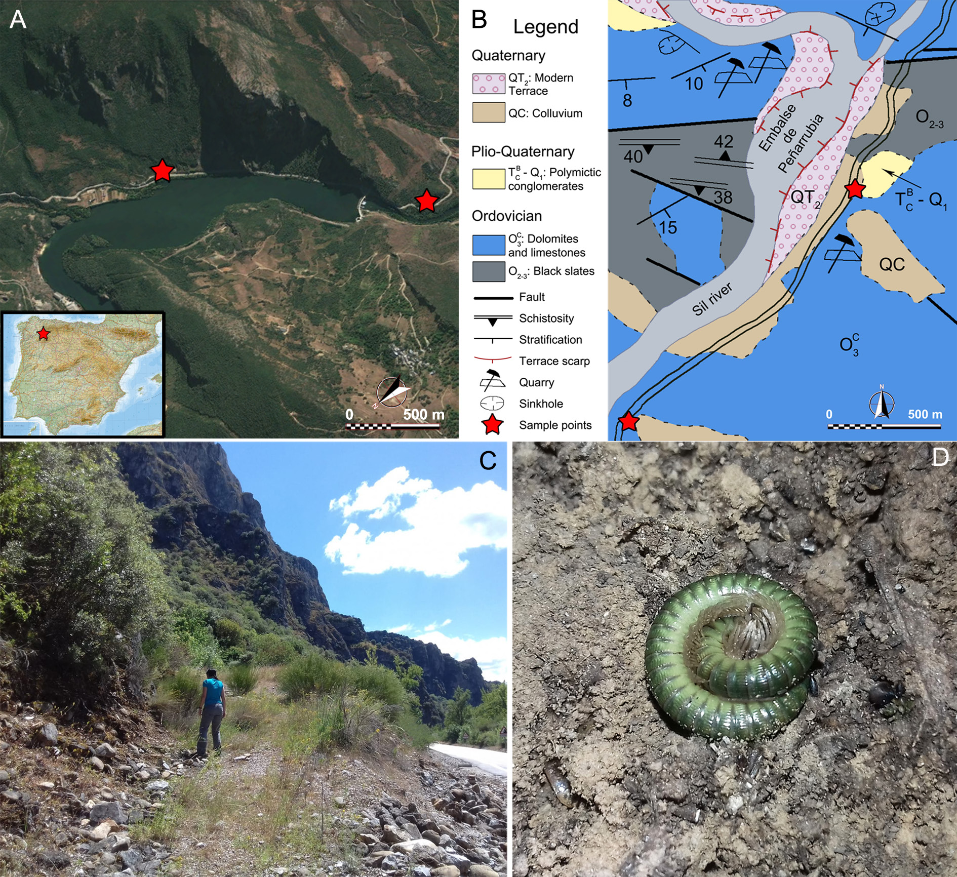
SPAIN • ♂; León province, el Bierzo, Peñarrubia ; 42°27 44.2̎ N, 6°48 32.6̎ W; 405 m a.s.l.; 21 Jun. 2019; José D. Gilgado and Virginia Martínez-Pillado leg.; slope beside the road parallel to the Peñarrubia reservoir; under stones; MNCN 20.07 About MNCN /2069.
Paratypes
SPAIN • 1 ♀, same collection data as for holotype; MNCN 20.07/2070 • 1 ♂, León province, el Bierzo, Peñarrubia ; 42°27 19.2̎ N, 6°49 6.9̎ W; 430 m a.s.l.; 12 Oct. 2006; Carlos E. Prieto leg; entrance of small cave without name, close to the dam; MNCN 20.07/2071 .
Description
BODY SIZE. Holotype male: 60 body rings including collum, telson; two apodous rings before telson. Body approximately 50 mm long; 2.5 mm high; 2.3 mm wide at mid-body section. Paratype male: 62 body rings (two apodous); approximately 39.5 mm long; 2.2 mm high; 2.05 mm wide at mid-body section. Female: 63 rings (two apodous); 53 mm long; 2.8 mm high; 2.45 mm wide at mid-body section.
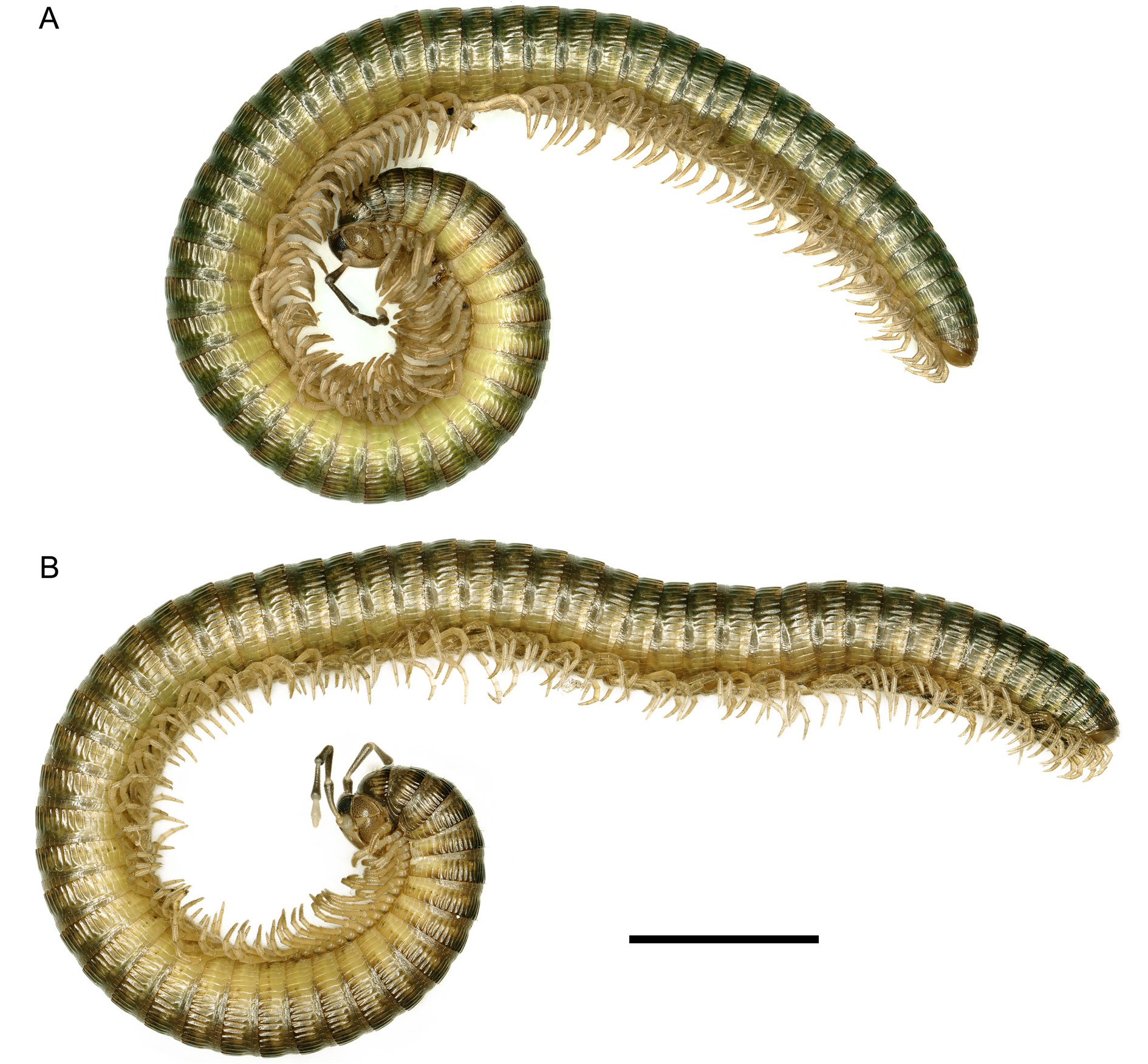
COLOUR. Living specimens: intense green colour, dorsally darker, ventrally lighter ( Fig. 1D View Fig ). Dorsal median light stripe absent. In first body rings, prozonites lighter, posterior half of metazonites darker than anterior part, darker (brownish) crests than intercrests. Colour pattern subtly, progressively changing towards end of body, getting more even, mostly in prozonites; metazonites having more intense darker green colour ( Fig. 2 View Fig ). Legs pale beige ( Fig. 2 View Fig ). Antennomeres 2–5 pigmented in greyish colour, more in distal extremes; antennomeres 6–7 little or no pigmentation ( Figs 2 View Fig , 3B View Fig ). Colour fading to paler brownish or greyish tone ( Fig. 3A View Fig ) after long-term storage in ethanol.
HEAD. Convex, without flattened area or bumps of Cyphocallipus ( Figs 2 View Fig , 3A, C View Fig ), covered with small setae, larger on forehead than posterior part of head. Posterior region having lighter colour than forehead ( Fig. 3A, C View Fig ). Eyes with ca 59 ommatidia distributed in eight rows ( Fig. 3A View Fig ). Tömosvary organ dorsally between eyes, insertion of antennae, closer to former, being only slightly larger than largest ommatidia. Antennomeres of similar shape, proportions to L. alternans , with 6–7 lacking pigmentation ( Fig. 3B View Fig ). Antennomere 5 distally, laterally with sensory region full of short, dense sensillae, also present in L. alternans and other Dorypetalidae species as Dorypetalum helenae ( Stoev & Enghoff 2006) .
TRUNK. Collum: colour pattern as in Fig. 3C View Fig , not very marked crests. Five setae on each side of median half, with setae a,d–e in anterior position; b slightly more posterior than a; c slightly more posterior than b ( Table 1 View Table 1 ). Chaetotaxy of first seven rings as in Table 1 View Table 1 , all setae posterior from body ring 6. Prozonites with scale-like microsculpture, narrow, not very marked crest-like lines, not always continuous with crests of metazonite ( Fig. 3D View Fig ). Crests of metazonites equal in size, unlike L. alternans . Pleurotergite 7 with 18 crests on each side. In male holotype, ozopores present from ring 5 (very inconspicuous on this ring) up to ring 57. Ozopores placed between anterior extremes of 5 th – 6 th crest, except first one: closer to 5 th crest.
LEGS. First pair of smaller size than following, with bump on coxae. Second pair slightly larger, showing gonopores. Two first pairs having one macroseta on inner part of prefemora, bundles of macrosetae in inner side of femora, postfemora; in tarsus: brush-like row of setae on inner side ( Fig. 4 View Fig A–B). Leg 3 not having these bundles of macrosetae, instead: fields of small aggregated setae on inner side of prefemur, femur, postfemur. Leg 3 showing, under optic microscopy, brush-like row of setae on tarsus, papillae ( Fig. 4C View Fig ). Leg 3 with coxal sacs, conspicuous from legs 3–16, missing or not visible in following ones ( Fig. 4 View Fig C–D). Leg 4 seemingly without brush-like row of setae but with papillae, aggregated inner fields of setae on inner side of trochanter, prefemur, femur, postfemur ( Fig. 4 View Fig D–E). Same pattern repeated on all legs, except for last 15, lacking these papillae, fields of setae. Claws of all legs well developed, two basal minor spines ( Fig. 4E View Fig ).
TELSON ( Fig. 3F View Fig ). Epiproct with one spinneret, four setae at each side of sagittal plane, two most proximal setae in middle, two most distal ones close to posterior margin. Paraproct bipartite, with macroseta in each division, as in L. alternans . Hypoproct tripartite with 1+2+1 setae.
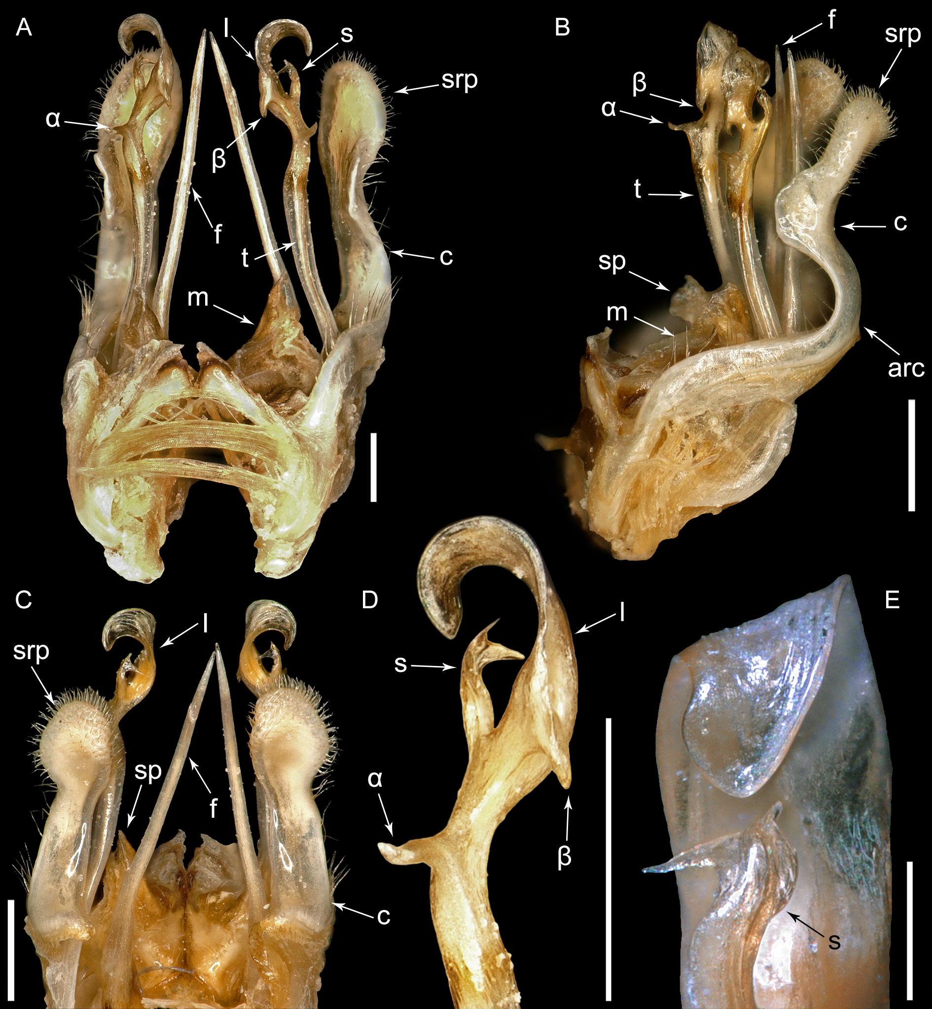
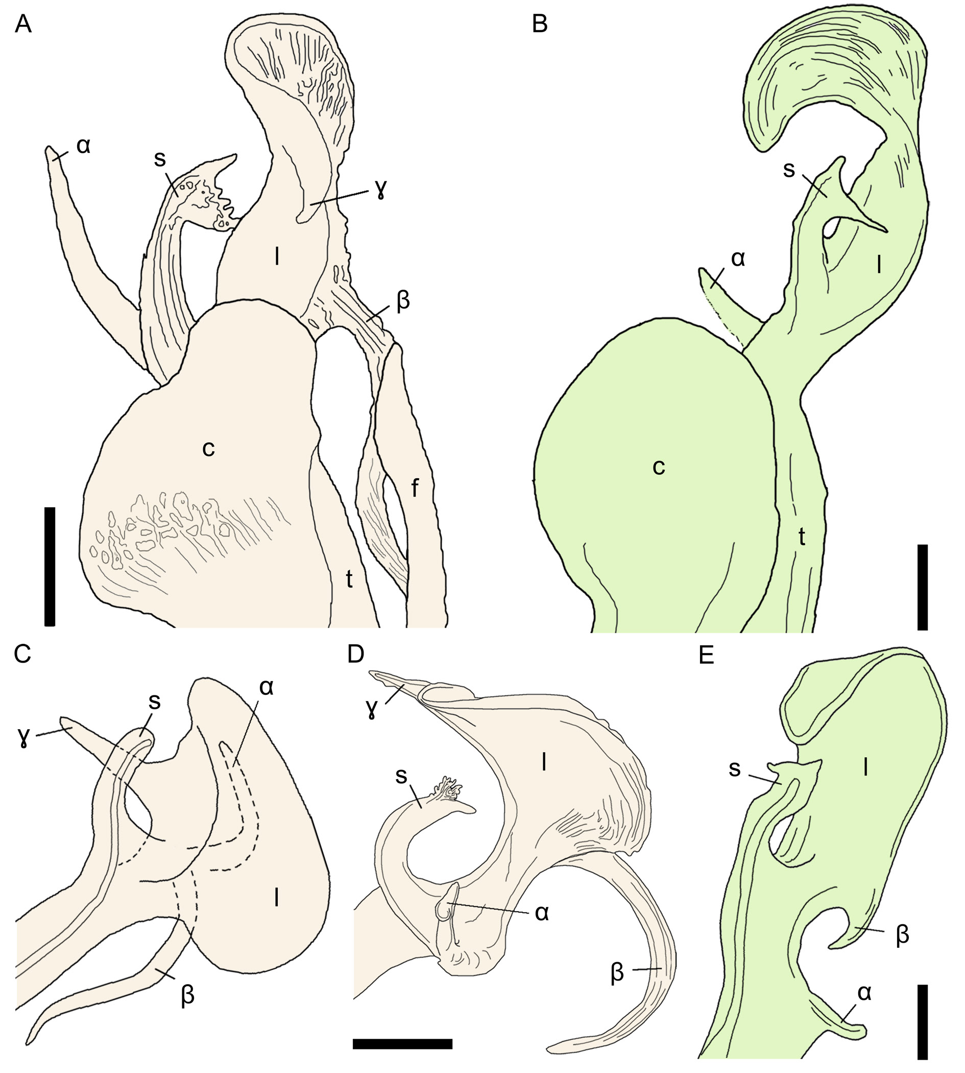
MALE GONOPODS ( Fig. 5 View Fig ). Gonocoxite (c) simple, narrow, with almost semi-circular arc (arc) in middle part, row of setae on posterior margin, as seen in lateral view ( Fig. 5B View Fig ). Arc of gonocoxite ending in blunt semi-rectangular process (srp), almost as long as arc, narrower in central part, pilose on distal margins ( Fig. 5 View Fig A–C). Process flat on interior side, without “spoon-like” shape to accommodate telopodite. Pair of membranous structures (m) of small size interiorly placed between bases of gonocoxites (c), posteriorly to pseudoflagellum (f), telopodites (t). Membranous structures (m) seemingly triangular shaped in posterior view, however in lateral view: subrectangular projection (sp) in margin. Homologous structure present in L. alternans in fig. 5A of Reboleira & Enghoff (2015), with similar but slightly different shape. Pseudoflagellum (or hornflagellum) (f) straight, as in L. alternans , almost as long as telopodite (t). Telopodite ending in complex structure similar to L. alternans , with several differences ( Figs 5 View Fig D–E, 6): tip of telopodite (l) flat (as a lamella), curved in way that tip projecting towards solenomerite (s), similarly to hook ( Figs 5D View Fig , 6B View Fig ). Same processes as L. alternans present, except for γ, having several differences ( Fig. 6 View Fig ): α shorter, less twisted (only slightly curved in tip), placed more basally; β short, not twisted, shape of solenomerite (s) slightly different: narrow pointy process pointing anteriorly, shorter pointy tip projecting towards tip of telopodite.
FEMALE. First pair of legs of smaller size compared to third pair of legs. Second pair of legs reduced ( Fig. 3E View Fig ). Second pleurotergite without conspicuous ventrolateral posteriad process as Cyphocallipus excavatus (see Hoffman 2009), margin describing subtle tentative insinuation of process in that position ( Fig. 3E View Fig ). Everted coxal sacs visible from third pair of legs, as male ( Fig. 3E View Fig ).


No known copyright restrictions apply. See Agosti, D., Egloff, W., 2009. Taxonomic information exchange and copyright: the Plazi approach. BMC Research Notes 2009, 2:53 for further explanation.
|
Kingdom |
|
|
Phylum |
|
|
Class |
|
|
Order |
|
|
Family |
|
|
Genus |