
Alonopsis aureolata Doolittle, 1912
|
publication ID |
https://doi.org/ 10.5281/zenodo.203597 |
|
DOI |
https://doi.org/10.5281/zenodo.5671947 |
|
persistent identifier |
https://treatment.plazi.org/id/3A2BA70A-FF8E-F840-FF45-FA7D1F7E7F6E |
|
treatment provided by |
Plazi |
|
scientific name |
Alonopsis aureolata Doolittle, 1912 |
| status |
|
Alonopsis aureolata Doolittle, 1912
Doolittle, 1912: 561–565, Pls. 42–43; Kubersky, 1977: 656–663, figs 3–20 ( americana ).
Type locality. Lake Sunapee, Sullivan County, New Hampshire, USA.
Holotype: immature female on a slide, U. S. National Museum (cat. 4# 44366), from Lake Sunapee, Sullivan County, New Hampshire, USA collected 22.04.1910.
Material. 37 parthenogenetic females from Sebago Lake, Maine, USA, 0 9.07.1966, coll. D. G. Frey, National Museum of Natural History (Washington DC, USA), Frey’s collection sample 1912.
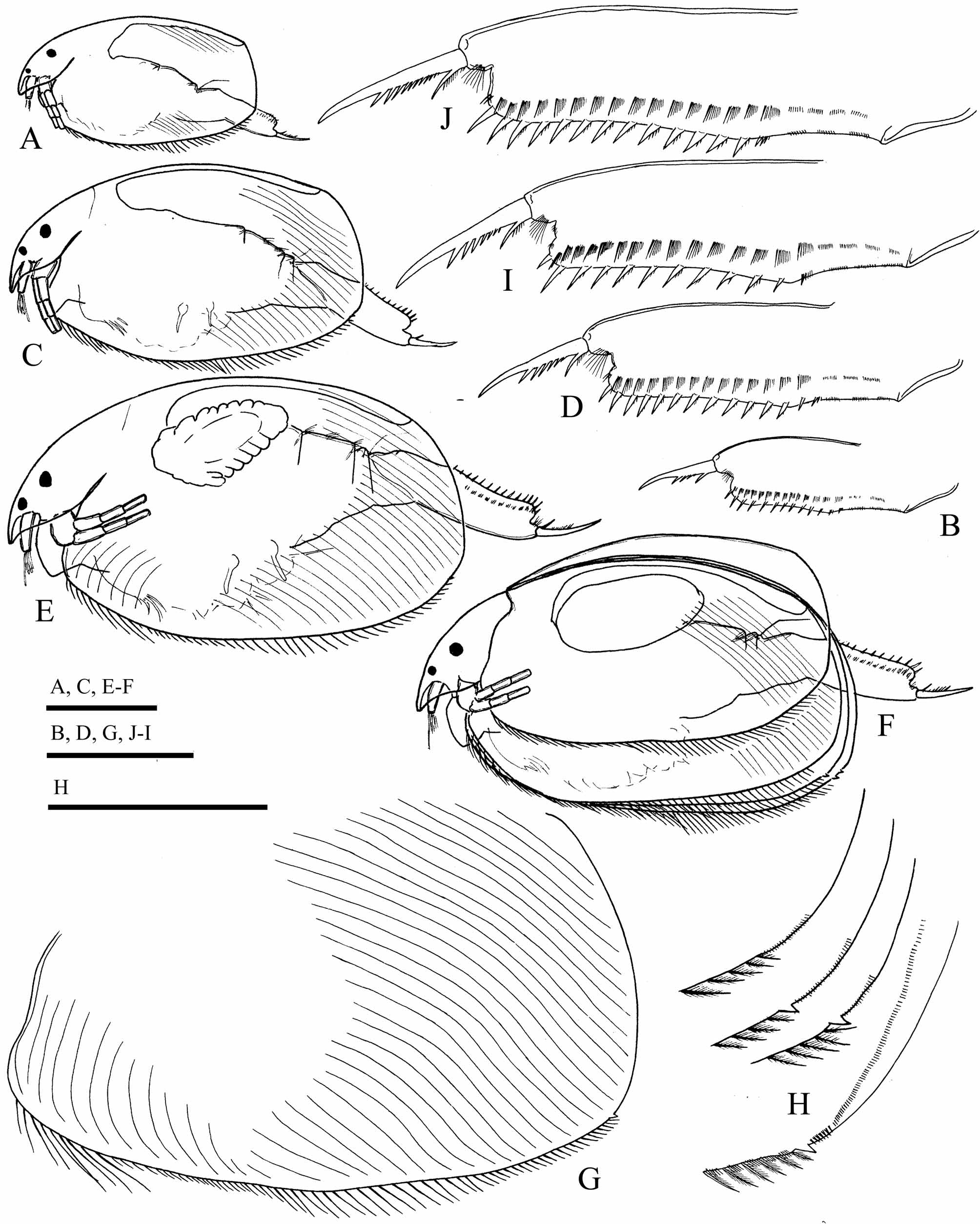
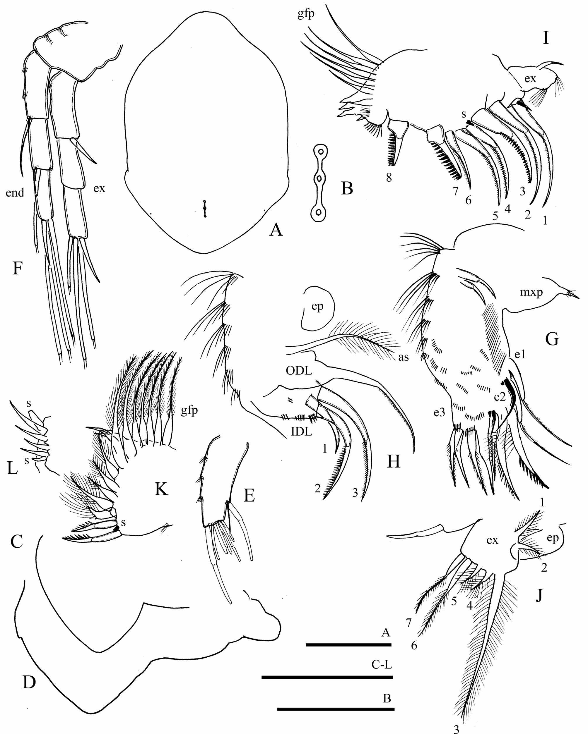
Description. Parthenogenetic female. Body ( Fig. 6 View FIGURE 6 A, C, E–F) height-length ratio about 0.59–0.63. Valves ( Fig. 6 View FIGURE 6 G–H) and head as for genus. Head shield ( Fig. 7 View FIGURE 7 A) with very short, broadly rounded rostrum. Head pores as for genus ( Fig. 7 View FIGURE 7 B), IP/PP ratio about 1.5–2 in adult. Labrum as for genus ( Fig. 7 View FIGURE 7 C–D). Thorax, abdomen and postabdomen ( Fig. 6 View FIGURE 6 B, D, I–J) as for genus, same as in the previous species.
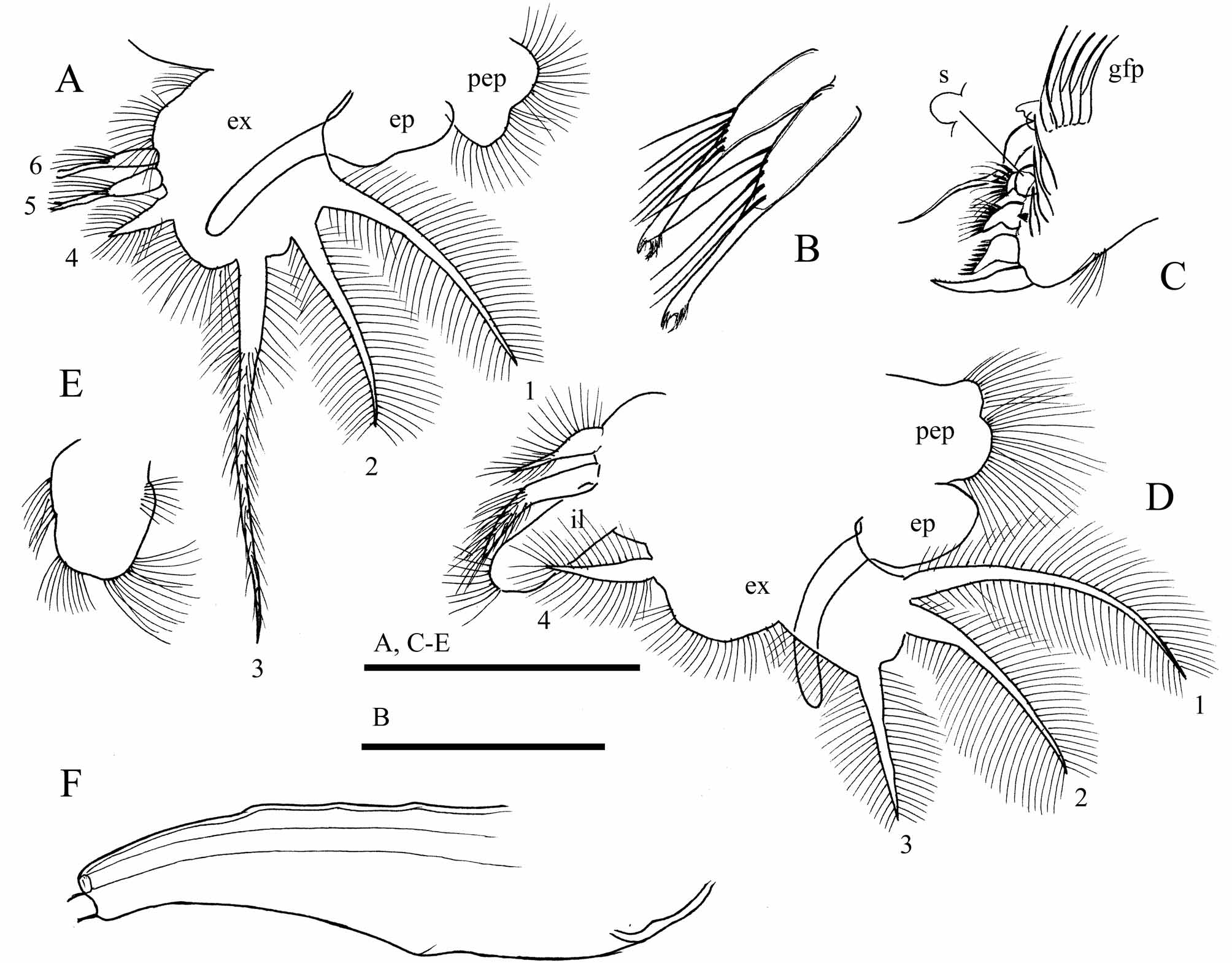
Antennule ( Fig. 7 View FIGURE 7 F), antenna ( Fig. 7 View FIGURE 7 D) and limb I ( Fig 7 View FIGURE 7 G–H) same as in the previous species. Limb II ( Fig. 7 View FIGURE 7 I) with scrapers 7 and 8 of similar size and shape, armed with 12–15 long, thick denticles. Limbs III ( Fig. 7 View FIGURE 7 J–L) and limb IV ( Fig. 8 View FIGURE 8 A–C) same as in the previous species. Limb V ( Fig. 8 View FIGURE 8 D) as for genus, incursion between exopodite lobes as obtuse angle. Limb VI ( Fig. 8 View FIGURE 8 E) with clusters of marginal setules in studied specimens of different proportions compared to the previous species.
Male. According to description of Kubersky (1977), body shape is similar to that of the previous species. Postabdomen ( Fig. 8 View FIGURE 8 F) narrowing distally, but curved, with irregularly curved ventral margin and concave postanal margin, postanal angle defined, obtuse. Position of sperm ducts, armament of postabdomen, postabdominal claws similar to the previous species. Descriptions and drawings of antennule and limb I in description of Kubersky (1977) are somewhat vague. These structures appear to be similar to these of the previous species, but some differences possibly remain overlooked.
Size. In studied material, length of female of juvenile instar I— 0.41–0.46 mm; juvenile instar II— 0.50–0.57 mm; adult female— 0.62–0.89 mm.
No known copyright restrictions apply. See Agosti, D., Egloff, W., 2009. Taxonomic information exchange and copyright: the Plazi approach. BMC Research Notes 2009, 2:53 for further explanation.
|
Kingdom |
|
|
Phylum |
|
|
Class |
|
|
Order |
|
|
Genus |
|
Kingdom |
|
|
Phylum |
|
|
Class |
|
|
Order |
|
|
Genus |