
Ursoconnus Franz
|
publication ID |
https://doi.org/10.11646/zootaxa.4294.5.3 |
|
publication LSID |
lsid:zoobank.org:pub:62D89C4D-72CB-4CF5-91D7-A828D0BD8C69 |
|
persistent identifier |
https://treatment.plazi.org/id/3B39879C-FF82-FFE1-FF1E-FB99E201F91F |
|
treatment provided by |
Plazi |
|
scientific name |
Ursoconnus Franz |
| status |
|
3. Morphological structures and taxonomic status of Ursoconnus Franz
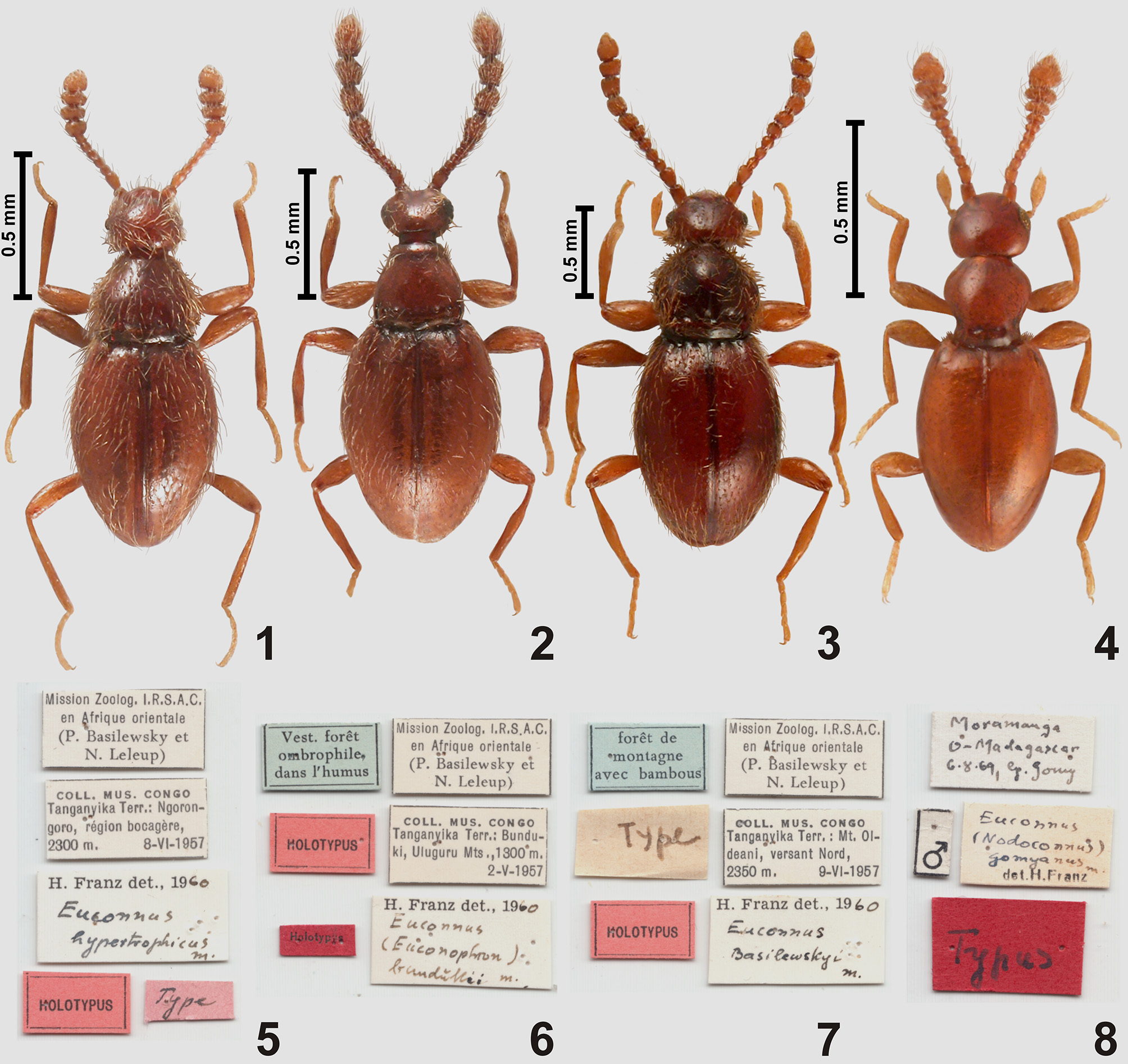
The general body shape of Euconnus ( Ursoconnus) basilewskyi ( Fig. 3 View FIGURES 1 – 8 ) resembles that of Euconophron (morphological structures of the latter were illustrated in Jałoszyński (2017c)), except for the pronotum, and several important diagnostic characters of Euconophron are present in Ursoconnus .
The antennae in Ursoconnus are gradually thickened distally and slightly modified in males (antennomeres VI–X in E. basilewskyi are slightly asymmetrical; Fig. 3 View FIGURES 1 – 8 ); those in Euconophron have indistinctly (often barely discernibly) demarcated club composed of four terminal antennomeres.
The head capsule in Ursoconnus and Euconophron is relatively short, it is weakly elongate in Euconophron and broader than long in Ursoconnus ( Figs 20–21 View FIGURES 19 – 22 ). In the type species of both subgenera the tempora bear thick bristles ( Figs 20–21 View FIGURES 19 – 22 ); the frontoclypeal groove is absent or indistinct ( Fig. 19 View FIGURES 19 – 22 ); the hypostomal ridges ( Fig. 21 View FIGURES 19 – 22 ; hr) complete, strongly convergent posteriorly and reaching posterior tentorial pits; the median area of the labrum is covered with a coarse microsculpture (this character was not described in Jałoszyński (2017c) for Euconophron , but reexamination of SEM micrographs revealed a similar structure of the labrum in both subgenera); and the mandibles are extremely long and slender, each with one mesal tooth located near middle ( Fig. 19 View FIGURES 19 – 22 ; md). The latter character was regarded as a unique diagnostic character of Euconophron ( Jałoszyński 2017c) .
The pronotum, although in E. basilewskyi ( Fig. 20 View FIGURES 19 – 22 ) at the first sight appears different in shape compared to that of E. promptus , the type species of Euconophron , is in fact very similar, and the observed differences can be regarded as resulting from a general broadening and flattening of the prothorax (in a similar way as reported for species of Plaumanniola Costa Lima, 1962; see Jałoszyński (2016e)). In Ursoconnus and Euconophron the anterior and posterior pronotal corners are distinct; the anterior pronotal margin is much shorter than the posterior margin; the broadest site is located near middle (typically slightly behind middle in Euconophron and slightly in front of middle in Ursoconnus ); and the pronotal base bears an uneven number of antebasal pits ( Fig. 20 View FIGURES 19 – 22 ; abp) and very short, barely discernible sublateral carinae ( Fig. 20 View FIGURES 19 – 22 ; slc). The presence of the median antebasal pit was previously ( Jałoszyński 2017c) regarded as a unique diagnostic character of Euconophron .
The prosternum in Ursoconnus and Euconophron has a very short, rudimentary basisternal part ( Fig. 21 View FIGURES 19 – 22 ; bst); its intercoxal area lacks a process or carina; and the anterior margin of prosternum is deeply emarginate, so that lateral portions of basisternum form subtriangular lobes projecting anteriorly. The latter character was also previously ( Jałoszyński 2017c) regarded as unique for Euconophron . Additionally, the inner parts of prothoracic hypomera in Ursoconnus and Euconophron are rapidly narrowed anteriorly; the adcoxal margin of hypomeron is distinctly concave; and the sides of prothorax are covered with thick bristles. The only major difference is the longitudinal median groove ( Fig. 20 View FIGURES 19 – 22 ; mg) present in Ursoconnus and absent in Euconophron .
Meso- and metaventral structures, and the elytral base in Ursoconnus and Euconophron do not show any important differences.
The aedeagus of E. basilewskyi ( Figs 31–32 View FIGURES 27 – 34 ) is clearly Euconophron -like, i.e., the median lobe is relatively stout, with its apical region distinctly bifurcate.
Conclusions. The only morphological differences between the type species of Ursoconnus and Euconophron are the shape of pronotum (broader in Ursoconnus ), the median pronotal groove (present in Ursoconnus and absent in Euconophron ), and the antennal modification in males (present in Ursoconnus and absent in Euconophron ). Compared to many important similarities listed above, these are minor differences. The shape of pronotum in Euconophron and Ursoconnus is variable; species from Afrotropical region were seen with an intermediary shape between that in the type species of each subgenus; also species with a broad and subpentagonal pronotum but lacking the median groove are known to the author. The antennal modifications in males can be regarded as an apomorphy of a species group, insufficient to define a subgenus. Moreover, species from various tropical areas placed in Euconophron are known to the author, which have the clypeal tooth, found in Ursoconnus ( Figs 19, 21 View FIGURES 19 – 22 ). Consequently, Ursoconnus is placed as a junior synonym of Euconophron .


No known copyright restrictions apply. See Agosti, D., Egloff, W., 2009. Taxonomic information exchange and copyright: the Plazi approach. BMC Research Notes 2009, 2:53 for further explanation.
|
Kingdom |
|
|
Phylum |
|
|
Class |
|
|
Order |
|
|
Family |
|
|
SubFamily |
Scydmaeninae |