
Nodoconnus Franz, 1986
|
publication ID |
https://doi.org/10.11646/zootaxa.4294.5.3 |
|
publication LSID |
lsid:zoobank.org:pub:62D89C4D-72CB-4CF5-91D7-A828D0BD8C69 |
|
persistent identifier |
https://treatment.plazi.org/id/3B39879C-FF83-FFEA-FF1E-FAF2E45FFC92 |
|
treatment provided by |
Plazi |
|
scientific name |
Nodoconnus Franz |
| status |
|
4. Morphological structures and taxonomic status of Nodoconnus Franz
Nodoconnus seems to have the general body form and a combination of characters different from those of all previously reviewed subgenera of Euconnus . For this reason, a detailed redescription based on the type species is given.
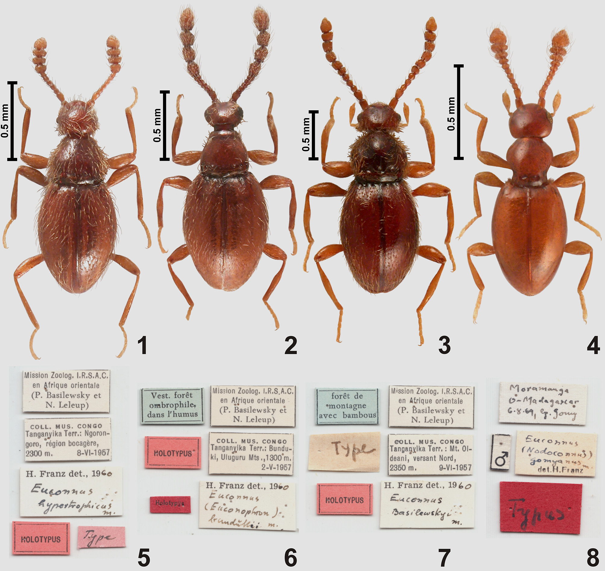
General body shape ( Fig. 4 View FIGURES 1 – 8 ) strongly elongate and flattened, body deeply constricted between head and pronotum and between pronotum and elytra.
Head capsule ( Figs 23–24 View FIGURES 23 – 24 ) large in relation to pronotum; divided by occipital constriction into large anterior and small posterior part ('neck' region), posterior part retracted into pronotum. 'Neck' region slightly broader than half width of head, short and broadening posteriorly; the narrowest place of occipital constriction about as wide as half width of head. Anterior part of head capsule flattened and slightly broader than long, with eyes distant both from occipital constriction and from anterior margin of clypeus. Tempora much longer than compound eyes; vertex ( Fig. 23 View FIGURES 23 – 24 ; vt) transverse and weakly convex, not bulging posterodorsally, anteriorly vertex confluent with subtrapezoidal frons ( Fig. 23 View FIGURES 23 – 24 ; fr); supraantennal tubercles feebly marked; frontoclypeal groove absent. Vestiture of head capsule composed of thin setae distributed on genae, postgenae and tempora; vertex and frons glabrous. Ventral side of anterior part of head ( Fig. 24 View FIGURES 23 – 24 ) flattened; gular plate ( Fig. 24 View FIGURES 23 – 24 ; gp) broad and with barely discernible gular sutures; posterior tentorial pits ( Fig. 24 View FIGURES 23 – 24 ; ptp) strongly elongate, slot-shaped and located anteriorly to transverse impression separating 'neck' region from anterior part of head.
Mouthparts ( Fig. 24 View FIGURES 23 – 24 ) only partly visible in the studied specimen. Submentum ( Fig. 24 View FIGURES 23 – 24 ; smn) very short and strongly transverse; mentum ( Fig. 24 View FIGURES 23 – 24 ; mn) subtrapezoidal; prementum small and transverse, largely membranous; labial palps with short palpomere I and strongly elongate palpomeres II and III. Maxillae generalized, as in all subgenera of Euconnus ; mandibles only partly visible, subtriangular and evenly curved, mesal margins not visible. Posteriorly and laterally mouthparts demarcated by distinct hypostomal ridges ( Fig. 24 View FIGURES 23 – 24 ; hr) which are strongly bent mesally and connected at middle, forming one slightly arcuate transverse ridge largely perpendicular to the long axis of the head and located very close to the posterior margin of mentum.
Antennae ( Figs 4 View FIGURES 1 – 8 , 25 View FIGURES 25 – 26 ) relatively short, with slender proximal portion and strongly thickened and flattened antennomeres VIII–XI forming indistinctly demarcated club; antennomere XI slightly asymmetrical and in dorsal view rhomboidal in shape.
Prothorax ( Figs 4 View FIGURES 1 – 8 , 23–24 View FIGURES 23 – 24 ) flattened and only slightly broader than head, but much narrower than elytra. Pronotum bell-shaped, broadest in front of middle; anterior margin weakly arcuate; anterior corners weakly marked, broadly obtuse-angled; lateral margins in anterior half strongly rounded, behind the broadest site strongly convergent and in front of base slightly divergent posteriorly. Pronotum lacking lateral carinae or edges, base with one pair of very shallow and diffuse antebasal pits ( Fig. 23 View FIGURES 23 – 24 ; abp) connected by shallow transverse impression; sublateral carinae absent. Pronotal disc glabrous, only anteroventral areas of each hypomeron covered with thick bristles directed anteriorly ( Fig. 24 View FIGURES 23 – 24 ). Prosternum about half as long as pronotum, with basisternal part ( Fig. 24 View FIGURES 23 – 24 ; bst) much shorter than coxal part and not demarcated posteriorly from procoxal cavities; intercoxal area lacking process or carina; procoxal sockets closed. Hypomera elongate, with inner (mesal) margins concave, each divided by incomplete, anteriorly obliterated hypomeral ridge ( Fig. 24 View FIGURES 23 – 24 ; hyr) into narrow and long subtriangular inner (adcoxal) part and large outer part confluent laterally with side of pronotum; notosternal sutures ( Fig. 24 View FIGURES 23 – 24 ; nss) entire.
Mesothorax. Mesoscutellum in intact specimens entirely covered by posterior pronotal margin, its posterior tip not exceeding posterior margin of elytral articulating lobe. Mesoventrite ( Fig. 26 View FIGURES 25 – 26 ) with a pair of transverse impressions functioning as procoxal rests ( Fig. 26 View FIGURES 25 – 26 ; pcr) in anterior region, impressions filled with sparse setae and separated at middle by anterior portion of mesoventral intercoxal process; the latter ( Fig. 26 View FIGURES 25 – 26 ; msvp) narrow and strongly elevated; mesocoxal projections with thick bristles directed anteriorly and well-visible in dorsal view ( Fig. 23 View FIGURES 23 – 24 ).
Metathorax. Metaventrite ( Fig. 26 View FIGURES 25 – 26 ) much longer than mesoventrite, approximately subquadrate; lateral (admetacoxal) parts of posterior margin strongly concave; intermetacoxal area weakly expanded posteriorly and forming short metaventral intercoxal process ( Fig. 26 View FIGURES 25 – 26 ; mtvp) as broad as about fourth of metaventral width, with strongly concave posteromedian margin.
Elytra ( Fig. 4 View FIGURES 1 – 8 ) oval, with rounded apices and concave base which is distinctly broader than pronotal base; humeral calli distinct and elongate; each elytron with two barely discernible and asetose rudiments of basal foveae; elytral disc glabrous.
Metathoracic wings present.
Legs ( Figs 4 View FIGURES 1 – 8 , 24 View FIGURES 23 – 24 , 26 View FIGURES 25 – 26 ) moderately long and slender, lacking any peculiar structures.
Abdomen elongate, gradually narrowing posteriorly, suture between two terminal visible sternites (i.e., VII and VIII) less distinct than between remaining sternites.
Aedeagus ( Figs 33–34 View FIGURES 27 – 34 ) with symmetrical median lobe, moderately elongate, with large 'collar' surrounding basal foramen; endophallic structures sclerotized and symmetrical; parameres long and slender, free (i.e., not fused with median lobe), with apical setae.
Remarks. Franz (1986) described several 'varieties' of Euconnus gomyanus , based on differences in the antennal club, which can be strongly or weakly broadened. True taxonomic status of these forms remains to be clarified. Franz (1986) included the sharply demarcated, 3-segmented antennal club in his diagnosis of Nodoconnus . This is not only insufficient to define Nodoconnus , but also misleading. The antennal club in E. gomyanus ( Fig. 25 View FIGURES 25 – 26 ) is composed of four antennomeres and is not sharply demarcated; also the weakly broadened club illustrated by Franz (1986: p. 260) for Euconnus ( Nodoconnus) lambomakandroi Franz, 1986 seems to be four-segmented and indistinctly demarcated.
Conclusions and emended diagnosis of Nodoconnus . Nodoconnus is maintained as a valid name and a subgenus of Euconnus , based on the following diagnostic characters. Apomorphy: submentum very short and broad, demarcated posteriorly by complete hypostomal ridges forming one continuous ridge perpendicular to the long axis of the head (character not known in any other subgenus of Euconnus ); and a unique set of synapomorphies, of which some occur in other subgenera, but not in the same combination: antennae with club composed of four terminal antennomeres (distinctly or indistinctly demarcated); head capsule short and broad, with occipital constriction subequal in width to half width of head; pronotum bell-shaped, broadest in anterior half and with lateral margins sinuate posteriorly, with only traces of paired antebasal pits; basal elytral foveae rudimentary, connected by shallow transverse impression; body covered with very sparse setae, pronotum and elytra nearly glabrous, thick bristles present only on anteroventral area of each prothoracic hypomeron, prosternum and lateral regions of mesocoxal projections; hypomeral ridges incomplete, obliterated anteriorly.
Lectotype designation. Franz (1986) described Euconnus gomyanus on the basis of two specimens, male and female, collected in the same place and deposited in his personal collection (currently at NHMW). The only data concerning the specimens given by Franz (1986) are: "Es liegen mir 2 Exemplare ( ♂♀) vor, die von J. Gomy am 6.8.1969 bei Moramanga gesammelt wurden. Beide Exemplare sind in meiner Sammlung verwahrt". Franz did not refer to them as holotype and paratype, even though one of them is labeled " Typus ", and the other one " Paratypus ". This is not a valid holotype fixation and both specimens have the status of a syntype. The male with labels shown in Fig. 8 View FIGURES 1 – 8 is here designated as lectotype.


| NHMW |
Naturhistorisches Museum, Wien |
No known copyright restrictions apply. See Agosti, D., Egloff, W., 2009. Taxonomic information exchange and copyright: the Plazi approach. BMC Research Notes 2009, 2:53 for further explanation.
|
Kingdom |
|
|
Phylum |
|
|
Class |
|
|
Order |
|
|
Family |
|
|
SubFamily |
Scydmaeninae |