
Traumatomutilla diophthalma ( Klug, 1821 )
|
publication ID |
https://doi.org/ 10.5252/zoosystema2021v43a1 |
|
publication LSID |
urn:lsid:zoobank.org:pub:6A6C06FA-2A60-41F1-8F6D-92EAE415087D |
|
DOI |
https://doi.org/10.5281/zenodo.4450674 |
|
persistent identifier |
https://treatment.plazi.org/id/3C595D3D-FFE1-FFB7-FF22-FC8AFD85F92E |
|
treatment provided by |
Felipe |
|
scientific name |
Traumatomutilla diophthalma ( Klug, 1821 ) |
| status |
|
Traumatomutilla diophthalma ( Klug, 1821)
( Figs 7 View FIG ; 8 View FIG )
Mutilla diophthalma Klug, 1821: 318 .
Ephuta (Traumatomutilla) diophthalma – André 1902: 55.
Traumatomutilla diophthalma – André 1904: 40.
Traumatomutilla diopthalma [sic] – Williams et al. 2017: 8.
TYPE MATERIAL. — Holotype. Brazil [Brasilien] • ♀; Bahia; Freireyss S. leg.; ZMB 141/6 ; ZMB (examined).
ADDITIONAL MATERIAL EXAMINED. — Panama • 2 ♀; Panama Prov. [Province], Chorrera, corregimiento Playa Leona, orilla río Perequete ; 19−20.III.1991; R. Cambra leg.; MIUP • 2 ♀; Panama Prov. [Province], Chorrera, Llano largo; 26−29.III.1990; A. Mena leg.; INPA, MIUP • 1 ♀; Panama Prov. [Province], Parque Nacional Soberania, Camino de Cruces ; 17.II.1998; R. Cambra & A. Santos leg.; MIUP • 1 ♂, reared in laboratory; MIUP • 1 ♂; Chica ; 1-25.X.2013; Malaise trap; Y. Cheng leg.; INPA • 1♀; Cocle Prov. [Province], Valle de Anton ; 6.VII.1991; R. Contreras leg.; MIUP • 1 ♀; Cocle Prov. [Province], Valle de Anton ; 13.VII.1991; J. Coronado leg.; MIUP • 1 ♀; Cocle Prov. [Province], Valle de Anton ; 9-10.I.1991; J. Coronado leg.; INPA • 1 ♂; Colon Prov. [Province], Gamboa ; 16-30.IV.2016; Malaise trap; Lezcano & Estrada leg.; INPA .
Colombia • 1 ♂; Magdalena, P.N. N. [Parque Nacional Natural] Tayron, Neguanje ; 11°20’N, 74°02’W; 10m [sic]; 28.VII.2001; R. Henriquez leg.; IAvH GoogleMaps .
Venezuela • 1♀; Falcon, Yaracal ; 19.VII.1986; L. Joly leg.; MIUP • 1 ♀, Cojedes, Hato Piñero, cr. [circa] El Baul ; 3-10.IX.1994; J. Lattke leg.; INPA .
Brazil • 1 ♀; BMNH .
Pará • 1 ♀; BMNH • 1♀; Pará, Ponta de Pedras ; 27.X.1982; M. F. Torres leg.; MPEG • 1 ♀; Pará, Santarém ; IV.1919, S. M. Klages leg.; (Label: Traumatomutilla diophthalma (Cresson, nec Klug, det. Mickel 1953); MIUP • 1♂; Pará, E. [east of] Araguaia; 19-31.I.1983; J. A. Rafael leg.; MIUP • 1 ♂; Pará, Serra Norte, Manganês; 06-09. IX.1985; Márcio Zanuto leg.; MPEG • 1 ♀; Mato Grosso do Sul, Cotriguaçú, -9.84’31.39’’S, -58.26’26.29’’W, 245 [sic]; 20.IX.206; G. Araújo leg.; UFMT • 1 ♀; Goiás, Serranópolis, S-21 J0799057 TEC1 [sic]; 10-15.I.2007; INPA .
Paraguay • 1♀; Caaguazu, Ypau Señorita ; 13.XII.2001; U. Dreschel leg.; FSCA • 1 ♀; Caaguazu, Zudañez ; XII.1948; UMMZ • 1♀; Concepción, Parque Nacional Paso Bravo, Santa Sofia ; 22°19’20”S, 57°10’12”W; 28.X.2002; B. Garcete leg.; MIUP GoogleMaps • 1 ♀; Paraguari, Parque Nacional Ybycui ; 151 m [above sea level]; 26°04’ S, 56°50’ W; 4-16.X.2004; B. Garcete leg.; MIUP GoogleMaps • 1 ♀; Canindeyu, R.N. B. [Reserva Nacional Bosque] Mbaracayu, Jejui-mi ; 10-14.I.1997; B. Garcete leg.; MIUP .
DIAGNOSIS. — Female. T2 with subcircular pair of integumental spots; lateral face of propodeum usually with sculpture sparse anterodorsad, lacking micropunctures; dorsal face of propodeum sloping posterad, conspicuously elevated posteromedially, sharply angulate into posterior face.
Male. Pronotum clothed with sparse setae, integument visible; body setae with overall silvery-white tone; dorsum of propodeum with dense areas of appressed silvery-white setae at least anterolaterally.
DISTRIBUTION. Panama ( Panama, Cocle, and Colon), Colombia (Magdalena), Venezuela (Falcon and Cojedes), Brazil (Pará, Bahia, Mato Grosso do Sul, and Goiás), and Paraguay (Caaguazu, Concepción, Paraguari, and Canindeyu).
DESCRIPTION
Female
Body length. 13 mm.
Head ( Fig. 7A, B View FIG ). Posterior margin almost straight. Occipital carina evenly wide throughout. Vertex width 0.85 × pronotal width. Eye almost circular, its height in frontal view 1.6 × the distance from its ventral margin to mandibular condyle. Head densely coarsely and confusedly, areolate-punctate to foveolate-punctate; with conspicuous broad smooth intervals on front; sculpture denser and coarser on vertex. Genal carina present. Mandible oblique, tapering slightly apicad, conspicuously bidentate, unarmed ventrally. Dorsal scrobal carina well defined, not reaching antennal tubercles; lateral scrobal carinae present, connected to dorsal carina. Antennal tubercles irregularly rugose. Flagellomere 1: 2.0 × pedicel length; flagellomere 2: 1.55 × pedicel length.
Mesosoma ( Fig. 7A, B View FIG ). Dorsal thoracic length almost as long as mesosomal width. Mesosomal dorsum densely and coarsely areolate-punctate with smooth rounded intervals; sculpture overall slightly larger laterally on pronotum. Humeral carina present, narrowly disconnected from slightly produced subangulate epaulet; anterolateral corners of pronotum angulate in dorsal view. Anterior face of pronotum defined, short, shorter than pronotal collar, vestigially and coarsely striated longitudinally basad, with coarse dense punctures dorsad; dorsal face roundly angulate into anterior face in lateral view. Pronotal spiracle almost flat against lateral margin of pronotum. Lateral face of pronotum sparsely foveolate-punctate with interspersed micropunctures; mesopleuron sculpture, micropunctate anteriorly, sparsely and vestigially foveolatepunctate along mesopleural ridge; metapleuron sculpture almost completely concealed by dense setation, except dorsal fourth smooth, unsculptured; with dense coarse longitudinal rugosities on dorsal margin anterior to propodeal spiracle. Lateral face of propodeum sparsely foveolate-punctate,intervals smooth and shining predominantly as wide as surrounding sculpture. Ratios of widths of mesosoma at humeral angles, pronotal spiracles, widest point of mesonotum, narrowest point of mesonotum and propodeum immediately posterior to propodeal spiracles. Lateral margin of mesonotum marginally constricted anterior to propodeal spiracle, slightly diverging anterad. Propodeal spiracle inconspicuously pronounced from lateral margin of mesosoma; post-spiracular area present. Scutellar scale and anterolateral carinae absent; scabrous intervals absent on scutellar area. Propodeum conspicuously elongate, dorsal face much longer than and well differentiated from posterior face.
Metasoma ( Fig. 7A, B View FIG ). Ratios of width of T1, width of T2 and length of T2, 30:68:67. Disc of T2 densely and coarsely foveolate-punctate to punctate with dense interspersed micropunctures; foveolae sparser and micropunctures absent laterally and over integumental spots. T3–6 sculpture, except pygidial plate, predominantly concealed by dense setation, sparsely and coarsely foveolate-punctate to simply punctate with dense interspersed micropunctures where visible; micropunctures sparser on T5, absent on T6; pygidial plate subpyriform, defined by strong, projected, flange-like lateral carinae at apical fourth of plate; surface predominantly with irregular longitudinal rugosities; interstice apparently granulose. S1 sparsely punctured, surface wedge-like, ending in a rounded longitudinal slightly concave carina. S2 sparsely foveolatepunctate, sculpture sparser posterad; anteromedial crest-fold almost absent. S3–6 densely and coarsely foveolate-punctate with sparse micropunctures at S3–4; sculpture denser and micropunctures absent on S6.
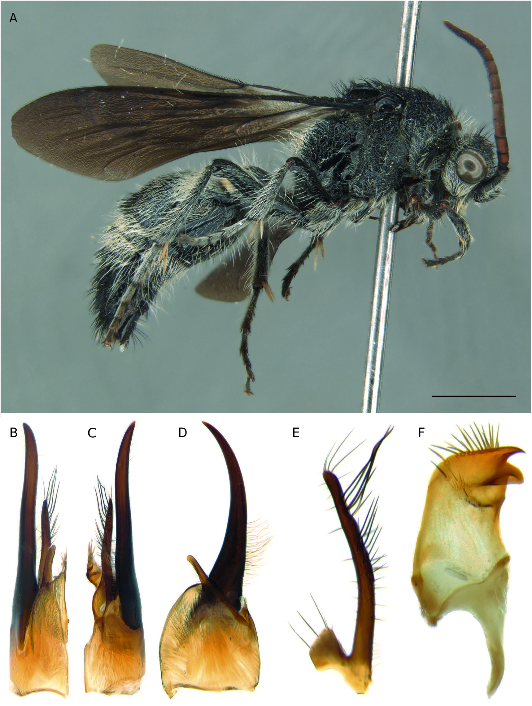
Male (hitherto unknown).
Body length. 12-12.5 mm.
Head ( Fig. 8A View FIG ). Transversely subrectangular with posterolateral angles rounded in dorsal view. Width 0.88 × pronotal width. Eye almost circular. Ocelli small; OOD 3.4 × DLO, IOD 1.5 × DLO. Occipital carina distinct. Head surface densely and coarsely punctate. Gena ecarinate. Antennal scrobe concave to eye margin, with prominent transverse dorsal scrobal carina. Clypeus concave laterally immediately below antennal insertion, conspicuously convex medially; coarsely and densely punctate to micropunctate; apical medial margin slightly concave, with a small denticle on each side of the concavity. Scape bicarinate. Flagellomere 1: 2.05 × pedicel length; flagellomere 2: 2.3 × pedicel length. Mandible obliquely tridentate apically, inner tooth slightly larger than middle tooth; lacking dorsal or ventral projections.
Mesosoma ( Fig. 8A View FIG ). Epaulets well defined, slightly projected from anterior margin of pronotum, broadly separated from humeral carina, anterolateral angles of pronotum rounded. Anterior face of pronotum sparsely punctate with interspersed micropunctations laterally, with a conspicuous smooth unsculptured area. Tegula convex, mostly glabrous and impunctate except for dense coarse punctures on anterior third and along inner margin. Mesoscutum densely and coarsely foveolate-punctate, notaulus absent, parapsis vestigial, reduced to posterior half of mesoscutum. Scutellum convex, densely and coarsely areolate-punctate to foveolate-punctate; with longitudinal irregular carina medially formed by aligned intervals. Axilla produced posterolaterally as acute projections in dorsal view, with conspicuous flat coarsely and densely foveolate-punctate dorsal surface. Metanotum slightly wider laterally, its surface obscured by dense setation. Propodeal dorsum convex, partially concealed by dense setation, densely areolate where visible; lateral face densely and coarsely areolate, areolations less defined anterad; dorsal face rounded into and poorly distinguished from posterior face. Lateral face of pronotum densely coarsely and confusedly punctate to micropunctate; mesopleura slightly swollen on dorsal half, without any or projections; mesopleural sculpture densely and coarsely areolate-punctate to foveolate-punctate with interspersed micropunctures anteriorly. Metapleuron foveolate-punctate ventrally, micropunctate to smooth dorsally.
Wings ( Fig. 8A View FIG ). Fore wing with elongate sclerotized pterostigma; marginal cell elongated and with Rs convex, not truncate apically; three submarginal cells; dark brown, slightly but conspicuously lighter on basal third.
Legs ( Fig. 8A View FIG ). Simply setose, no strong spines discernible dorsally; spurs finely serrate on margins.
Metasoma ( Fig. 8A View FIG ). T1 0.48 × as wide as T2.T2 length 0.72 × its width. Dorsal metasomal sculpture partially concealed by dense setation, densely and coarsely punctate with interspersed micropunctures where visible; pygidial plate irregularly and vestigially rugose, weakly defined by parallel carinae apicolaterally. S1 longitudinally elevated medially, slightly pronounced carina lower medially. S2 sparsely foveolate-punctate to punctate, interspersed micropunctations present anterolaterally, foveolae conspicuously sparser and larger posterad; with vestigial longitudinal anteromedial crest-fold; sternal pit absent. S3–5 sparsely and coarsely foveolate-punctate with interspersed micropunctures; S6–7 sparsely foveolate-punctate. S7 longer than broad, posterior margin projected laterally and medially, medial projection terminating in a pair of very small subacute closely spaced tooth-like on posterior margin.
Genitalia ( Fig. 8 View FIG B-F). Parapenial lobe not at all pronounced posteriorly, simply rounded. Ratios of free length of paramere, cuspis and digitus, 53:31:14; paramere slightly sinuous in dorsal view, upcurved posteriorly in lateral view; with dense setae ventrally at anterior half; cuspis short, stout, slightly swollen medially and narrower posterad in lateral view; narrower posterad and almost straight in dorsal view; abruptly curved dorsally in wide angle at anterior third; with dense conspicuous, strongly sinuous setae on ventral surface, except at anterior third with simple short setae; dorsal surface with overall inconspicuous simple short setae; paracuspis welldeveloped, not sessile, slightly elongate vertically, subrounded at posterior margin, densely setose along posterodorsal margin, setae predominantly shorter than or as short as paracuspis; digitus short, slightly curved inward in dorsal view and slightly upcurved in lateral view, inconspicuously setose basodorsally; penis valve strongly concave on inner surface, with closely spaced pair of short teeth posteroventrally; posterior tooth acute, subposterior tooth rounded, with lateral pocket present on outer surface; apical distance between teeth 0.1 × length of valve; dense setae present along posterior margin and inconspicuous short setae present at base of subposterior tooth on outer surface.
Coloration and variations
Female ( Fig. 7A, B View FIG ). Integument black, except mandibles and antennal flagella partially reddish-brown and T2 with a pair of orange subcircular integumental spots which vary slightly in size. Body setae predominantly silvery-white varying in density, except the following areas with black setae varying in density: front, genae; pronotal dorsum, mesonotum medially, scutellar area, propodeal dorsum medially; T1 medially, disc of T2 (except integumental spots), fringe of T2–5 sublaterally, T6 laterally, and S6.
Male ( Fig. 8A View FIG ). Integument black, with mandibles and flagella partially reddish-brown. Head mostly with dense white setae except sparse, erect and large, black setae on postero-lateral areas of vertex and near inner eye margins; pronotum dorsum, mesoscutum, axillar projections, scutellum and tegula with black setae; dorsum of propodeum with sparse white setae, dense and decumbent antero-laterally; pronotum lateral face with white setae; mesopleura with sparse black setae near tegula other area with sparse white setae; propodeum lateral face with sparse white setae; legs with white setae except apex of meso and metafemora dorsally with black setae, T1 with white setae, dense and decumbent on dorsal face; T2 to T7 with black setae except anterior third and narrow lateral area of T2, narrow apical fringe of T2, narrow lateral areas of T3–4 with white setae; S1 to S4 with white setae; S5–S6 with white and black setae, S7 with black setae.
HOSTS
Hymenoptera : Apoidea Latreille, 1802: Sphecidae Latreille, 1802 : Podium sp. (in laboratory); Hymenoptera : Apoidea: Crabronidae Latreille, 1802 : Trypoxylon sp. (in situ). Host association: wooden trap nests were placed in a forested area in Panama by RAC for two weeks (17-31.II.1998), after which one of the nests was found to be occupied and the entrance closed with resin. The occupied trap was taken to the lab at MIUP and both halves of the nest were separated revealing a Podium Fabricius, 1804 larva. The nest was once again closed so the larvae could pupate. Once the larvae reached the pupa stage, a female of T. diophthalma was placed inside the nest using forceps and the entrance was once again closed leaving the mutillid locked inside the nest for 24 hours, after which the female was removed. Approximately 30 days later a male of T. diophtalma emerged. Additionally, one female of T. diopthalma emerged from a species of Trypoxylon Latreille, 1797 in trap-nests placed at Amazonian forest areas in Cotriguaçú, Mato Grosso state, Brazil from August to July of 2017 by Gustavo Júnior de Araújo as part of his PhD fieldwork.
REMARKS
This species is, to the best of our knowledge, the most widely distributed in the genus, being found from Paraguay and Mid-western Brazil to Panama. Its distribution, however, is remarkably “patchy” in between these extremes. It is not clear whether this is due to lack of sampling or its range being indeed disjunct, interrupted near the Amazon and resumed further South. The latter would be a novel distribution for Traumatomutilla , since most species seem to be widespread and common across large areas – e.g. T. ocellaris ( Klug, 1821) which is found from Argentina to the southern edges of the Amazonian Forest – or restricted to certain types of environments like T. bifurca ( Klug, 1821) in the Caatinga and T. guarata Casal, 1969 in the Atlantic Forest.

No known copyright restrictions apply. See Agosti, D., Egloff, W., 2009. Taxonomic information exchange and copyright: the Plazi approach. BMC Research Notes 2009, 2:53 for further explanation.
|
Kingdom |
|
|
Phylum |
|
|
Class |
|
|
Order |
|
|
Family |
|
|
Genus |
Traumatomutilla diophthalma ( Klug, 1821 )
| Bartholomay, Pedro R., Williams, Kevin A., Cambra, Roberto A. & Oliveira, Marcio L. 2021 |
Traumatomutilla diopthalma
| WILLIAMS K. A. & BARTHOLOMAY P. R. & OLIVEIRA M. L. 2017: 8 |
Traumatomutilla diophthalma
| ANDRE E. 1904: 40 |
Ephuta (Traumatomutilla) diophthalma
| ANDRE E. 1902: 55 |
Mutilla diophthalma
| KLUG J. C. F. 1821: 318 |